MALDI TOF Analysis of the Enrichment of Phosvitin Peptides using... Microchromatography Techniques.
advertisement
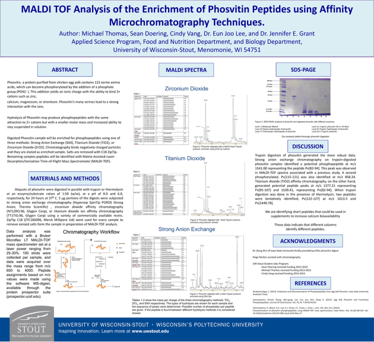
MALDI TOF Analysis of the Enrichment of Phosvitin Peptides using Affinity Microchromatography Techniques. Author: Michael Thomas, Sean Doering, Cindy Vang, Dr. Eun Joo Lee, and Dr. Jennifer E. Grant Applied Science Program, Food and Nutrition Department, and Biology Department, University of Wisconsin-Stout, Menomonie, WI 54751 ABSTRACT Phosvitin, a protein purified from chicken egg yolk contains 123 serine amino acids, which can become phosphorylated by the addition of a phosphate group (PO42- ). This addition yields an ionic charge with the ability to bind 2+ cations such as zinc, SDS-PAGE MALDI SPECTRA 200 kDa 116 kDa 97.4 kDa Zirconium Dioxide 66 kDa Table 1 45 kDa calcium, magnesium, or strontium. Phosvitin’s many serines lead to a strong interaction with the ions. 31 kDa 21.5 kDa 14.4 kDa 1 Hydrolysis of Phosvitin may produce phosphopeptides with the same attraction to 2+ cations but with a smaller molar mass and increased ability to stay suspended in solution. 3 4 5 6 7 8 9 Figure 5. SDS-PAGE analysis of phosvitin and digested phosvitin with different enzymes. Lane 1) Molecular Marker Lane 5) Pepsin Hydrolysate of phosvitin Lane 7) Thermolysin Hydrolysate of phosvitin Lane 3) 1mg/mL phosvitin (34 or 45 kDa) Lane 6) Trypsin Hydrolysate of phosvitin Lane 9) 0.1mg/mL phosvitin. Our protocol yields thorough phosvitin digestion Digested Phosvitin sample will be enriched for phosphopeptides using one of three methods: Strong Anion Exchange (SAX), Titanium Dioxide (TiO2), or Zirconium Dioxide (ZrO2). Chromatography binds negatively charged particles and they are eluted as enriched sample. Salts are removed with C18 ZipTip. Remaining samples peptides will be identified with Matrix-Assisted-LaserDesorption/Ionization Time-of-Flight Mass Spectrometer (MALDI-TOF). Figure 2: Phosvitin digested with a NaOH Heat Trypsin protocol, enriched using ZrO2 tip method Titanium Dioxide Table 2 MATERIALS AND METHODS (a) Aliquots of phosvitin were digested in parallel with trypsin or thermolysin at an enzyme/substrate ration of 1:50 (w/w), at a pH of 8.0 and 6.8, respectively, for 24 hours at 37⁰ C. 5 µg portions of the digests were subjected to strong anion exchange chromatography (Hypersep SpinTip POROS Strong Anion, Thermo Scientific) , zirconium dioxide affinity chromatography (NT1ZRO.96, Glygen Corp), or titanium dioxide ion affinity chromatography (TT1TIO.96, Glygen Corp) using a variety of commercially available resins. ZipTip C18 (ZTC18S096, Merck Millipore Ltd) were used for every sample to remove ionized salts form the sample in preperation of MALDI-TOF analysis. Data analysis was performed with a Bruker Microflex LT MALDI-TOF mass spectrometer set at a laser power ranging from 25-30%. 100 shots were collected per sample, and data were acquired over the mass range from m/z 600 to 4000. Peptide assignments based on m/z values were made using the software MS-digest, available through the protein prospector suite (prospector.ucsf.edu). 2 (b) (c) DISCUSSION Trypsin digestion of phosvitin generated the more robust data. Strong anion exchange chromatography on trypsin-digested phosvitin samples identified a potential phosphopeptide at m/z 1541.00 representing the peptide Pv[82-94]. This peak was observed in MALDI-TOF spectra associated with a previous study. A second phosphorylated, Pv[115-121] was also identified at m/z 858.24. Titanium dioxide (TiO2) affinity chromatography, on the other hand, generated potential peptide peaks at m/z 1377.21 representing Pv[95-107] and 1539.41, representing Pv[82-94]. When trypsin digestion was done in the presence of thermolysin, two peptides were tentatively identified; Pv[122-127] at m/z 1013.5 and Pv[1449.78]. We are identifying short peptides that could be used in supplements to increase calcium bioavailability Figure 3: Phosvitin digested with Heat Trypsin protocol, enriched using titanium dioxide Strong Anion Exchange These data indicate that different columns identify different peptides. Table 3 ACKNOWLEDGMENTS Dr. Dong Ahn of Iowa State University Kindly provided purifies phosvitin digest. Paige Mullen assisted with chromatography UW-Stout Student Jobs Program: -Sean Doering received funding 2012-2015 -Michael Thomas received funding 2013-2015 -Cindy Vang received funding 2014-2015 REFERENCES Figure 4: Phosvitin digested with a Heat Trypsin protocol, enriched using SAX tips Tables 1-3 show the mass per charge of the three chromatography methods: TiO2, ZrO2, and SAX respectively. The types of hydrolysis are shown for each sample and the sequence of peaks were determined. Possible number of phosphates per peptide are given. If the peptide is found between different hydrolysis methods it is considered shared. Mudiyanselage, C. (2012). Production and Characterization of Phosphopeptides from Egg Yolk Phosvitin. Iowa State University Graduate Thesis Samaraweera, Himali; Zhang, Wan-gang, Lee, Eun Joo; Ahn, Dong U; (2011). Egg Yolk Phosvitin and Functional Phosphopeptides. Journal of Food Science. Vol. 76, Nr. 7 (R143-R150) Samaraweera, H., Moon, S.H., Lee, E.J., Grant, J.E., Fouks, J., Choi, I., Suh, J.W., Ahn, D.U. (2014) Characterisation of phosvitin phosphopeptides using MALDI-TOF mass spectrometry. Food Chem. Dec 15;165:98-103. doi: 10.1016/j.foodchem.2014.05.098. Epub 2014 May 27.

