Absorption of X-rays
TEP
5.4.11
-01
Related topics
Bremsstrahlung, characteristic X-radiation, Bragg scattering, law of absorption, mass absorption coefficient, absorption edges, half-value thickness, photoelectric effect, Compton effect, and pair production
Principle
The polychromatic X-radiation that is emitted by an X-ray tube is filtered in terms of its energy with the
aid of a monocrystal. The resulting monochromatic radiation is used as the primary radiation source for
examining the absorption behaviour of various metal foils of different thicknesses.
Equipment
1
1
1
1
1
1
1
1
1
XR 4.0 X-ray expert unit
X-ray goniometer
X-ray plug-in unit with a Cu X-ray tube
Counter tube, type B
X-ray lithium fluoride crystal, mounted in a holder
X-ray diaphragm tube, 2 mm
X-ray absorption set for X-rays
measure XRm 4.0 X-ray software
Data cable USB, plug type A/B
09057-99
09057-10
09057-50
09005-00
09056-05
09057-02
09056-02
14414-61
14608-00
Additional equipment
PC, Windows® XP or higher
This experiment is included in the “XRP 4.0 X-ray solid state” and “XRC 4.0 X-ray characteristics” upgrade sets. Optionally, a tungsten tube (09057-80) can be used for the experiment.
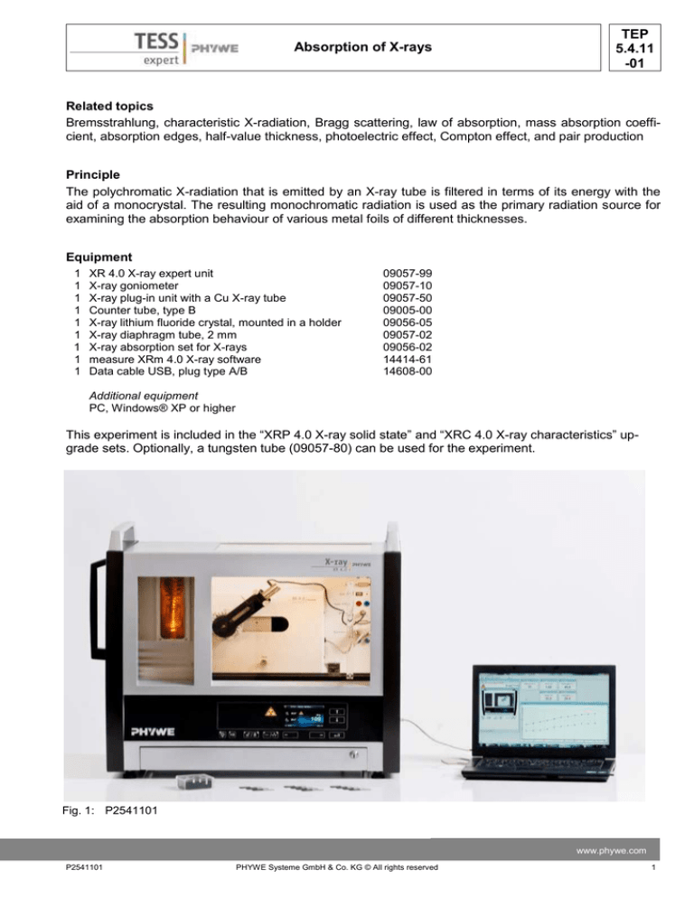
Fig. 1: P2541101
www.phywe.com
P2541101
PHYWE Systeme GmbH & Co. KG © All rights reserved
1
TEP
5.4.11
-01
Absorption of X-rays
Tasks
1. Determine the attenuation of the X-radiation by aluminium and zinc foils of different thicknesses and
at two different wavelengths of the primary radiation.
2. Determine the mass absorption coefficient μ/ρ for aluminium, zinc, and tin absorbers of constant
thickness as a function of the wavelength of the primary radiation. Prove the validity of μ/ρ = f(λ3) in a
graphical manner.
3. Determine the absorption coefficients μ for copper and nickel
as a function of the wavelength of the primary radiation. Determine the energy values of the corresponding K shells based
on the graphical representation. Prove the validity of μ/ρ = f(λ3).
Set-up
Connect the goniometer and the Geiger-Müller counter tube to
their respective sockets in the experiment chamber (see the red
markings in Fig. 2). The goniometer block with the analyser crystal
should be located at the end position on the right-hand side. Fasten the Geiger-Müller counter tube with its holder to the back stop
of the guide rails. Do not forget to install the diaphragm in front of
the counter tube (see Fig. 3). Insert a diaphragm tube with a diameter of 2 mm into the beam outlet of the tube plug-in unit for the
collimation of the X-ray beam.
Note
Details concerning the operation of the X-ray unit and goniometer
as well as information on how to handle the monocrystals can be
Fig. 2: Connectors in the experiment
found in the respective operating instructions.
chamber
GM-counter
tube
Goniometer at
the end position
Diaphragm tube
Counter tube
diaphragm
Mounted
crystal
Fig. 3: Set-up of the goniometer
2
PHYWE Systeme GmbH & Co. KG © All rights reserved
P2541101
TEP
5.4.11
-01
Absorption of X-rays
Procedure
- Connect the X-ray unit via USB cable to the
USB port of your computer (the correct port of
the X-ray unit is marked in Fig. 4).
- Start the “measure” program. A virtual X-ray unit
will be displayed on the screen.
- You can control the X-ray unit by clicking the
Fig. 4: Connection of the computer
various features on and under the virtual X-ray
unit. Alternatively, you can also change the parameters at the real X-ray unit. The program will
automatically adopt the settings.
- Click the experiment chamber (see the red
marking in Fig. 5) to change the parameters for
the experiment.
- If you click the X-ray tube (see the red marking
in Fig. 5), you can change the voltage and current of the X-ray tube (see Fig. 6).
Start the measurement by clicking the red cir-
-
cle:
After the measurement, the following window
appears:
For setting the
X-ray tube
For setting the
goniometer
Fig. 5: Part of the user interface of the software
-
-
Select the first item and confirm by clicking OK.
The measured values will now be transferred directly to the “measure” software.
At the end of this manual a short introduction to
the evaluation of the resulting spectra is given.
Note
- Never expose the Geiger-Müller counter tube to
the primary X-radiation for an extended period
of time.
Fig 6: Voltage and current settings
www.phywe.com
P2541101
PHYWE Systeme GmbH & Co. KG © All rights reserved
3
TEP
5.4.11
-01
Absorption of X-rays
Task 1: Absorption of X-rays as a function of the
Overview of the settings of the goniometer and
material thickness.
X-ray unit:
The absorption set includes aluminium and tin foils
Task 1
of various different thicknesses. They are fastened
Anode voltage UA = 35 kV; anode current
to the Geiger-Müller counter tube by pushing them
IA = 1 mA
into the diaphragm that is installed in front of the
Task 2
counter tube. Manually select two different glancing
2:1 coupling mode
angles for which the intensity is determined first
Integration time 50 s (gate timer); angle step
without an absorber (I0) and then with an absorber
width: 1-2°
(I). In the case of copper, suitable angular positions
Scanning range: 6°-16°
are, for example, 20.4° (Kβ line) (or 21.5° in the
Anode voltage UA = 35 kV; anode current
case of tungsten, α1/2 line) and approximately 10°
IA = 1 mA
(in the range of the bremsstrahlung). Then, note
Task 3
down the corresponding pulse rates without an absorber and with the zinc and aluminium absorber of
2:1 coupling mode
the “absorption set for X-rays”. In order to vary the
Integration time 50 s (gate timer); angle step
thickness of the absorbers, it is also possible to
width: 1°
use two foils at the same time.
Scanning range: 6°-25°
In order to keep the relative errors of the measAnode voltage UA = 20-35 kV; anode current
urement values as small as possible, the measIA = 1 mA
urement should be performed up to an intensity of
I ≥ 1000 pulses-1. In general, this requires measurement times of at least 50 s (integration time, gate timer).
The recording of the X-ray spectrum of the copper
anode is described in greater detail in P2540101.
Task 2: Determination of the mass absorption coefficient for a constant material thickness and as a
function of the wavelength of the X-radiation.
For this task, you will need the aluminium foil
d = 0.08 and the tin foil d = 0.025 mm. Now, while
using only one of the foils at a time, record a spectrum at an interval of 6°< ϑ <16° and in steps of Δϑ
= 1°- 2°. The measuring time should be 50 s minimum (integration time, gate timer). Goniometer settings: see Figure 7. In order to determine I0, record
a third spectrum without any absorber at all. Following the conversion of the glancing angle into the
associated wavelength λ, you will obtain the abFig. 7: Goniometer settings for task 2
sorption as a function of λ.
Task 3: Determination of the absorption coefficient μ for copper and nickel as a function of the wavelength of the primary radiation.
Proceed as described for task 2 with the copper and nickel foils with a diameter of
d = 0.025 mm. Record the spectra at an interval of 6° < ϑ < 25° and in steps of Δϑ = 1°. The measuring
time should be 50 s minimum (integration time, gate timer). In the area of the absorption edges, it is also
possible to work with smaller angle step widths in order to better reproduce the behaviour. This part of
the experiment can be performed with an anode voltage of 35 kV. An anode voltage of 20 to 25 kV,
however, provides better results. On the other hand, the integration time must be increased considerably
in this case.
4
PHYWE Systeme GmbH & Co. KG © All rights reserved
P2541101
TEP
5.4.11
-01
Absorption of X-rays
Theory and evaluation
If X-rays with intensity I0 penetrate matter of the layer thickness d, then the intensity I of the radiation
that passes through the matter is:
I I 0 e , z d
(2)
The absorption coefficient μ [cm-1] is dependent on the wavelength λ (energy) of the X-radiation and on
the atomic number Z of the absorber. This relationship enables the direct determination of the absorption
coefficient:
𝐼
ln 𝐼
− 0= 𝜇
𝑑
In order to be able to directly compare the absorption behaviour of various materials, it is advantageous
to use the so-called half-value thickness d1/2. Absorbers of this thickness reduce the intensity of the primary radiation by half.
d1 / 2 0,69
1
(2)
Since the absorption is proportional to the mass of the absorber, the mass absorption coefficient μ/ρ
(density ρ [gcm-3] is often used instead of the linear absorption coefficient μ.
The following processes are responsible for the absorption:
1. photoelectric effect
2. scattering (Compton effect)
3. pair production
Pair production, however, requires a certain threshold energy that corresponds to twice the amount of
the electron rest energy (2E0 = 2m0c2 = 1.02 MeV). As a result, the absorption coefficient only comprises
two components:
photoelectriceffekt scattering
(3)
In addition, the following applies to the available energy range of the radiation: τ > σ
The dependence of the mass absorption coefficient on the primary radiation energy and on the atomic
number Z of the absorber is described with sufficient precision by the following (empirical) relationship:
k 3 Z 3
(4)
The numerical value of the constant k in equation (4) only applies to the wavelength range λ < λK,
whereby λK is the wavelength corresponding to the energy of the K level. For the range λK < λ, another k
value applies.
In accordance with (4), the absorption increases drastically with the wavelength as well as the atomic
number of the absorbing element.
The X-ray tube emits polychromatic radiation. A monocrystal is used to transform this radiation into
monochromatic primary radiation for the absorption experiments. When X-rays impinge on the lattice
planes of the monocrystal, they will only be reflected if the Bragg condition (5) is fulfilled.
2d sin n ; (n = 1, 2, 3, …)
(5)
ϑ = glancing angle
www.phywe.com
P2541101
PHYWE Systeme GmbH & Co. KG © All rights reserved
5
TEP
5.4.11
-01
Absorption of X-rays
0 Curve 3:
0.05
0.1λ = 139 0.15
Zn (Z = 30);
pm
n = 1, 2, 3,...
d = 201.4 pm; interplanar spacing LiF (200)
0.2
1
In the case of lower intensities, the background radiation must be taken into consideration at UA = 0
kV. At high counting rates, the true pulse rate N
results from the measured rate N* if the dead time τ
of the Geiger-Müller counter tube is taken into consideration.
N
N*
(with τ = 90 μs)
1 N *
0.1
(1)
Task 1: Absorption of X-rays as a function of the
material thickness.
Figure 8 shows the pulse rate ratio I/I0 for different
thicknesses d of the absorbers aluminium and zinc.
Curves 1 and 2 apply to aluminium (Z = 13, ρ = 2.7
g/cm3) and curve 3 to zinc (Z = 30, ρ = 7.14 0.01
g/cm3). When the absorber thickness increases, Fig. 8: Semi-logarithmic representation of the quotient
the intensity that is let through decreases exponenI/I0 as a function of the absorber thickness d
tially (1). It is also apparent that
UA = 35 kV, IA = 1 mA
- for the same primary radiation energy (waveCurve 1: Al (Z = 13); λ = 139 pm
length), the absorption increases when the
Curve 2: Al (Z = 13); λ = 70 pm
atomic number of the absorber increases
(curves 1 and 3).
- for an increasing primary radiation energy, the absorption decreases in the same absorber material
(curves 1 and 2).
The results of Figure 8, which can be obtained from equations (1) and (5), are listed in table 1. For aluminium, the dependence of the absorption on the
wavelength (μ/ρ = f(λ3)) is confirmed in an exemTable 1: Dependence of the absorption on the
plary manner:
wavelength
μ1/ρ / μ2/ρ = 7.98; (λ1/λ2)3 = 7.83
6
μ / cm-1
d1/2 / cm
μ/ρ / cm2g-1
Al (Z = 13)
ρ = 2.7 g/cm-3
λ = 139 pm
λ = 70 pm
112
14.1
6.2∙10-3
20.4
41.5
5.2
Zn (Z = 30)
ρ = 7.14 g/cm-3
λ = 139 pm
280
2.5∙10-3
39.2
The Z dependence of the mass absorption coefficient in accordance with (4) cannot be determined here, since the primary radiation energy
lies within the K level of zinc. Equation (4) is only
valid outside of the absorption edges.
PHYWE Systeme GmbH & Co. KG © All rights reserved
P2541101
TEP
5.4.11
-01
Absorption of X-rays
Task 2: Determination of the mass absorption coefficient for a constant material thickness and as a function of the wavelength of the X-radiation.
Following the conversion of the glancing angle to the associated wavelength λ in accordance with (5),
you will obtain the absorption as a function of λ. Formula (1) can now be used to determine μ and – as a
3
next step – the mass absorption coefficient 𝜇⁄𝜌. If one plots √𝜇⁄𝜌 as a function of the wavelength in pm,
straight lines, like the ones shown in Figure 9, will result. These straight lines represent the correlation
μ/ρ = f(λ3).
Figure 9 shows the course of μ/ρ = f(λ3) for aluminium and tin (Z = 50, ρ = 7.28 gcm-3).
Task 3: Determination of the absorption coefficient μ for copper and nickel as a function of the wavelength of the primary radiation.
Figures 10 and 11 show the absorption behaviour of copper (Z = 29, ρ = 8.96 gcm-3) and nickel (Z = 28,
ρ = 8.99 gcm-3). In both cases, the correlation μ/ρ = f(λ3) is shown in the areas where λ ≠ λK. However, at
the so-called absorption edges where λ = λK, the absorption changes abruptly since now the associated
primary radiation energy can ionise the relevant atoms on the K shell.
Both curves deviate from the linearity of the absorption edge at λ < 70 pm. This is due to an increase in
intensity of the primary radiation because of second-order interferences. The short-wave onset of the
bremsspectrum is given by the acceleration voltage UA (see P2540901). If it is 35 kV, for example, the
following applies to the shortest wavelength in accordance with (7): λc = 35.4 pm. In accordance with the
Bragg condition, radiation of this wavelength is reflected under the angle ϑ = 10.1° with n = 2. Under this
angle, however, the wavelength of the first-order radiation portion (n = 1) is λ = 70.8 pm so that – as of a
glancing angle of ϑ > 10.1° (ϑ > 70.8 pm) – the primary radiation that impinges on the absorber comprises a radiation portion with shorter wavelengths.
As a consequence, the absorber appears to be
3 /
more “transparent” than it really is at this wavein g-1/3 cm2/3
length. Nevertheless, the absorption edge can
3
be determined with a sufficient level of accuraSn
cy.
2.5
Equation (7) can be used to determine the energy levels of the K levels.
2
hc
EK
e K
1.5
where
Planck’s constant
h =6.6256∙10-34 Js
Velocity of light
c =2.9979∙108 m/s
Elementary charge e =1.6021∙10-19 C
Al
(7)
1
0.5
0
λ in pm
40
60
80
100
With λK = 138 pm of Figure 10, the following
results for copper: EK = 8.98 keV (literature value 8.98 keV).
3 / for aluminium and tin as a function of the
Fig. 9:
Correspondingly, with λK = 149 pm of Figure
primary radiation energy; UA = 25 kV.
11, the following results for nickel: EK = 8.32
keV (literature value 8.33 keV).
www.phywe.com
P2541101
PHYWE Systeme GmbH & Co. KG © All rights reserved
7
TEP
5.4.11
-01
Absorption of X-rays
Since Z(Ni) < Z(Cu), EK(Ni) < EK(Cu) and correspondingly also λK(Ni) > λK(Cu).
3
/
3
in g-1/3 cm2/3
/
in g-1/3 cm2/3
λ in pm
λ in pm
Fig. 10: Absorption edge of copper; UA = 25 kV;
λK = 138 pm
Fig. 11: Absorption edge of nickel; UA = 25 kV;
λK = 149 pm
Note
Nickel filters are used to monochromatise the radiation of copper X-ray tubes.
In this case, only the intensity of the characteristic Kα radiation is slightly reduced (EKα= EK-EL2,3 = (8.98 –
0.95) keV = 8.03 keV), while the Kβ radiation is reduced very strongly due to the edge absorption of
nickel (EKβ= EK-EM2,3 = (8.98 – 0.074) keV = 8.9 keV) (see also P2541201).
8
PHYWE Systeme GmbH & Co. KG © All rights reserved
P2541101


