Unit 8: Ch. 8: Digestive System p. 242 Function of the Digestive
Unit 8: Ch. 8: Digestive System p. 242
Function of the Digestive System
1.
Mechanical and chemical digestion of food
2.
Absorption of nutrient molecules
3.
Elimination of solid wastes
Organs of the Gastrointestinal tract
Oral cavity
Pharynx
Esophagus
Stomach
Small intestine
Colon
Accessory Organs
Pancreas
Liver
Gallbladder
Salivary glands
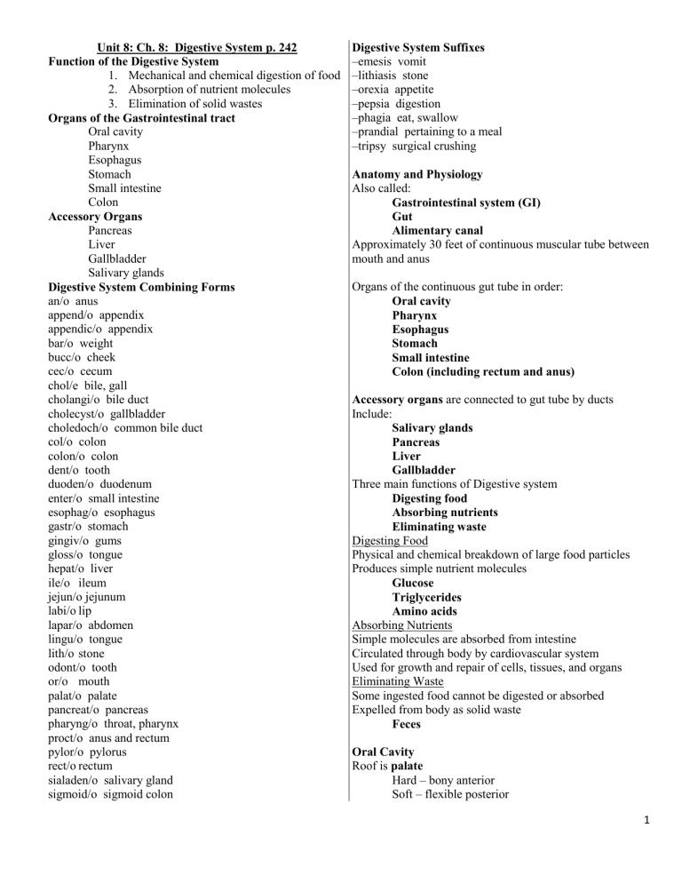
Digestive System Combining Forms an/o anus append/o appendix appendic/o appendix bar/o weight bucc/o cheek cec/o cecum chol/e bile, gall cholangi/o bile duct cholecyst/o gallbladder choledoch/o common bile duct col/o colon colon/o colon dent/o tooth duoden/o duodenum enter/o small intestine esophag/o esophagus gastr/o stomach gingiv/o gums gloss/o tongue hepat/o liver ile/o ileum jejun/o jejunum labi/o lip lapar/o abdomen lingu/o tongue lith/o stone odont/o tooth or/o mouth palat/o palate pancreat/o pancreas pharyng/o throat, pharynx proct/o anus and rectum pylor/o pylorus rect/o rectum sialaden/o salivary gland sigmoid/o sigmoid colon
Digestive System Suffixes
–emesis vomit
–lithiasis stone
–orexia appetite
–pepsia digestion
–phagia eat, swallow
–prandial pertaining to a meal
–tripsy surgical crushing
Anatomy and Physiology
Also called:
Gastrointestinal system (GI)
Gut
Alimentary canal
Approximately 30 feet of continuous muscular tube between mouth and anus
Organs of the continuous gut tube in order:
Oral cavity
Pharynx
Esophagus
Stomach
Small intestine
Colon (including rectum and anus)
Accessory organs are connected to gut tube by ducts
Include:
Salivary glands
Pancreas
Liver
Gallbladder
Three main functions of Digestive system
Digesting food
Absorbing nutrients
Eliminating waste
Digesting Food
Physical and chemical breakdown of large food particles
Produces simple nutrient molecules
Glucose
Triglycerides
Amino acids
Absorbing Nutrients
Simple molecules are absorbed from intestine
Circulated through body by cardiovascular system
Used for growth and repair of cells, tissues, and organs
Eliminating Waste
Some ingested food cannot be digested or absorbed
Expelled from body as solid waste
Feces
Oral Cavity
Roof is palate
Hard – bony anterior
Soft – flexible posterior
1
Hanging down from soft palate is uvula
Speech production
Location of gag reflex
Cheeks are lateral walls
Lips are anterior opening
Entire cavity lined with mucous membrane
Figure 8.1 – Anatomy of the oral cavity.
Digestion begins when food enters mouth
Mechanically broken up by chewing
Tongue moves food within mouth
Mixes with saliva
Digestive enzymes
Lubricates
Taste buds on tongue surface
Detect bitter, sweet, salty, sour flavors
Teeth
Cutting teeth
Bite
Tear
Cut
Incisors
Cuspids (canines)
Teeth
Grinding teeth
Bicuspids (premolars)
Molars
Third molar is wisdom tooth
Tooth Structure
Gums
Mucous membrane + connective tissue
Seals off teeth in socket
Tooth is divided into:
Crown
– above gum
Root
– below gum
Enamel
Outer covering
In crown only
Hardest substance
Dentin
Under enamel
In crown and root
Bulk of tooth
Pulp cavity
In crown and root canal
Blood vessels, nerves
Cementum and periodontal ligaments
Anchors root in jawbone
Humans Have 2 Sets of Teeth
Deciduous teeth
First set, baby teeth
20 teeth erupt between ages 6 and 28 months
Permanent teeth
Second set, adult teeth
About 6 years of age, baby teeth fall out
Replaced by 32 permanent teeth
Process continues until 18-20 years of age
Pharynx
Swallowed food enters oropharynx
Proceeds down pharynx into laryngopharynx
Epiglottis
Covers larynx and trachea
Shunts food away from lungs & into esophagus
Figure 8.2 – Structures of the oral cavity, pharynx, and esophagus.
Esophagus
10-inch long muscular tube
Food enters from pharynx
Delivered to stomach
Propelled along by wavelike muscular movements
Called peristalsis
Pushes food through entire gut tube
The Stomach
J-shaped muscular organ
Collects & churns food
Mixes it with hydrochloric acid (HCl)
Forms chyme
Watery mix of food and digestive juices
Three regions
Fundus
– upper
Body – main
Antrum – lower
Rugae are folds in stomach lining
Stretch out to allow stomach to expand with food
Sphincters
Muscular valves
Control flow of food
Lower esophageal (cardiac) sphincter
Keeps food from backing up into esophagus
Pyloric sphincter
Allows highly acidic chyme to enter small intestine
Small Intestine
Longest portion of alimentary canal
Averages 20 feet
Between pyloric sphincter and colon
Site of:
Completion of digestion
Majority of absorption
Three Sections of Small Intestine
Duodenum
First section – about 10-12 inches long
2
Starts at pyloric sphincter
Jejunum
Second section – about 8 feet long
Ileum
Third section – about 12 feet long
Connects to colon at ileocecal valve
Colon
5 feet long
Extends from ileocecal valve to anus
Fluid that remains after digestion and absorption enters colon
Most is water and is reabsorbed into body
Solid waste left over is feces
Evacuated in bowel movements
Regions of the Colon
Cecum
Appendix
Ascending colon
Transverse colon
Descending colon
Sigmoid colon
Rectum and Anus
Rectum is area for storage of feces
Leads to anus
External opening of alimentary canal
Feces are evacuated
Called defecation
Accessory Organs
Generally function by producing substances necessary for chemical breakdown of food
Salivary glands
Liver
Gallbladder
Pancreas
Salivary Glands
Produce saliva
Allows food to be swallowed without choking
Saliva + food = bolus
Contains amylase
Begins digestion of carbohydrates
Three pairs
Parotid glands
Sublingual glands
Submandibular glands
Liver
Located in right upper quadrant of abdomen
Processes nutrients
Detoxifies harmful substances
Produces bile
Emulsification
Breaks up large fat globules into smaller droplets
Gallbladder
Lies under liver
Stores bile produced by liver
Hepatic duct
Cystic duct
Common bile duct carries bile to duodenum
Pancreas
Digestive juices include:
Buffers – neutralize acidic chyme
Enzymes – digest carbohydrates, lipids, and proteins
Word Building
Digestive System Vocabulary
Pathology
Clinical Laboratory Tests
Diagnostic Imaging
Procedures
Digestive System Pharmacology
Digestive System Abbreviations
3
