Bio1-RBCs met
advertisement
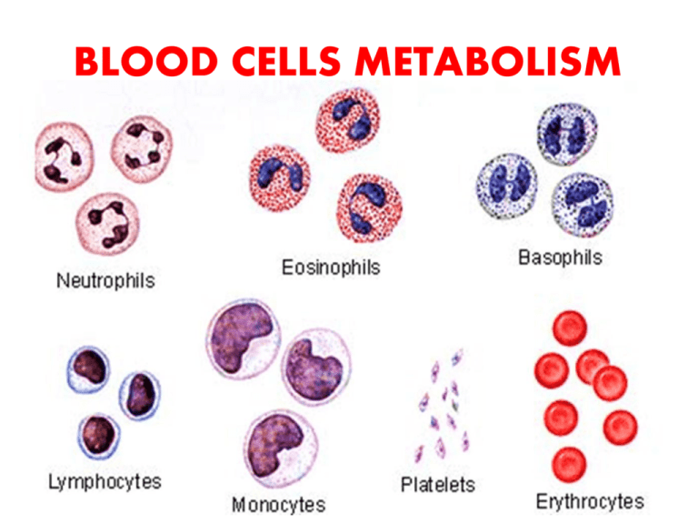
BLOOD CELLS METABOLISM Objectives of the Lecture 1- Understanding the general structural & functional features of red blood cells (RBCs). 2- Recognizing the main metabolic pathways occurring in RBCs with reference to their relations to functions of RBCs. 3- Identifying some of the main & common diseases of RBCs as implication of defects of RBCs metabolism. 4- Understanding the relation of characteristic features of structure of membrane of RBCs. 5-Recognizing the main functions of other blood cells and their metabolism RBCs Metabolism & Functions Introduction: • RBCs contain no mitochondria, so there is no respiratory chain, no citric acid cycle, and no oxidation of fatty acids or ketone bodies. • Energy in the form of ATP is obtained ONLY from the glycolytic breakdown of glucose with the production of lactate (anaerobic glycolysis). • ATP produced being used for keeping the biconcave shape of RBCs & in the regulation of transport of ions & water in and out of RBCs. Red Blood Cells (erythrocytes) 1. Function erythrocyte as a bag for hemoglobin 2. O2 → transport, reactive oxygen species (ROS) CO2 → transport, formation of HCO3- H+ → transport, maintaining pH (35% of blood buffering capacity) Structure large surface cytoskeletal proteins membrane as an osmometer (for diffusion of gases) (for elasticity) (Na+/K+-ATPase) RBCs membrane structure • RBCs must be able to squeeze through tight spots in microcirculation (capillaries). For that RBCs must be easily & reversibly deformable. Its membrane must be both fluid & flexible . • About 50% of membrane is protein, 40% is fat & up to 10% is carbohydrate. • RBCs membrane comprises a lipid bilayer (which determine the membrane fluidity), proteins (which is responsible for flexibility) that are either peripheral or integral penetrating the lipid bilayer & carbohydrates that occur only on the external surface. Defects of proteins may explain some of the abnormalities of shape of RBCs membrane as hereditary spherocytosis & elliptocytosis. • The membrane skeleton is four structural proteins that include & spectrin, ankyrin, protein 4.1 & actin • • • • • Spectrin is major protein of the cytoskeleton & its two chains ( & ) are aligned in an antiparallel manner . & chains are loosely interconnected forming a dimer, one dimer interact with another, forming a head to head tetramer. Ankyrin binds spectrin & in turn binds tightly to band 3 securing attachment of spectrin to membrane. band 3 is anion exchange protein permits exchanges of Cl- for HCO3+. Actin binds to the tail of spectrin & to protein 4.1 which in turn binds to integral proteins, glycophorins A, B & C. Glycophorins A,B,C are transmembrane glycoproteins What happens to red blood cells when placed in hypotonic, hypertonic, and isotonic solutions? • osmolarity (0.9%NaCl) • acanthocytes • hemolysis Red Blood Cells (erythrocytes) 3. membrane transporters Na+/K+-ATPase (active transport) GLUT-1 (insulin independent) anion exchanger = band 3 protein (Cl-/HCO3-) 4. membrane antigens blood groups: ABO system Differ in antigen (glycoprotein) Over the surface of RBCs Red Blood Cells (erythrocytes) 5. metabolism glucose is the main fuel 90% anaerobic glycolysis (ATP, lactate: Cori cycle; 2,3-BPG) 10% hexose monophosphate pathway (NADPH) enzyme defects : * glucose-6-P dehydrogenase * pyruvate kinase → hemolytic anemia ??? ATP is generated by anaerobic glycolysis → ATP is used for ion transport across the cell membrane glycolysis produces 2,3-BPG and lactate approx. 5 to 10% of Glc is metabolized by hexose monophosphate pathway → production of NADPH → it is used to maintain glutathione in the reduced state Red Blood Cells (erythrocytes) 6. Enzymes carbonate dehydratase (= carbonic anhydrase, CA) CO2 + H2O HCO3- + H+ The red cell also contain rhodanase responsible for the detoxication of cyanides. methemoglobin reductase superoxide dismutase catalase glutathione peroxidase glutathione reductase antioxidative enzyme system Red Blood Cells (erythrocytes) 6. Erythropioesis White Blood Cells (leukocytes) Classification • granulocytes neutrophils (phagocytosis) eosinophils (allergy, parasites) basophils (allergy) • agranulocytes monocytes → macrophages lymphocytes (B, T) → immunity Reactive oxygen ROS and nitrogen RNS species in blood elements ERYTHROCYTES: enzymes for deactivation of ROS formed from high content of oxygen found in the cells PHAGOCYTES:enzymes for production of ROS and RNS to destroy particles in phagosomes White Blood Cells (leukocytes) Neutrophils (microphages) high content of lysosomes (hydrolytic enzymes) few mitochondria glucose dependent: NADPH production NADPH is used for production of reactive oxygen species → they kill bacteria Basofils contain heparin and histamine B-lymphocytes produce antibodies (= immunoglobulins, -globulins) Platelets(thrombocytes) participate in hemostasis Platelets(thrombocytes)
