File - Ms. Drozd - Human Body systems
advertisement

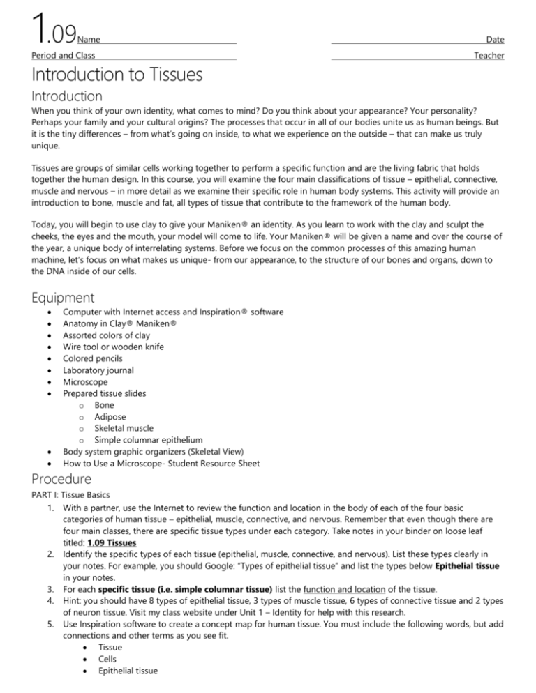
1.09 Name Period and Class Introduction to Tissues Date Teacher Introduction When you think of your own identity, what comes to mind? Do you think about your appearance? Your personality? Perhaps your family and your cultural origins? The processes that occur in all of our bodies unite us as human beings. But it is the tiny differences – from what’s going on inside, to what we experience on the outside – that can make us truly unique. Tissues are groups of similar cells working together to perform a specific function and are the living fabric that holds together the human design. In this course, you will examine the four main classifications of tissue – epithelial, connective, muscle and nervous – in more detail as we examine their specific role in human body systems. This activity will provide an introduction to bone, muscle and fat, all types of tissue that contribute to the framework of the human body. Today, you will begin to use clay to give your Maniken® an identity. As you learn to work with the clay and sculpt the cheeks, the eyes and the mouth, your model will come to life. Your Maniken® will be given a name and over the course of the year, a unique body of interrelating systems. Before we focus on the common processes of this amazing human machine, let’s focus on what makes us unique- from our appearance, to the structure of our bones and organs, down to the DNA inside of our cells. Equipment Computer with Internet access and Inspiration® software Anatomy in Clay® Maniken® Assorted colors of clay Wire tool or wooden knife Colored pencils Laboratory journal Microscope Prepared tissue slides o Bone o Adipose o Skeletal muscle o Simple columnar epithelium Body system graphic organizers (Skeletal View) How to Use a Microscope- Student Resource Sheet Procedure PART I: Tissue Basics 1. With a partner, use the Internet to review the function and location in the body of each of the four basic categories of human tissue – epithelial, muscle, connective, and nervous. Remember that even though there are four main classes, there are specific tissue types under each category. Take notes in your binder on loose leaf titled: 1.09 Tissues 2. Identify the specific types of each tissue (epithelial, muscle, connective, and nervous). List these types clearly in your notes. For example, you should Google: “Types of epithelial tissue” and list the types below Epithelial tissue in your notes. 3. For each specific tissue (i.e. simple columnar tissue) list the function and location of the tissue. 4. Hint: you should have 8 types of epithelial tissue, 3 types of muscle tissue, 6 types of connective tissue and 2 types of neuron tissue. Visit my class website under Unit 1 – Identity for help with this research. 5. Use Inspiration software to create a concept map for human tissue. You must include the following words, but add connections and other terms as you see fit. Tissue Cells Epithelial tissue 1.09 Name Date Period and Class Teacher Connective tissue Muscle tissue Nervous tissue Neurons Cartilage Blood Tendons Ligaments 6. Add images or photographs to your concept map to help reinforce your words. 7. For each of the four main categories, assign one word that summarizes the role this type of tissue plays in the body. Using a different color, write this word in the box/bubble for that particular tissue type. 8. In this activity, focus on three specific types of tissue- bone, skeletal muscle and fat (adipose). Find a logical place for these three words on your concept map and add them to your organizer. 9. Search the web for slides of simple columnar epithelium, bone, adipose and skeletal muscle. Sketch in your notebook what these look like under the microscope. Label each picture in your notes. 10. Answer conclusion questions 1 and 2. PART II: Building Identity- Giving Your Maniken® A Face Throughout the course, you will be asked to mark specific bones and structures on your Maniken®. For each bone, you will to identify and find the structure on your model. Use a pencil to number the bone (starting with #1). On a blank body system graphic organizer (skeletal view), assign the same number to the bone on the diagram and write a key at the bottom of the page (or next to the actual bone). Title your diagram “Skeletal System.” 11. Use the skull anatomy tutorial presented by GateWay Community College at http://www.gwc.maricopa.edu/class/bio201/skull/skulltt.htm to identify the following bones of the skull. Mark these bones on your Maniken® and on the skeletal system organizer. o Mandible o Maxilla o Zygomatic Process o Frontal Bone o Temporal Bone o Occipital Bone o Parietal Bone 12. Use the tutorial of the head and neck muscles presented by GateWay Community College at http://www.gwc.maricopa.edu/class/bio201/head/head1.htm and other Internet sources to find the location of the following muscles. o Orbicularis Oculi o Orbicularis Oris o Temporalis 13. In your laboratory journal, take notes on the basic function or action of each muscle. 14. Obtain another copy of the body system graphic organizer (skeletal view). 15. Sketch the muscles you researched in Step 12 in pencil on the organizer. Label the top of the diagram “Muscular System.” Label each muscle you add to the diagram. Make sure to add a brief statement of function. Alternatively, you can create a key on a separate sheet of paper. 16. Using your knowledge of directional terms, tissues, and the bones and muscles of the face, follow your teacher’s instructions to build the face of your Maniken®. You will use bone landmarks to apply muscle and fat to the face. 17. Take a look at what you have created. Is your Maniken® going to be male or female? With your partner, decide on a name for your Maniken®. Remember, he or she is going to be with you the entire year! 18. Answer the remaining conclusion questions. 1.09 Name Period and Class Date Teacher Conclusion 1. What do you notice is the main difference between the structure of the connective tissues and the structure of the epithelium? Make sure to note the organization of cells in these two tissue types. 2. Explain how the structure of epithelium and the structure of connective tissue, specifically bone, relate to the function of the tissue. 3. How does the distribution of tissues contribute to our appearance and to our identity? 4. Describe the role of fat in our cheeks and behind our eyes. 5. Think about the action of the muscles you have built on your Maniken. Describe specific motions that you would not be able to complete if you damaged your temporalis, your orbicularis oculi or your orbicularis oris. How would this affect your ability to communicate? 6. Reflect on your own identity. What do you think helps make you, you?


