Lecture 5: Skeletal Muscle
advertisement
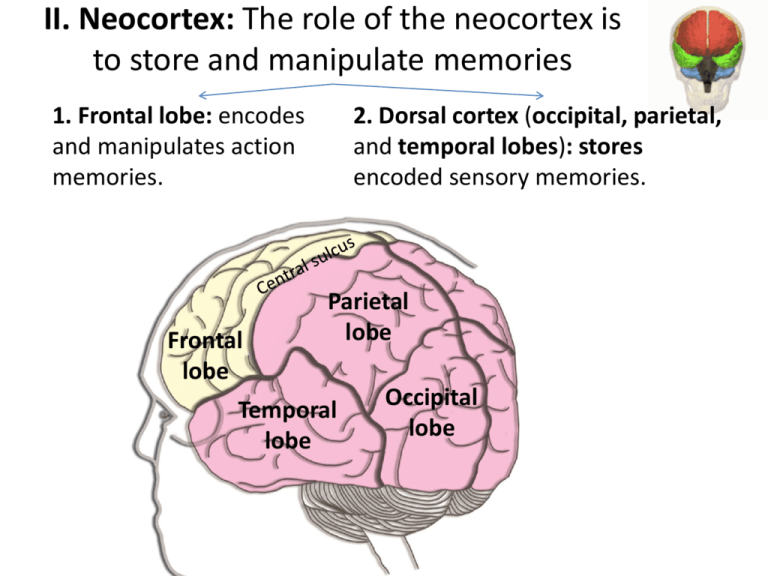
II. Neocortex: The role of the neocortex is to store and manipulate memories 1. Frontal lobe: encodes and manipulates action memories. Frontal lobe 2. Dorsal cortex (occipital, parietal, and temporal lobes): stores encoded sensory memories. Parietal lobe Temporal lobe Occipital lobe 1. Frontal lobe: encodes and manipulates action memories Dorsal Ventral or crossover • A single skeletal muscle cell is called muscle fiber (MF) – electrically continuous inside, – electrically isolated from outside • Each MF is formed during development by the fusion of a number of undifferentiated mononucleated cells, known as myoblasts • Myoblasts fuse into multinucleated cylindrical MF; completed by around the time of birth One = cell • Single MF is 10µm to 300µm in diameter and may extend up to 30cm in length (whole length of biceps muscle) and contain >1,000 nuclei • DEFINITION: muscle is a number of MFs bound together by connective tissue (collagen) • Muscles are connected to bones by tendons (triple helix of protein collagen* - stronger than steel of the same diameter) • Tendons can be very long: some of the muscles that move fingers are located in the forearm**. • Why finger muscles are away from the hand? • What is the relationship between a motor neuron and muscle fibers? A. B. C. D. one motor neuron innervates one muscle fiber several motor neurons innervate one muscle fiber one motor neuron innervates two or three muscle fibers one motor neuron innervates around 100 muscle fibers • Definition: Motor unit = motor neuron and MFs it controls (usually several hundred MFs) Ventral root • Motor unit 1 = motor neuron 1 and blue MFs • Motor unit 2 = motor neuron 2 and red MFs • The muscle fibers of a motor unit are dispersed randomly within the area of the muscle • Muscle fibers innervated by the same motor neuron are not contiguous The number of motor units vary greatly among muscles: • 10 in extraocular muscles • 100 in intrinsic muscles of the hand • 5,000 in a large leg muscle The number of muscle fibers (cells) per muscle: • 1000 MFs in extraocular muscles (1000 MF / 10 motor units 100 MF in one motor unit) • 1,000,000 MF in large leg muscle (1,000,000 MFs / 5,000 motor units 200 MFs in one motor unit) The strength of muscle contraction depends on: 1. The number of motor units recruited 2. The frequency of motor unit discharge 3. The speed of contraction of muscle fibers in the motor unit 4. The nature of motor unit (whether it is fatigue-resistant or fatigue –prone) Contraction duration: • Snail muscle contraction can last for 1 second • Honey bee contraction lasts for 3 millisecond (300 twitches / second) In all skeletal muscles: • Contractile elements: Actin, myosin • ATP provides energy • Increase in calcium concentration triggers contraction Single cell (electrically isolated) Sarcomere 2um 1um • Sarcomere (Greek: sarco- (“flesh”) + -mere (“component”)) • Sarcomere shortens during contraction like a collapsing telescope • Stack many sarcomeres together longitudinally myofibril • A single myofibril has the length of a muscle fiber • Muscle fibers look striated under the light microscope Inside a sarcomere Myosin cross-bridges are pulling on the actin Actin is attached to Z-line discs Z-line disc Actin is attached to Z-line discs Actin= thin filament Myosin is suspended by titin titin Myosin = thick filament • three sarcomeres stuck longitudinally • sarcomere cross section Range of sarcomere contraction Cannot contract any further No tension Range of sarcomere contraction Cannot form crossbridges No tension Actin is found in a any cell actin Fibroblast labeled with probes for actin and the nucleus. The actin of cytoskeleton is visualized Tropomyosin is held in place by Troponin • Myosin tails bind to form the Calciumtogether binding protein thick filament Myosin head 120nm • Myosin head interacts with actin • Hydrolyzes ATP Cross-bridge cycle in 5 frames 1. Actin (A) bound to myosin (M): A· M 2. Binding of ATP dissociates myosin: A· M + ATP A + M· ATP fast reaction 3. ATP hydrolysis to ADP and Pi energizes myosin: A + M· ATP A + M · ADP · Pi fast reaction 4. Cross-bridge binds to actin, now ADP and Pi can be released: A + M · ADP · Pi A · M · ADP · Pi 5. As soon as ADP + Pi are released, the head region is glad to bend back and pulls actin filaments: A · M · ADP · Pi A · M + ADP + Pi 1. Actin (A) bound to myosin (M): A· M Muscle is stiff. Few hour after death no ATP Rigor mortis (Lat. Stiff death) 2. Binding of ATP dissociates myosin: A· M + ATP A + M· ATP Muscle is flexible 3. ATP hydrolysis to ADP and Pi energizes myosin: A + M· ATP A + M · ADP · Pi this myosin head position is unstable. It wants to bend back, but it cannot until it releases ADP and Pi. Without binding to actin, myosin releases ADP and Pi very slowly: half life is 14 seconds Even at rest muscle continues to burn ATPs producing heat 4. Cross-bridge binds to actin 5. As soon as ADP + Pi are released, the head region is glad to bend back and pulls actin Two distinct roles of ATP in crossbridge cycle 1. The energy released by ATP hydrolysis provides the energy for cross-bridge movement. 2. The binding of ATP to myosin allows myosin to dissociate from actin – Several hours after death ATP concentration decreases skeletal muscle become stiff (Rigor mortis) – The stiffness disappears in 48h to 60h after death as a result of disintegration of muscle tissue • Only 50% of cross-bridges are bound at any moment • One stroke of cross-bridge produces only a very small movement (~10nm) • As long as muscle remains ‘on’ cross-bridges repeat their swiveling motion • What keeps muscle from continuous contractile activity? What keeps muscle from continuous contractile activity? Tropomyosin is held by Troponin in resting muscle in the position that does not allow myosin to bind to actin 33 Muscle tension 100nM 1µM Calcium concentration 10µM • Muscle tension is a function calcium concentration +30mV 1. Motor neuron action potential Excitation-contraction coupling -80mV +30mV -90mV EPSP 2. EPSP is amplified Muscle fiber action potential Depolarization produced by influx of Na (through ACh channels) is amplified by muscle’s own voltageactivated channels 4. Calcium concentration inside 5. Muscle tension (one twitch) 3. Muscle fiber action potential is conducted from neuromuscular junction located in the middle of a fiber to both ends of a muscle fiber • Where does calcium come from? Where does calcium come from in a neuron? • From ECF. • Since concentration gradient is huge (1mM outside vs. 0.0001mM inside), it is enough to just open calcium channels calcium will be pouring into the axonal terminal • It works for axon terminals, since axon terminals are small (1µm) • muscle fibers on the other hand are huge (up to 300µm) it would take forever for calcium from ECF to diffuse to central myofibrils Solution surround each myofibril with calcium storage • AP opens calcium channels on SR • After the end of AP, calcium is sucked away by a number of ATP driven pumps: 30,000 pumps/ µm2 How to transmit AP to all s. reticula simultaneously? Solution T-tubules (local plumbing) - electrically continuous with ECF, just like power lines in a house are used to transmit electricity Temporal summation and Tetanus isometric contraction == constant length • Maximum tension that this muscle can produce under this length • In a lab we can manipulate muscle length measure maximum tension developed by the muscle at that length isometric contraction == constant length • Conclusion: there is some optimal length at which the developed tension is maximal • mechanism? - consider myosin and actin arrangement: isometric contraction == constant length • In the body muscles are close to optimal length (a) in the extended state. • Bones prevent muscles from extending beyond this length isometric contraction == • How much muscle will shorten with the weight of 50 gram • With a load of 50gram this muscle will only shorten to E • each sarcomere will shorten by 0.5µm (from 2.1µm to 1.6µm) 50 gram 50 gram (gram) isotonic contraction == constant load E Types of skeletal muscles Two types of tasks: – Some tasks involve continuous muscle activity (tonus, walking) muscles are not allowed to fatigue. Movements involved in these tasks are usually slow. – Other tasks normally last only short time (running away from a predator). The movements involved in these tasks are usually fast. To address these two very different tasks, we have two different types of muscles fibers: – slow MF that are optimized to never fatigue: you can walk for 2 miles (using slow muscles) without getting tired – fast MF that are optimized for speed, but fatigue quickly: if you run two miles (using fast muscles, you will be very tired) Contraction velocity Slow (Type I) twitch waveform Fast (Type IIx) 30ms 300ms Rate of fatigue never quickly Why fast/slow? Myosin ATPase activity is slow (slowly split ATP) Myosin ATPase activity is fast (split ATP fast) Source of ATP production Oxidative phosphorylation (aerobic) Glycolysis (anaerobic) Mitochondria Many Few Myoglobin content High (red muscle) Low (white muscle) Fiber diameter small large Motor unit size small large Capillaries around MF Large number of capillaries Few capillaries Motor unit firing rate Continuously at a low rate High firing rate Athletes Extreme endurance athletes World-class sprinter Tension fine control by slow MF gross control by fast MF time Find the slow-twitch MF, fast? fast MF (big and white) slow MF (small and red) Sensors • Close your eyes move your hand forward, back • How do you know the position of the muscles? • Two types of sensors: • Length sensors inside the muscle: muscle spindle • Tension sensors: Golgi tension organ Golgi tendon organ • How sensory information is encoded by neurons? • Information is encoded in frequency of action potentials (firing frequency = number of action potentials per second) • How motor information is encoded by a motor neuron? • Again, information is encoded in frequency of action potentials: the greater the firing rate the greater the tension • What is the range of firing rate? • Is every motor neuron action potential followed by a muscle contraction? • In addition CNS can recruit more motor units Amyotrophic lateral sclerosis • Also known as Charcot disease, Lou Gehrig's disease in US and motor neuron disease in UK. • Lou Gehrig (1903 – 1941) was a baseball player who played 17 for the New York Yankees, from 1923 through 1939. In 1939 he was diagnosed with ALS and died two years later. • ALS is characterized by gradually worsening muscle weakness. This results in difficulty speaking, swallowing, and eventually breathing. • The defining feature of ALS is the death of both upper and lower motor neurons in the motor cortex of the brain, the brain stem, and the spinal cord. Prior to their destruction, motor neurons develop protein-rich inclusions in their cell bodies and axons. • In 90% of case the cause is unknown. In 10% ALS is due to inherited mutation. • Stop here


