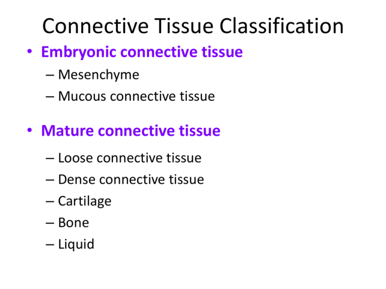
Connective Tissue Classification
• Embryonic connective tissue
– Mesenchyme
– Mucous connective tissue
• Mature connective tissue
– Loose connective tissue
– Dense connective tissue
– Cartilage
– Bone
– Liquid
Embryonic Connective Tissues
• There are 2 Embryonic Connective Tissues:
– Mesenchyme gives rise to all other connective tissues.
– Mucous C.T. (Wharton's Jelly) is a gelatinous substance
within the umbilical cord and is a rich source of stem cells.
Mature Connective Tissues
• Loose Connective Tissues
– Areolar Connective Tissue is the most widely distributed in
the body. It contains several types of cells and all three fiber
types.
• It is used to attach skin and underlying tissues, and as a
packing between glands, muscles, and nerves.
– Adipose
– Reticular
Mature Connective Tissues
• Loose Connective Tissues
– Loose areolar
– Adipose tissue is located in the subcutaneous layer
deep to the skin and around organs and joints.
• It reduces heat loss and serves as padding and as an energy
source.
– Reticular
Mature Connective Tissues
• Loose Connective Tissues
– Loose areolar
– Adipose
– Reticular connective tissue is a network of interlacing
reticular fibers and cells.
• It forms a scaffolding used by cells of lymphoid tissues such as
the
spleen and
lymph nodes.
Mature Connective Tissues
• Dense Connective Tissues
– Dense Irregular Connective Tissue consists
predominantly of fibroblasts and collagen fibers
randomly arranged.
• It provides strength when forces are pulling from many
different directions.
– Dense regular
– Elastic
Mature Connective Tissues
• Dense Connective Tissues
– Dense Irregular
– Dense regular Connective Tissue comprise
tendons, ligaments, and other strong attachments
where the need for strength along one axis is
mandatory (a muscle pulling on a bone).
– Elastic
Mature Connective Tissues
• Dense Connective Tissues
– Dense Irregular
– Dense regular
– Elastic Connective Tissue consists predominantly of
fibroblasts and freely branching elastic fibers.
• It allows stretching of certain tissues like the elastic
arteries (the
aorta).
Mature Connective Tissues
• Cartilage is a tissue with poor blood supply that
grows slowly. When injured or inflamed, repair is
slow.
– Hyaline cartilage is the most abundant type of
cartilage; it covers the ends of long bones and parts of
the ribs, nose, trachea, bronchi, and larynx.
• It provides a smooth surface for joint movement.
– Fibrocartilage
– Elastic cartilage
Mature Connective Tissues
• Cartilage
– Hyaline cartilage
– Fibrocartilage, with its thick bundles of collagen
fibers, is a very strong, tough cartilage.
• Fibrocartilage discs in the intervertebral spaces and the
knee joints support the huge loads up and down the long
axis
of the body.
– Elastic cartilage
Mature Connective Tissues
• Cartilage
– Hyaline cartilage
– Fibrocartilage
– Elastic cartilage consists of chondrocytes located in a
threadlike network of elastic fibers.
• It makes up the malleable part of the external ear
and the
epiglottis.
Mature Connective Tissues
• Bone is a connective tissue with a calcified
intracellular matrix. In the right circumstances,
the chondrocytes of cartilage are capable of
turning into the osteocytes that make up bone
tissue.
– We will study bone in detail in Chapter 6.
Mature Connective Tissues
• Blood and lymph are atypical liquid connective
tissues that we will study in Chapters 19 and 22.
As we have seen, blood has many cells. It also
has fibers (such as fibrin that makes blood clot).
Summary of Mature Connective Tissues
Muscle and Nerve Tissues
• Muscles and nerve tissues are the last of the 4 basic
tissue types. Neurons and muscle fibers are considered
excitable cells because they exhibit electrical
excitability, the ability to respond to certain stimuli by
producing electrical signals such as action potentials.
– Action potentials can propagate (travel) along the plasma
membrane of a neuron or muscle fiber due to the presence
of specific voltage-gated ion channels.
• Each will be studied in depth in upcoming chapters.
Muscle and Nerve Tissues
Epithelial Membranes
• Combining two tissues creates an organ.
However, most of the organs and all of the
organs systems studied this year contain all 4
basic types of tissues.
– Epithelial membranes are the simplest organs in the
body, constructed of only epithelium and a little bit
of connective tissue.
Epithelial Membranes
• Epithelial membranes = epithelium + connective tissue
– Mucous membranes
– Serous membranes
– Cutaneous membrane = skin
• Skin is not a simple organ. We will study the integument
as our first organ system in the next chapter.
Epithelial Membranes
• Mucous membranes line “interior” body surfaces
open to the outside:
– Digestive tract
– Respiratory tract
– Reproductive tract
• Serous membranes line some internal surfaces:
– Parietal layer next to body wall
– Serous fluid between layers
– Visceral layer next to organ
Epithelial Membranes
• Skin as a cutaneous membrane is studied in Chapter 5.
Synovial Membranes
• Synovial membranes enclose certain joints
and are made of connective tissue only.
Glands
• Epithelial glands are another example of simple
organs
– Glands that secrete their contents directly into the
blood are called endocrine glands.
– Glands that secrete their contents into a lumen or
duct are called exocrine glands.
• We will look at some common types
of exocrine glands (endocrine glands
are studied in Chapter 18.)
Exocrine Glands
• Exocrine glands secrete substances through
ducts to the surface of the skin or into the
lumen of a hollow organ.
– Secretions of the exocrine gland include mucus,
sweat, oil, earwax, saliva, and digestive enzymes.
• Examples of exocrine glands are sudoriferous
(sweat) glands.
Exocrine Glands
• The two criteria for categorizing multicellular
glands according to structure:
– Whether the ducts are branched or unbranched…
• In a simple gland the duct does not branch.
• In a compound gland the duct branches.
– … and the shape of the secretory portion of the
gland
• Tubular glands have tubular secretory parts.
• Acinar glands have rounded secretory parts.
• Tubuloacinar glands have features of both.
Exocrine Glands
unbranched
duct
(simple)
branched duct
(compound)
Exocrine Glands
tubular shape in
secretory portion
Exocrine Glands
acinar or alveolar
(grape-like) shape
in secretory
portion
Exocrine Glands
• The criteria for categorizing multicellular glands
according to function is based on the manner in
which the gland secretes its product from inside
the cell to the outside environment.
– Merocrine
– Apocrine
– Holocrine
Exocrine Glands
• Merocrine secretion is the most common
manner of secretion.
– The gland releases its product by exocytosis and no
part of the gland is lost or damaged .
Exocrine Glands
• Apocrine glands “bud” their secretions off
through the plasma membrane, producing
membrane-bound vesicles in the lumen of the
gland.
– The end of the cell breaks off by “decapitation”,
leaving a milky, viscous odorless fluid.
– This type of sweat only develops a strong odor
when it comes into contact with bacteria on the skin
surface.
Exocrine Glands
• Holocrine secretions are produced by rupture
of the plasma membrane, releasing the entire
cellular contents into the lumen and killing the
cell (cells are replaced by rapid division of stem
cells.)
– The sebaceous gland is an example
of a
holocrine gland, because its secretion (sebum) is
released with remnants of
dead cells.
Tissue Repair
• A convenient way to refer to certain cells when
discussing a tissue is Parenchyma or Stroma.
– The parenchymal cells of an organ consist of that
tissue which conducts the specific function of the
organ. Cells of the stroma are everything else—
connective tissue, blood vessels, nerves.
• For example: The parenchyma of the heart is
cardiac muscle cells. The nerves, intrinsic blood
vessels, and connective tissue of the heart
comprise the stroma.
Tissue Repair
• Parenchyma is interesting. Because organspecific function usually centers on
parenchymal cells (“how’s your heart
working?”), histological and physiological
descriptions of the tissues of an organ often
emphasize parenchyma.
• Unfortunately, stroma is commonly ignored as
just boring background tissue. No organ,
however, can function without the mechanical
and nutritional support provided by the stroma.
Tissue Repair
• When tissue damage is extensive, return to
homeostasis depends on active repair of both
parenchymal cells and stroma.
– Fibroblasts divide rapidly.
– New collagen fibers are manufactured.
– New blood capillaries supply materials for healing.
• All of these processes create an actively growing
connective tissue called granulation tissue.
Aging and Tissues
• Tissue heals faster in young adults.
• Surgery of a fetus normally leaves no scars.
• Young tissues have a better nutritional state,
blood supply, and higher metabolic rate.
• Extracellular components also change with
age.
• Changes in the body’s use of glucose,
collagen, and elastic fibers contribute to the
aging process.
End of Chapter 4
Copyright 2012 John Wiley & Sons, Inc. All rights reserved.
Reproduction or translation of this work beyond that permitted in
section 117 of the 1976 United States Copyright Act without
express permission of the copyright owner is unlawful. Request for
further information should be addressed to the Permission
Department, John Wiley & Sons, Inc. The purchaser may make
back-up copies for his/her own use only and not for distribution or
resale. The Publisher assumes no responsibility for errors,
omissions, or damages caused by the use of these programs or
from the use of the information herein.


