Respiratory System Mink Dissection
advertisement
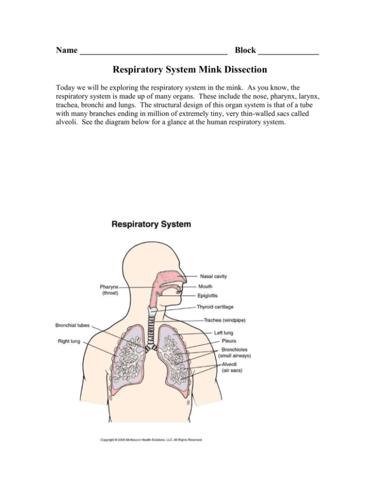
Name __________________________________ Block ______________ Respiratory System Mink Dissection Today we will be exploring the respiratory system in the mink. As you know, the respiratory system is made up of many organs. These include the nose, pharynx, larynx, trachea, bronchi and lungs. The structural design of this organ system is that of a tube with many branches ending in million of extremely tiny, very thin-walled sacs called alveoli. See the diagram below for a glance at the human respiratory system. 1. HOW TO VIEW THE PHARYNX, TRACHEA, LARYNX & VOCAL CORDS: In order to observe the regions of the pharynx it will be necessary to cut through the bones on the lower portion of the jaw. The caudal boundary of the oral cavity is formed by the pillars of the fauces, a pair of connective tissue and muscular folds that arch from the base of the tongue to the palate. The region between the pillars and the esophagus is the PHARYNX. In the pharynx the air passage moves from its dorsal position in the nasal cavity to its ventral position in the TRACHEA, thereby crossing the food passage. The pharynx is divided into THREE REGIONS: the nasopharynx lies above the soft palate. It transmits air. In its walls are the slit-like openings of the auditory tubes (Eustachian tubes). The oropharynx lies between the pillars of the fauces and basihyal. In its walls are the flap-like palantine tonsils. The laryngopharynx is the pharyngeal region lying just above the LARYNX. Notice that the esophagus is normally collapsed and the trachea, on the other hand, is held open by a series of cartilaginous rings. Cut the LARYNX midventrally and spread the two halves apart. The larynx is formed by the large THYROID CARTILAGES. The smaller CRICOID CARTILAGES, and the minute ARYTENOID CARTILAGES are on its dorsal surface. On its inner wall can be seen the two pairs of VOCAL CORDS. The caudal pair are the true vocal cords and are thought to produce sound while the cranial pair, the false vocal cords, are not for sound production. Make sketches of pharynx (all three portions indicated): Sketch the larynx and vocal cords here: Using a dissecting microscope, make a close up sketch of the trachea rings in the thoracic cavity: Dissect out a small segment of the trachea cartilage and make a sketch of a cross section view (looking down the windpipe): 2. HOW TO VIEW THE PLEURAL CAVITY, LUNGS, DIAPHRAGM AND BRONCHI: Open the thoracic cavity by cutting through the muscles and rib cartilages on the LEFT SIDE of and parallel to the sternum. Keep the scissors pointed toward you to avoid damaging the structures in the cavity. Pull the walls of the cavity apart, breaking the ribs. The lungs lie in the PLEURAL CAVITES. The right lung has 3 major lobes and a 4th smaller lobe more dorsal in position. The left lung has two lobes. Follow the trachea and esophagus as they enter the thorax. Dorsal to the heart, the trachea divides into left and right BRONCHI. These are responsible for carrying air to and from the lungs. The esophagus continues dorsal to the heart and penetrates the muscular DIAPHRAGM to enter the abdominal cavity. Sketch the lungs and bronchi here: 3. HOW TO VIEW THE INNER BRONCHI, BROCHIOLES AND ALVEOLI: The last part of this dissection will require use of the dissecting microscope. In order to view the INNER BRONCHI, BRONCHIOLES, and ALVEOLI you will need to cut a longitudinal section of the lung. Dissect a lung completely out of the mink. Place it on a tray and carefully cut a longitudinal section. Observe the internal structure of the lung. If you cannot make out any bronchioles or bronchi branches, you may need to make further cuts. If you look carefully under the microscope you may see some ALVEOLI SACS. Make sketches of inner bronchi and bronchioles here: While looking under the dissecting microscope, find an alveolus for sketching. The alveoli look like bumps in the lungs that are arranged like a bunch of grapes. It may be necessary to cut while looking through the dissecting microscope. QUESTIONS FOR RESPIRATORY SYSTEM DISSECTION: 1. What is the function of the alveoli? 2. How are the alveoli structured to support their function? (name three) 3. What part of the pharynx contains the Eustachian tubes? 4. Where are the vocal cords located? 5. How many lobes does each side of the lungs have? 6. Using correct anatomical language, where is the larynx in regards to the pharynx? 7. What is the function of the diaphragm? 8. What is the function of the cartilaginous rings in the walls of the trachea?
