The Hanging Drop Slide and Bacterial Motility
advertisement
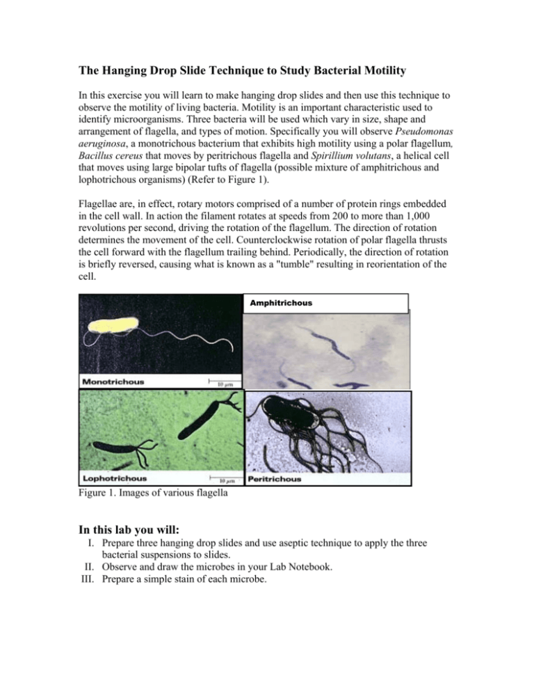
The Hanging Drop Slide Technique to Study Bacterial Motility In this exercise you will learn to make hanging drop slides and then use this technique to observe the motility of living bacteria. Motility is an important characteristic used to identify microorganisms. Three bacteria will be used which vary in size, shape and arrangement of flagella, and types of motion. Specifically you will observe Pseudomonas aeruginosa, a monotrichous bacterium that exhibits high motility using a polar flagellum, Bacillus cereus that moves by peritrichous flagella and Spirillium volutans, a helical cell that moves using large bipolar tufts of flagella (possible mixture of amphitrichous and lophotrichous organisms) (Refer to Figure 1). Flagellae are, in effect, rotary motors comprised of a number of protein rings embedded in the cell wall. In action the filament rotates at speeds from 200 to more than 1,000 revolutions per second, driving the rotation of the flagellum. The direction of rotation determines the movement of the cell. Counterclockwise rotation of polar flagella thrusts the cell forward with the flagellum trailing behind. Periodically, the direction of rotation is briefly reversed, causing what is known as a "tumble" resulting in reorientation of the cell. Amphitrichous Figure 1. Images of various flagella In this lab you will: I. Prepare three hanging drop slides and use aseptic technique to apply the three bacterial suspensions to slides. II. Observe and draw the microbes in your Lab Notebook. III. Prepare a simple stain of each microbe. Materials: 24-48 hour tryptic soy broth cultures of Pseudomonas aeruginonas, Bacillus cereus, Spirillulm volutans Microscope slides Coverslips Plumber’s tape Bunson burner Striker Microscope Lens paper, Kleenex and lens cleaner Immersion Oil Methylene blue dye Sterile transfer pipette s Inoculating loop Procedures: I. Prepare three hanging drop slides a. Place four very small balls of plumber’s tape on a clean microscope slide, creating a box shape using the balls as the four corners (Refer to Figure 2). b. After thoroughly mixing one of the cultures, use aseptic technique to place a small drop of bacterial suspension, using a sterile transfer pipette, onto the center of a coverslip. c. Lower the slide onto the coverslip such that the balls of plumber’s tape match up to the four corners of the coverslip. Press gently using a piece of lens paper. d. Turn the handing drop slide over quickly, keeping the drop suspended from the coverslip. e. Repeat procedure for the remaining two cultures. Step c. Balls ● ● Step a. Edge of slide Coverslip Culture drop ● ● Step b. Culture Drop Step d. Prepared Hanging Drop Slide Figure 2. Diagram representing the Steps of the Hanging Drop Method. II. Observe and draw the microbes in your Lab Notebook a. Place the slide on the microscope stage, coverslip up, so the drop is over the light hole b. Examine the drop by first locating its edge under low power and focusing on the drop. Switch to the higher, dry objectives and observe the culture. c. Using immersion oil, switch to the 100X objective. In order to see the bacteria clearly, close the diaphragm as much as possible for increased contrast. d. Note and draw in your Lab Notebook the bacterial shape, size, arrangement and motility. III. Prepare a simple stain of each microbe a. Once you have finished observing the live bacteria, prepare a Simple Stain for each and re-examine the organisms. Record your observations in your Lab Notebook.. b. When preparing the Discussion section in your Lab Notebook, discuss the differences you observed between the wet mount and the stained slides.
