(TGTX-1101), A Novel Anti-CD20 Monoclonal
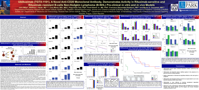
Ublituximab (TGTX-1101), A Novel Anti-CD20 Monoclonal Antibody, Demonstrates Activity in Rituximab-sensitive and
Rituximab –resistant B-cells Non-Hodgkin Lymphoma (B-NHL) Pre-clinical in vitro and in vivo Models
John Miller
1
, Matthew J. Barth, MD
1,2,3
, Cory Mavis, BSc, MSc
4
, Ping-Chiao Tsai, PhD
5
, Pavel Klener Jr., MD, PhD
6
, Francisco Hernandez-Ilizaliturri, MD
4,5
and Myron S. Czuczman, MD
4,5
1
Department of Pediatrics, Roswell Park Cancer Institute, Buffalo, NY;
2
Pediatric Hematology/Oncology, Women and Children's Hospital of Buffalo, Buffalo, NY;
3
Department of Pediatrics, University at Buffalo,
Buffalo, NY; Departments of
4
Medicine and
5
Immunology, Roswell Park Cancer Institute, Buffalo, NY;
6
Department of Internal Medicine-Dept. of Hematology, First Faculty of Medicine and General Teaching
Hospital, Prague, Czech Republic
Abstract
Ublituximab induced higher levels of ADCC than rituximab in RR and AraCR B-cell NHL cell lines
Ublituximab and rituximab reduced viability by a similar degree in MCL lines
Ublituximab and rituximab had no impact on the cell cycle in various lymphoma cell lines
Ublituximab caused a higher degree of CDC lysis in patient-derived tumor cells than rituximab
MZL Follicular Lymphoma The addition of rituximab to chemotherapy regimens utilized in treating B-cell non-Hodgkin lymphoma (B-NHL) has resulted in significant improvement in treatment response and clinical outcomes. On the other hand, the use of rituximab is changing the biology and response to second-line therapy in patients with relapsed/refractory disease. Novel anti-CD20 mAbs continue to be developed that may offer additional treatment options for relapsed/refractory rituximab-pre-treated patients. Ublituximab (TGTX-1101) is a novel, chimeric mAb targeting a unique epitope on the CD20 antigen. Ublituximab has been glycoengineered to enhance affinity for all variants of Fc γRIIIa receptors. To further characterize the activity of ublituximab, we evaluated its anti-tumor activity in a panel of rituximab-sensitive (RSCL), rituximab
–resistant
(RRCL) cell lines, primary tumor cells isolated from patients with B-NHL by negative selection using magnetic beads, and in lymphoma SCID mice xenograft models. RSCL (Raji, RL, U2932, Granta, HBL-2, Jeko-1, Mino, Rec1 and Z-138), RRCL (Raji-2R, Raji 4RH, RL-4RH, and
U2932-4RH); and cytarabine-resistant (AraCR) mantle cell lymphoma cell (MCL) lines (Granta-AraCR, HBL-2-AraCR, Jeko-AraCR, Mino-
AraCR and Rec1-AraCR) were labeled with 51 Cr. Subsequently, cells were exposed to ublituximab, rituximab or isotope control and human serum (25%) for complement dependent cytotoxicity (CDC) assays or to effector cells isolated from healthy volunteers (effector:target ratio
40:1) for antibody dependent cellular cytotoxicity (ADCC) assays, respectively. Antibody-induced direct anti-proliferative effects and induction of apoptosis were determined by alamar blue reduction assay and Annexin-V and propidium iodide staining, respectively. Primary tumor cells
(n=11) were exposed to ublituximab, rituximab or isotype control +/- pooled human serum for 48 hr. Changes in ATP content were determined using the CellTiterGlo assay. For in vivo studies, 6-8 week old SCID mice were inoculated via tail vein injection with 1x10 6 Raji cells on day 0 and assigned to rituximab (10mg/kg/dose), ublituximab (10mg/kg/dose) or control group. MAb was given via tail vein injection on days +3, +7,
+10 and +14. Differences in survival were analyzed by Kaplan-Meier curves and p values calculated using log rank test. Ublituximab induced significantly higher ADCC when compared to rituximab in 11 out of 17 cell lines tested (including all RRCL and cytarabine resistant MCL cells):
(Raji 44.4% vs. 19.8%; Raji 4RH 17.5% vs. 8.3%; Raji 2R 28.2% vs. 12%; RL 40.9% vs. 17.8%; RL-4RH 33.5% vs. 17.2%; U2932 46.9% vs.
28.8%; U2932-4RH 40.2% vs. 22.1%; HBL-2AraCR 30.7% vs. 16.6%; Jeko 34.8% vs. 18.4; Jeko-AraCR 23.8% vs. 9.6; Mino 47.4% vs.
11.6%; Mino-AraCR 32.5% vs. 15.5; Rec1 30.9% vs. 0%; p-values <0.05). There was no significant difference between ublituximab and rituximab in terms of CMC (including studies performed in primary tumor cells) or direct signaling (i.e. apoptosis or cell proliferation). While ublituximab therapy prolonged the survival of lymphoma-bearing SCID mice when compared to controls, the anti-tumor activity in vivo was similar to rituximab. Our results suggest that ublituximab exhibits higher ADCC than rituximab in vitro, including in RRCL and elicits similar
CDC and direct anti-tumor effects. Despite this enhanced ADCC activity, initial in vivo experiments did not result in improved survival compared to rituximab, however additional in vivo experiments investigating the activity of ublituximab in RRCL and MCL mouse models, testing alternative dose/schedule regimens and/or in combination with other anti-lymphoma agents are planned.
Ublituximab is a glycoengineered anti-CD20 monoclonal antibody with enhanced ADCC activity
(a) (b)
*
*
*
*
*
*
*
*
*
* *
*
DLBCL
*
*
Black : Ublituximab
White : Rituximab
De Romeuf C, et al. Br J Haematol. 2008;14
:635-43
Figure 1. (a) Anti-CD20 mAbs work through ADCC, CDC and direct induction of apoptosis. Ublituximab is glycoengineered to increase affinity for Fc γRIIIa receptors. (b) This advanced glycosylation pattern has been shown to increase ADCC induced cell lysis in CLL patient tumor cells exposed to ublituximab (filled bar) versus rituximab (open bar).
*
*
*
*
*
*
*
*
*
*
Materials and Methods
*
Cell lines: Experiments were performed on a wide variety of B-cell NHL cell lines. Rituximab-sensitive cell lines (RSCL): Raji, RL, U2932, Z-
138, Jeko, Mino, Rec-1, HBL-2 and Granta; rituximab-resistant cell lines (RRCL): Raji 2R, Raji 4RH, RL 4RH and U2932 4RH; and cytarabine-resistant (AraCR) cell lines: Jeko AraCR, Mino AraCR, Rec-1 AraCR, HBL-2 AraCR and Granta AraCR. RRCL’s were developed according to Czuczman et al, Clin Cancer Res.; 2008 14:1561-70.
Patient Samples: Malignant B-cells were isolated by MACS sorting from pre-treatment biopsy tissue obtained from B-cell lymphoma patients treated at Roswell Park Cancer Institute. Tissue was mechanically disrupted and the cells were passed through a 100 µm cell strainer.
Lymphocytes were then enriched by density centrifugation. B-cells were isolated from enriched lymphocytes using a human B-cell Isolation Kit
II (Miltenyi Biotec). 51Cr Release Assay (CDC and ADCC experiments): Cells were incubated with 51Cr for 2 hours and 100 µl of 51 Crlabeled cell suspension (1x10 6 cells/ml for CDC studies, 1x10 5 cells/ml for ADCC studies) were incubated in 96 well plates. Cells were treated with rituximab (10
g/ml), ublituximab (10
g/ml) or trastuzumab isotype (10
g/ml) and human serum (CDC, 1:4 dilution) or PBMC’s (ADCC,
40:1 effector: target ratio) for 6 hours at 37 o C and 5% CO
2
. 51 Cr release was measured from the supernatant by standard gamma counter.
Percent lysis was calculated as follows: % Lysis = (Sample cpm —background cpm)/(Maximum cpm—background cpm).
In vivo mouse survival: Six- to eight-week old severe combined immunodeficiency mice from the Department of Laboratory Animal
Resources at Roswell Park Cancer Institute were inoculated with 1x10 6 Raji cells by tail vein injection on day 0 and separated into rituximab, ublituximab or control groups. On days +3, +7, +10 and +14, the mice were treated with rituximab (10mg/kg/dose) or ublituximab
(10mg/kg/dose) by tail vein injection. The mice were monitored for a period of 100 days following completion of therapy. Using development of limb paralysis as the survival point, Kaplan Meier analysis was used to measure differences in survival.
Apoptosis: Tumor cells (0.5x10^6/mL) were incubated with rituximab, ublituximab or isotype (10ug/mL) for 24 and 48 hours. At each time point, 1x10^6 cells were washed with PBS and then incubated with Annexin-V and propidium iodide. Induction of apoptosis was then determined using flow cytometry.
Cell cycle: Tumor cells (0.5x10^6/mL) were incubated with rituximab, Ublituximab or isotype (10ug/mL) for 24 and 48 hours. At each time point, 1x10^6 cells were washed with PBS, fixed in 70% ethanol then incubated with ribonuclease. Cells were then incubated with propidium iodide and cell cycle determined using flow cytometry.
Viability Assay (Direct killing experiments): Cells (2x10 5 ) were treated with rituximab (10
g/ml), ublituximab (10
g/ml) or trastuzumab isotype (10
g/ml) and rabbit anti-mouse antibody (1
g/ml) and incubated in a 96 well plate at 37 o C and 5% CO
2
. After 24, 48 or 72 hours, the cells were treated with 20µl of AlamarBlue (Invitrogen) and incubated for a further 3 hours. Viability was measured by a Multiskan Spectrum
(Thermo Scientific) at a wavelength of 590nm.
*
Bars show mean
Error bars show 95%
CI of mean
*P<0.05
Figure 2: Ublituximab caused significantly higher levels of cell lysis by ADCC in B-cell NHL tumor cell lines . NHL cell lines were tested for ADCC cell lysis using a 51 Cr release assay in the presence of effector cells at an effector to target ratio of 40:1. In 11 of 17 NHL lines (Raji, Raji 2R,
Raji 4RH, RL, RL 4RH, U2932 4RH, Jeko, Mino, Mino AraCR, Rec-1 and HBL-2 AraCR), ublituximab induced significantly (p<0.05) higher lysis than rituximab. In the remaining cell lines, ublituximab caused higher lysis, but the results were not significant.
*
*
Bars show mean
Error bars show 95%
CI of mean
Figure 5: Ublituximab and rituximab showed no significant effect on the cell cycle . Raji family cells were treated with 10µg/ml of isotype, rituximab or ublituximab and then incubated for 24 hrs. Cells were stained with propidium iodide and analyzed by flow cytometry for cell cycle information. Neither ublituximab or rituximab differed significantly from the isotype control group.
Ublituximab and rituximab had similar effects on survival in human B-cell lymphoma SCID mice
Bars show mean
Error bars show 95%
CI of mean
*P<0.05
Figure 3. Ublituximab and rituximab similarly decreased viability in mantle cell lymphoma (MCL) cell lines over 72 hours. Both cytarabine-sensitive and cytarabine resistant (AraCR) cell lines were treated with 10
g/ml of ublituximab, rituximab or trastuzumab isotype and 1
g/ml of goat anti-mouse mAb (X-link antibody) for 24, 48 or 72 hours and viability was measured by AlamarBlue colorimetry. Ublituximab and rituximab decreased viability by a similar level in 7/8 cell lines. Ublituximab had slightly more activity in
Mino AraCR at 48 and 72 hours.
Figure 7. In lymphoma cells collected from patient samples, ublituximab induced higher levels of CDC lysis than rituximab in 3 of 5 samples. B-cell lymphoma cells were isolated from patient biopsies, treated with 10µg/ml of ublituximab, rituximab or isotype and tested for CDC lysis using a 51 Cr release assay. Ublituximab caused greater lysis than rituximab in 3 of 5 samples.
Ublituximab and rituximab did not induce apoptosis in various DLBCL cell lines
Conclusions
Bars show mean
Error bars show ± 1 standard error of the mean
Figure 4: Ublituximab and rituximab did not induce significant apoptosis. Cells were treated with 10µg/ml of mAb for 24 hrs and then stained with annexins
V and PI and analyzed by flow cytometry. Neither ublituximab or rituximab had a significant effect.
Figure 6: Ublituximab and rituximab were equally effective at prolonging survival in lymphoma SCID mice. Mice were inoculated with 1x10 6 Raji cells by tail vein injection and treated on days 3, 7, 10 and 14 with
10mg/kg/dose of ublituximab or rituximab or a vehicle control. Mice were observed for the survival point of limb paralysis for up to 100 days. Kaplan-Meier survival curves and log-rank analysis were used to detect differences in survival. Both rituximab and ublituximab significantly lengthened survival compared to the control but they did not differ from each other.
Ublituximab induces higher antibody dependent cellular cytotoxicity than rituximab in vitro
Ublituximab and rituximab reduce viability equally in the presence of a goat anti-mouse X-linking antibody
Neither ublituximab or rituximab has significant effects on the cell cycle or apoptosis
Ublituximab and rituximab prolong survival to an equal extent in an in vivo
Burkitt’s lymphoma SCID mouse model
Ublituximab is more effective at causing complement dependent cytotoxicity in primary tumor cells than rituximab
Research, in part, supported by a NIH grant R01 CA136907-01A1 awarded to
Roswell Park Cancer Institute and the Eugene and Connie Corasanti
Lymphoma Research Fund
