THE EFFECTS OF ASEPTIC AND STERILE TECHNIQUE ON
advertisement
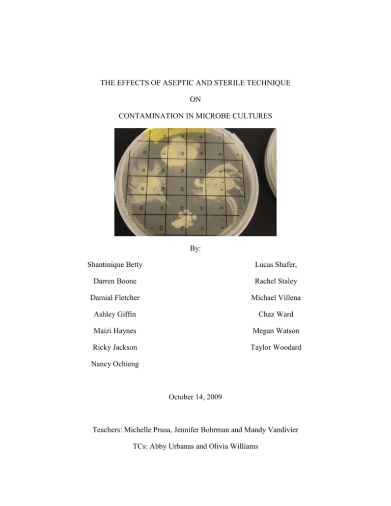
THE EFFECTS OF ASEPTIC AND STERILE TECHNIQUE ON CONTAMINATION IN MICROBE CULTURES By: Shantinique Betty Lucas Shafer, Darren Boone Rachel Staley Damial Fletcher Michael Villena Ashley Giffin Chaz Ward Maizi Haynes Megan Watson Ricky Jackson Taylor Woodard Nancy Ochieng October 14, 2009 Teachers: Michelle Prusa, Jennifer Bohrman and Mandy Vandivier TCs: Abby Urbanas and Olivia Williams INTRODUCTION Microbiology is the study of small life, or microbes. It is important that scientists study this small life for many reasons. For example, microbiology is essential to the medical field in combating disease. Harmful microbes that can make people sick are called pathogens (Pommerville, 2004). Microbiologists can study these pathogens and learn how to fight and prevent infection. Scientists make antibodies and vaccines from weakened forms of microbes to treat and prevent illness (DasSarma & DasSarma, 2005). Microbes can also be beneficial. Some of these microbes are important to digestive processes, and others can be used to make cheeses or yogurt (DasSarma & DasSarma, 2005). It is important to have a clean working environment in order to study microbes effectively in any laboratory setting, such as in research labs or medical labs. Contaminants in a laboratory can cause many problems (O’Brien, 2001). If a researcher inoculates or transfers bacteria to a culture improperly, then contaminating microbes could react with the bacteria being studied, which could compromise results. Contaminants can be pathogenic, and in an environment where they are able to grow in high quantities, the pathogens could cause a researcher to become ill. In medical laboratories, contamination can cause severe repercussions for not only the medical technicians performing the tests but also for the patients awaiting treatment. In the event of contamination in this setting, technicians might study the wrong organism. This could be costly to medical facilities to re-administer tests, and the time wasted with a misdiagnosis could lead to the patient’s condition becoming worse or fatal (Ryan, 2002). Researchers employ several sterile techniques to avoid contamination. Sterile technique is used to attempt to eliminate as much contamination as possible from all laboratory settings. 1 C:\Documents and Settings\adburns\Desktop\Microbes final group paper.doc October 14, 2009 Hands, equipment, and work surfaces must be sterile, or free of living organisms, to avoid contamination. Of course, nothing can ever be completely free of life. In order to limit contamination, researchers wash their hands according to the Centers for Disease Control (CDC) guidelines (CDC, 2002). Researchers also wear proper lab attire, such as long pants, closed toe shoes, gloves, and goggles. This is done to keep the contaminants from the skin from being released into the air. Researchers also tie hair back, if necessary, to keep the dust and other particles from their hair out of the cultures (CDC, 2002). While conducting an experiment, sterile work surfaces and equipment such as Petri dishes and swabs are important. Along with sterile techniques that all labs utilize, researchers also use aseptic techniques when studying bacteria and transferring a target species of bacteria to cultures. Major sources of contamination include airborne contaminants and the use of non-sterile equipment (Ryan, 2002). Aseptic technique is a list of guidelines explaining how to avoid contamination when transferring bacteria from one culture to another. If aseptic techniques are not followed, cultures can become contaminated with non-targeted species of bacteria, causing researchers to study the wrong organism or forcing them to repeat the experiment. A sterile technique experiment was conducted to test the effects hand cleansing procedures has on contamination of agar plates. For the sterile technique experiment, two handcleansing procedures (washed and sanitized) and two un-cleansed procedures (rinsed and unwashed) were studied to see the impact of sterile procedures on the growth of microbes. The hypothesis for this experiment was cultures inoculated with hands that have been washed or cleansed with hand sanitizer according to CDC guidelines will have less growth of microbes than 2 C:\Documents and Settings\adburns\Desktop\Microbes final group paper.doc October 14, 2009 cultures inoculated with unwashed or rinsed hands. The null hypothesis was neither washing hands nor using hand sanitizer will have any effect on the growth of microbes. An aseptic technique experiment was conducted to test the effects of aseptic technique on the transfer of target bacteria, Sarcina aurantiaca (S. aurantiaca). S. aurantiaca was transferred from stock plates to treatment plates using aseptic techniques and three improper variations of aseptic techniques to test the impact of aseptic techniques on contamination growth in culture plates. The hypothesis for this experiment was that cultures inoculated with S. aurantiaca using aseptic technique will have less growth of biological contaminants than cultures inoculated using a non-aseptic technique. The null hypothesis was that plates inoculated using aseptic technique will have the same amount of contamination as plates inoculated using non-aseptic technique. 3 C:\Documents and Settings\adburns\Desktop\Microbes final group paper.doc October 14, 2009 METHODS The sterile technique experiment took place on Monday July 13, 2009, at 1:00 p.m. The aseptic technique experiment was conducted on Thursday July 2, 2009, at 9:35 a.m. Both took place at Frostburg State University, in the Compton Science Center’s biology laboratory, room 231. STERILE TECHNIQUE The sterile technique experiment had five treatments with four replicates. The first treatment was unwashed hands. The second treatment was hand washing as suggested by the CDC guidelines, which recommends that hands be run under water, washed with soap by rubbing vigorously for at least 15 seconds, and rinsed and dried with a disposable towel. Hands were then allowed to dry for one minute. The third treatment was the use of an alcohol-based hand sanitizer according to the manufacturer’s recommendations. A small amount of hand sanitizer was applied to the Figure 1- Sterile Template palm of one hand and rubbed in with the other until dry. The fourth procedure was rinsed hands. Hands were placed in water and rubbed without soap for 15 seconds, dried with a paper towel, then air-dried for one minute. The fifth treatment was the control plate; this plate was immediately sealed so as not to be exposed to airborne contaminants. After the procedure for each treatment was conducted, plates, previously prepared with nutrient agar, were inoculated using four fingers, touching the plates in the same four corners following a template (Figure 1). 4 C:\Documents and Settings\adburns\Desktop\Microbes final group paper.doc October 14, 2009 Labeling tape was used to identify the treatment number and procedure. Tape was used so labels could be easily removed and observations on cultures could be made. The plates were sealed with Parafilm and placed upside-down in an incubator at 25 degrees Celsius. This was done to eliminate the possibility of condensation from the agar mixing with the culture. Cultures were allowed two days to grow before data were collected. ASEPTIC TECHNIQUE The aseptic technique experiment had four treatments with four replicates. This experiment was conducted using Sarcina aurantiaca (S. aurantiaca). This microbe was used because it grows rapidly, it is non-pathogenic, bright orange, and easy to distinguish from contamination. The work surface was cleaned with Roccal. Hands were washed and goggles and nitrile gloves were worn. Treatments were variations on the aseptic guidelines which include: keep open lids at a 45 degree angle, keep lids closed when possible, keep sterile equipment off work surface, replace lids as soon as possible, sterilize equipment before and after use, and keep skin and hair away from plates so dust from those surfaces do not contaminate the culture (Caprette, 2002). The first treatment procedure was to transfer S. aurantiaca and leave the lid open throughout the rest of the experiment to simulate airborne contamination. The second treatment was to open a sterile swab, leave it on the work surface, and transfer the stock bacteria at the end of the experiment to simulate the use of non-sterile equipment. The third treatment was to use a non-sterile plate from a previous experiment while following all aseptic techniques to transfer stock bacteria. The control followed all aseptic techniques. Once the bacteria were transferred to the plates, lids were replaced as soon as possible. They were also placed in an incubator at 25 degrees Celsius. This is the optimum temperature of 5 C:\Documents and Settings\adburns\Desktop\Microbes final group paper.doc October 14, 2009 growth for this species of bacteria. The cultures were allowed 11 days to grow before data was collected which allowed contaminants to grow enough to be observed. DATA ANALYSIS Petristickers were used to collect the data. Petristickers, which were clear stickers with a grid consisting of 32 squares, were placed on the bottom of the plate. If contamination was visible in any square, that square was marked and considered as one contaminated square. A oneway ANOVA was used to determine whether the number of contaminated squares from the treatments were significantly different. The alpha level was set at .05. A Tukey test showed where the variation occurred between treatments. 6 C:\Documents and Settings\adburns\Desktop\Microbes final group paper.doc October 14, 2009 RESULTS STERILE TECHNIQUE In the sterile technique experiment, there were no statistically significant differences in mean percent contamination among the unwashed hands (M = 46.09, SD = 24.79), washed hands (M = 59.38, SD = 11.12), hand sanitizer (M = 48.44, SD = 33.71), and rinsed hands treatments (M = 59.38, SD = 28.98). There were likewise no statistically significant differences in mean percent contamination among the unwashed hands treatment and the control (M = 1.56, SD = 3.13), and the hand sanitizer treatment and the control. The washed hands treatment and the rinsed hands treatment both had significantly higher mean percent contamination than the control, F(4,19) = 4.21, p = 4.21, one-tailed (Table 1, Fig. 2). Mean Percent Contamination between Sterile Technique Treatments Fig. 2. – This figure shows mean percent contamination in the sterile technique experiment for unwashed, washed, sanitized, and rinsed hands procedures, and for the control. 7 C:\Documents and Settings\adburns\Desktop\Microbes final group paper.doc October 14, 2009 Table 1. – Contamination of grid squares in the sterile technique plates Group 1 2 3 4 Average: Percentage: Unwashed Hands 23 5 19 12 14.75 46.09 Washed Hands 21 19 14 22 19 59.38 Hand Sanitizer 28 7 21 6 15.5 48.44 Rinsed Hands 10 17 32 17 19 59.38 Control 0 0 0 2 .05 1.56 ASEPTIC TECHNIQUE For the aseptic technique experiment, there were no statistically significant differences in mean percent contamination among the open plate (M = 10.16, SD = 9.33), non-sterile swab (M = 15.63, SD = 24.14), non-sterile plate (M = 19.53, SD = 8.22), and control treatments (M = 3.90, SD = 3.13, F[3,15] = 0.84, p = .50, one-tailed, Table 2, Fig. 3). Mean Percent Contamination between Aseptic Technique Treatments Fig. 3. – This figure shows mean percent contamination in the aseptic technique experiment for the open, non-sterile swab, and non-sterile plate procedures, and for the control. 8 C:\Documents and Settings\adburns\Desktop\Microbes final group paper.doc October 14, 2009 Table 2. – Contamination of grid squares in the aseptic technique plates Group Open Plate 1 2 3 4 Average: Percentage: 4 7 2 0 3.25 10.16 Non-sterile Swab 16 2 2 0 5 15.63 Non-sterile Plate 8 4 9 4 6.25 19.53 Control 2 1 2 0 1.25 3.90 9 C:\Documents and Settings\adburns\Desktop\Microbes final group paper.doc October 14, 2009 DISCUSSION AND CONCLUSIONS The hypothesis for the sterile technique experiment was that cultures inoculated with hands that have been washed or cleaned using hand sanitizer according to CDC guidelines and manufactures` recommendations will have less growth of microbes than cultures inoculated with unwashed hands. The null hypothesis was that washing hands, rinsing hands, or using alcohol based hand sanitizer would not affect the growth of microbes. Results did not support the hypothesis, but they did support the null hypothesis. Based on these data, it is not possible to conclude whether washing hands, using hand sanitizer, or rinsing hands reduces more contamination than not washing hands because there were no significant differences in the contaminants that grew among them. Also, there were some contaminants present in the control plate, meaning that the plates from the treatment groups might not have been sterile, potentially confounding the data. The greatest drawback in the sterile technique experiment was the fact that each Petri dish, except for the control, was touched by a different hand. The results for the sterile technique experiment could be related to the different species of bacteria on different hands (Fierer, Hamady, Lauber, & Knight, 2008). The different types of bacteria could have grown at different rates and quantities. Also, some could have been more difficult to eliminate from the surface of the skin through the hand cleaning processes. Another possible explanation for these results is that when washing hands, oils on the hands could have been washed away. This could expose a layer of bacteria underneath, allowing the bacteria to grow more easily and more quickly. However, there is little to no research to validate this hypothesis. Last of all, previous research has shown that moist hands are capable of transferring more bacteria than dry ones (Findon & 10 C:\Documents and Settings\adburns\Desktop\Microbes final group paper.doc October 14, 2009 Miller, 1997). This could be related to the fact that hands were in contact with water before touching the Petri dishes in the washed and rinsed hands procedures, and these procedures showed the most growth of bacteria. The hypothesis for the aseptic technique experiment was that cultures inoculated with S. aurantiaca using aseptic technique would have less growth of contaminants than cultures inoculated using a non-aseptic technique. The null hypothesis was that there will be no difference in using aseptic technique and non-aseptic technique. The results did not support the hypothesis, but did support the null hypothesis. Based on the data collected, it is not possible to conclude that following all aseptic technique procedures reduces biological contamination because there were no significant differences between the treatment group plates and the control plates. In the aseptic technique experiment, data collection was limited to what could be observed through the agar. It was difficult to observe the bacteria through the sealed plate and determine contamination. Also, some of the contaminants could have been close to the same color as S. aurantiaca. These could have been overlooked and not considered as contamination. There were limitations to both the aseptic and sterile technique experiments. First of all, there was little time to conduct the experiments and limited materials to work with. Also, there was no access to a microbiology lab with specialized equipment, so the contaminant species could not be identified. If identification of contaminant species could have been done, more specific conclusions may have been drawn from the experiment. For example, the difference between airborne contaminants and contaminants coming from hands could be noted. The difference between pathogenic and non-pathogenic bacteria could be examined as well. 11 C:\Documents and Settings\adburns\Desktop\Microbes final group paper.doc October 14, 2009 To improve the aseptic and sterile technique experiments, more trials could have been run for both experiments. This may have provided more accurate data. Also, for the sterile technique experiment, some of the variation that was observed between plates of the same procedure could be controlled by doing all procedures using the same hands. In obtaining results, the size of the colony had no effect on the amount of contamination counted with the petristickers. For example, a small colony could be counted the same as a large colony because any contamination in a square was counted as one. An improvement for gathering results would be to use smaller grids to have a more precise count of contamination. Finally, varieties in the different species of contamination could be noted, as well as how much contamination has grown over time. Although little can be said about how effective hand-washing and aseptic technique lab procedures are without significant results, scientists can still use these experiments to study ways of making sterile and aseptic techniques more effective. Perhaps researchers could study procedures such as washing hands and then using hand sanitizer or using different types of hand soaps. This could possibly help eliminate foreign bacteria from hands and reduce contamination. Any of these issues could be addressed through further research related to this study. 12 C:\Documents and Settings\adburns\Desktop\Microbes final group paper.doc October 14, 2009 REFERENCES Alpern, E.R., E.A. Alessandrini, L. Bell, L. Shaw, and K. Mcgowan. 2000. Occult Bacteremia from a Pediatric Emergency Department. Pediatrics, 106(s) pp. 505-511. Caprette, D.R. 2002. Laboratory Studies in Applied Microbiology. Accessed May, 2009. <http://www.ruf.rice.edu/~bioslabs/bios318maual.htm>. Carolina Biological Supply Company. 2005. Basic Microbiology Skills part I – Teacher’s Manual with Student Instructions (pp. S1-S2). Burlington, NC: DasSarma, P., and S. DasSarma. Centers for Disease Control and Prevention. 2002. Guideline for Hand Hygiene in Healthcare Settings. MMWR: Vol. 5. Corning Life Sciences. 2002. Understanding and Managing Cell Culture Contamination. Acton, MA: J. Ryan. Fierer, N., M. Hamady, C. Lauber, and R. Knight. 2008. The influence of sex, handedness, and washing on the diversity of hand surface bacteria. PNAS: 105(46) pp. 17,994-17,999 Findon, R and T. Miller. 1997. Residual moisture determines the level of touch-contactassociated bacteria transfer following hand-washing. Epidemiology and Infection: 119, pp.319-325. Hudson, B.K. and L. Sherwood. 1997. Streaking Plates for Isolated Colonies in Explorations in Microbiology. (pp.15-25). Upper Saddle River, NJ: Prentice Hall. O’Brien, S.J. 2001. Cell culture forensics. PNAS, 98(14), pp.7659-7658 Pommerville, J.C. 2004. Microbiology: Then and Now in Alcamo’s Fundamentals of Microbiology ( pp. 1-13). Sudbury, MA: Jones and Bartlett Publishers. 13 C:\Documents and Settings\adburns\Desktop\Microbes final group paper.doc October 14, 2009
