Cerebellar Anatomy and Connectivity Cerebellum = “Little Brain
advertisement
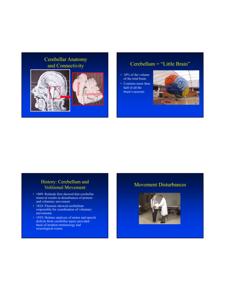
Cerebellar Anatomy and Connectivity Cerebellum History: Cerebellum and Volitional Movement • 1809: Rolando first showed that cerebellar removal results in disturbances of posture and voluntary movement • 1824: Fluorens showed cerebellum responsible for coordination of voluntary movements • 1939: Holmes analyses of motor and speech deficits from cerebellar injury provided basis of modern terminology and neurological exams Cerebellum = “Little Brain” • 10% of the volume of the total brain • Contains more than half of all the brain’s neurons Movement Disturbances Early Reports of Cognitive Disturbances Early reports of cognitive disturbance • Neuropsychological evaluation was primitive • Most reports were somewhat anecdotal • Consequently, cognitive contribution of cerebellum largely ignored Schmahmann, J.D. (1997) Int Rev Neurobiol 41, 3-27. Early Evidence of Cerebellar Link to Sensory & Associative Cortex • • • 1934: Abbie observed degeneration of human pontine nuclei following large lesions of parietal, temporal, occipital lobes 1942: Dow determined that dentate nucleus could be divided into older medial region and lateral neodentate 1942, 1958: Dow and Moruzzi, Snider & Stowell showed that proprioceptive, cutaneous, vagal, auditory and visual stimulation reaches the cerebellar cortex – Dow & Moruzzi: “…a hitherto unknown control may be exerted by the cerebellum in the sensory sphere and on autonomic functions.” – Snider: Concluded that cerebellum gets dual projections, one from sense organs and one from related sensory and motor cortical areas • The cerebellum is “a great modulator of neurologic function.” – They note that large cerebellar lesions, especially in lateral regions, produce little motor deficits Trends in Cerebellar Research • Pubmed Search: – Title contains *cerebell*, cerebrocerebell*, or cerebrocerebell* – Divide into Human and Animal – Cognitive content if title contains: • spatial, mental, emotion*, affective, reasoning, language, linguistic, planning, fluency, cognit*, memory, attention*, executive, nonmotor, or neuropsych* – Cognitive/Motor content if title contains: • timing, learning, conditioning, or speech – Motor content if title contains • motor, sensorimotor, oculomotor, movement*, postur*, balance, gait, gaze,saccad*, nystag*, locomot*, or walking Human 350 Number of Publications Number of Publications 300 Cognitive Motor/Cognitive Motor 250 200 150 100 50 0 1970 1975 1980 1985 1990 1995 Year 2000 2005 2010 Brain Development Animal 350 • The nervous system develops from ectoderm (outer layer) which forms a plate (~day 18) 300 Cognitive Motor/Cognitive Motor 250 200 150 100 50 0 1970 1975 1980 1985 1990 1995 Year 2000 2005 2010 – The edges of the plate curl and eventually fuse together forming a neural tube – By ~day 28, the rostral end of the neural tube has formed the ventricles and the tissue that surrounds these hollow chambers has formed three major divisions of the brain • Forebrain, midbrain, and hindbrain Desmond, J.E. (2010) Behav Neurol 23, 1-2. Brain Development ~ 4 weeks ~ 5-6 weeks Brain Development Neuroanatomy: Orientation Brain Development Superior/Dorsal Coronal Sagittal Section Posterior/Caudal axial Anterior/Rostral coronal Planes of Sectioning Superior View Inferior/Ventral Sagittal Anterior View Posterior View Axial Inferior View Cerebellum has 3 lobes 3 Lobes of the Cerebellum • Anterior lobe • Posterior lobe – The latter are divided by the primary fissure • Flocculonodular lobe – (most primitive, appears in fish) Flocculonodular lobe Superior View of Cerebellum Flocculus Nodulus Longitudinal Zones Longitudinally, there are Three Regions • Vermis (midline) • Intermediate zone • Lateral zone Vermis = “Worm” Note many parallel Convolutions, ie., “folia” 10 Lobules • There are 10 vermian and hemispheric “lobules” • The naming of the lobules has been variable across investigators and species studied Cerebellar Lobule Naming Human Examples Animal Human:Posterior Quadrangular Lobule Animal: Simplex Lobule Schmahmann name: Hemispheric lobule VI Human: Superior SemiLunar Lobule Animal: Crus I Schmahmann name: Crus I Brodal, A. (1981) Neurological Anatomy in Relation to Clinical Medicine, Third edn, Oxford University Press, New York. 10 Lobules Vermis Nomenclatures Schmahmann et al (1999) NeuroImage. 10, 233-60. • Lobules are separated by fissures Cerebellar Fissures Schmahmann et al Atlas Nomenclature Anterior View Superior View Schmahmann et al (1999) NeuroImage. 10, 233-60. Schmahmann et al (1999) NeuroImage. 10, 233-60. Cerebellar Fissures Posterior View Inferior View Left Lateral View Best view of the hemisphere lobules is on coronal sections Best view of vermis lobules is on sagittal section Cerebellar Deep Nuclei • Medial: Fastigial nuclei • Intermediate: Interposed nuclei – In humans: Emboliform and Globose nuc • Lateral: Dentate nuclei Deep Nuclei Visibility Deep Nuclei MRI Cryo section (coronal) T1-weighted Schmahmann et al (1999) NeuroImage. 10, 233-60. Y= -52 MRI T2-weighted Deep Nuclei – Children vs Adults Deep Nuclei Visibility Raw functional images from two subjects Nissl stain 29 years old 12 years old Courtesy of Dr. Dominic Cheng Longitudinal Zones Project to Different Deep Nuclei • Vermis Fastigial Nuclei • Intermediate Interposed Nuclei • Lateral Dentate Nuclei Y= -52 Myelin stain Schmahmann et al (1999) NeuroImage. 10, 233-60. Deep Cerebellar Nuclei Have Somatotopic Maps Inputs and Outputs: Big Picture Thalamus projects to neocortex Outputs from deep cerebellar nuclei via the superior cerebellar peduncle Pontine Mossy fiber inputs via middle cerebellar peduncle Inputs to Cerebellum • Mossy fibers – Come from (contralateral) pontine nuclei and spinal cord – Pontine fibers project through the middle cerebellar peduncle (aka: brachium pontis) – Spinal cord fibers project through inferior cerebellar peduncle (aka: restiform body) • Climbing fibers – Come from the (contralateral) inferior olivary nucleus – Project through the inferior cerebellar peduncle Spinal Mossy fiber inputs via inferior cerebellar peduncle Climbing fiber inputs via inferior cerebellar peduncle Cerebellar Peduncles Anterior View from 4th ventricle Cerebellar Peduncles Posterior View – Cerebellum Removed Superior Mossy fiber inputs Climbing fiber inputs Inferior Functional divisions • Vestibulocerebellum = flocculonodular lobe – – – – Eye movements, vestibulo-ocular reflexes, gait, balance Inputs from vestibular organs Projects to vestibular nuclei Evolution: oldest part (archicerebellum) • Spinocerebellum = vermis & intermed zone – Somatosensory inputs from spinal cord and trigeminal nerve, as well as auditory, visual and vestibular inputs – Projects to fastigial and interposed nuclei – Influences descending motor systems – Evolution: Newer (paleocerebellum) Mossy Fiber Inputs to Functional Divisions • Cerebrocerebellum = lateral zone – Inputs from cerebral cortex via the pons – Outputs to dentate nuc – Evolution: Newest (neocerebellum) Cortico-Ponto-Cerebellar Circuitry Decussation from dentate to thalamus Decussation from pons to cbl cortex Cortico-pontine projections • Note that cerebellar damage has ipsilateral motor effects, due to double decussation of inputs and outputs – E.g., left cerebellar damage causes problems in left limbs – In contrast left motor cortex damage causes problems for the right limbs Decussation=fibers (axons) cross midline Kandel, E.R. et al, eds. Principles of Neural Science , 3rd Ed. New York: Elsevier, 1991 Schmahmann, J. D. Human Brain Mapping 4:174-198 (1996). Ponto-Cerebellar Projections Anatomical Tracing in Animals • Old: Silver impregnation methods to view degenerating axons • Newer: Horseradish peroxidase and autoradiographical methods • Newest: Transneuronal tracing using viruses, e.g., Herpes, Rabies – Peter Strick & colleagues: Studies performed in monkeys Brodal, A. (1981) Neurological Anatomy in Relation to Clinical Medicine, Third edn, Oxford University Press, New York. Transneuronal transport of virus Rabies: Retrograde Neocortex Thalamus Injection Sites Herpes: Anterograde Neocortex First order Pons Dentate Nuc Second order Granule Cells Purkinje Cells Third order Purkinje Cells Kelly, R.M. and Strick, P.L. (2003) J Neurosci 23, 8432-44. Retrograde transneuronal transport of rabies virus M1: Nearly Identical Patterns for Anterograde & Retrograde Tracing Retrograde Tracing Anterograde Tracing Kelly, R.M. and Strick, P.L. (2003) J Neurosci 23, 8432-44. Dorsal Ventral Dorsal Dentate: Primary Motor Ventral Dentate: Prefrontal Very Significant: There is connectivity between cerebellum and cognitive neocortex! Kelly, R.M. and Strick, P.L. (2003) J Neurosci 23, 8432-44. Area 46: Nearly Identical Patterns for Anterograde & Retrograde Tracing Retrograde Tracing Anterograde Tracing Kelly, R.M. and Strick, P.L. (2003) J Neurosci 23, 8432-44. Implication: Closed Corticoponto-cerebello-thalamo Loops Kelly, R.M. and Strick, P.L. (2003) J Neurosci 23, 8432-44. Lobular Evolution Climbing Fiber Inputs: Inferior Olivary Nucleus Pct of cerebellum occupied by the lobule Balsters et al (2010) Neuroimage 49, 2045-52. Inferior Olive: Cross Section Inf. Olive and Pontine Nuc. Pontine Nuclei Inferior Olivary Nuclei https://www.msu.edu/~brains/brains/human/brainstem/select_cell.html Inf. Olivary Subdivisions • • • • Inf. Olivary Subdivisions • lPO (purple)=lamina of principal olive Three main subdivisions: Principal olive (PO) Dorsal accessory olive (DAO) Medial accessory olive (MAO) – Can be preceded by l (lateral), m (medial), d (dorsal), v (ventral) • DAO (red)=dorsal accessory olive • MAO (blue)=medial accessory olive • yellow: dc = subnuclei of MAO, vlo = ventrolat. outgrowth (of MAO) transverse sections showing left side only, numbers caudal to rostral. Top is dorsal. Inferior Olivary Inputs • Somatosensory input from spinal cord, trigeminal nuclei, dorsal column nuclei Cortico-rubroolivary projections – Mostly in cMAO and DAO • Vestibular and optokinetic input – dc, vlo • Visual information – cMAO • Cerebral Cortex via Red Nucleus – PO, rMAO • Direct projections from Deep Cerebellar Nuclei The neocortex can influence climbing inputs to the cerebellum through this projection, in addition to influencing the mossy fiber inputs in the pons via corticopontine projections. Outputs of Inf. Olive = “Climbing Fiber” Inputs to Cerebellum • Climbing Fibers project to: – Cerebellar Cortex – Cerebellar Deep Nuclei Outputs from Cerebellum • Come from the deep cerebellar nuclei • Project through the superior cerebellar peduncle (aka: brachium conjunctivum) – Most fibers cross the midline (decussation) Cerebellar Peduncles Outputs of Functional Divisions Cerebellar Outputs + more + more Vestibulocerebellar and Medial Spinocerebellar Outputs • Flocculonodular lobe receives vestibular input and projects directly to vestibular nuclei • Vermis receives input from neck, trunk, vestibular, retina and extraocular muscles. Output focuses on ventromedial descending motor systems (reticulospinal, vestibulospinal, and medial corticospinal) Zones of cerebellar cortex, deep nuclei and inferior olive Lateral Spinocerebellar and Cerebrocerebellar Outputs • Intermediate zone of spinocerebellum receives sensory input from limbs and influences dorsolateral descending motor systems (rubrospinal and corticospinal) acting on ipsilateral limbs • Cerebrocerebellum receives input from many cortical areas via the pontine nuclei and influences those areas via dentato-thalamo-cortical projections. There is also dentate red nucleus – inferior olive loop – Note: magnocellular RN involved – Note: parvocellular RN involved distal parts of limbs Sometimes referred to by its latin name, Nucleus Ruber Ruber = “red” Microzones & Microcomplex from red nuc. ~5000 microcomplexes in human cerebellum Microcomplex = microzone + related deep nucleus+nuclear target+olivary region Cerebellar Cortex Deep Nuclei Thalamus mossy fibers climbing fibers Cerebral Cortex Cerebral Cortex Cerebellar Cortex Deep Nuclei Thalamus Inferior Olive Red Nucleus Cortico-ponto-cerebello-thalamo loop Inferior Olive Red Nucleus Sensory Input Motor Execution Pontine Nuclei Deep Nuclei Inferior Olive Red Nucleus mossy fibers climbing fibers Cerebellar Cortex Olivary Loops Via Red Nucleus Cerebral Cortex Deep Nuclei Thalamus Sensory Input Cerebellar Cortex Inferior Olive Red Nucleus Motor Execution Pontine Nuclei mossy fibers climbing fibers Local Inferior Olive Loop Thalamus Sensory Input Motor Execution Pontine Nuclei Cerebral Cortex mossy fibers climbing fibers Overall Picture: Cerebellar Loops Sensory Input Motor Execution Pontine Nuclei Cerebellar Cortex Neuronal Organization • Five types of neurons – Inhibitory: • • • • Purkinje Golgi Basket Stellate – Excitatory • Granule Cerebellar Cortex Neuronal Organization • Three Layers of Cerebellar Cortex – Molecular Layer • Cell bodies of stellate and basket cells • Axons of granule cells, called parallel fibers • Dendrites of Purkinje Cells – Purkinje Cell Layer • Cell bodies of Purkinje cells – Granule Cell Layer • Granule cells, which receive inputs from the mossy fibers (Most numerous neurons in brain, 1010-1011) • Golgi cells, also receive mossy fiber input Cerebellar Cortical Cells Divergence and Convergence “Fractured somatotopy” One body part represented in Multiple regions due to divergence Parallel Fibers 200 million MF inputs diverge onto 40 billion granule cells PF fibers converge onto 15 million PC Further converge on Deep nuclei Excitatory and Inhibitory Connections Simple and Complex Spikes Recording from Purkinje Cell Types of Cerebellar Damage/Disorder • There are many conditions that can affect the cerebellum • In most human research studies, patients typically have – Tumor removal (beware of radiation) – Diffuse cerebellar degeneration (spino-cerebellar ataxias – inherited – multiple forms) – Stroke involving cerebellar vascular territory Cerebellar Vascular Supply medial branch SCA lateral SCA SCA SCA=superior cerebellar artery AICA=anterior inferior cerebellar artery PICA=posterior inferior cerebellar artery basilar artery AICA vertebral artery Brainstem PICA medial PICA lateral PICA Superior Cerebellar Arteries SCA Supply Zones PICA Supply Zones Example SCA Territory Infarct Example of PICA territory Infarct Signs of Cerebellar Damage Dysmetria Cerebellar damage videos • A lack of accuracy in voluntary movements – Hypermetria = overshoot of target – Hypometria = undershoot • Delay in initiation of movement is common • Occurs proximally and distally in upper and lower limbs • Affects both single-joint and multi-joint movements • Most pronounced in rapid movements • Often followed by corrective movements Balance/Walking Tests Finger-to-nose Test Abnormal - Finger-to-nose_WMV V9.wmv DWFC09ftnmov_57_WMV V9.wmv SPFSG04romb_57-1_WMV V9.wmv VSFGS02walkturn_57_WMV V9.wmv DWFSG7ambleturn_57_WMV V9.wmv VSFC11ftnmov_57_WMV V9.wmv youtube.com.Cerebellar ataxia_WMV V9.wmv Heel-to-shin Test Dysarthria: Impaired Articulation DWFMSiSpArtic_57_WMV V9.wmv Abnormal Coordination Exam ; Heel-to-shin_WMV V9.wmv VSFMSiSpArtic_57_WMV V9.wmv VSFC12heelkshin_57_WMV V9.wmv Impaired rapid alternating movement Ocular Dysmetria Dysdiadochokinesia Abnormal - Hand Rapid Alternating Movements_WMV V9.wmv VSFC07suppro_57_WMV V9.wmv DWFC11prosup_57_WMV V9.wmv youtube.com.Dysdiadochokinesia Song_WMV V9.wmv Dysmetria of Upper Limb: Pointing Movements Abnormal EMG in Cerebellar Patients Healthy Control Cerebellar Patient Normal triphasic pattern Concise elbow movement Elbow 2 bursts not demarcated Reduced rise rate Shoulder Comparable At slow speed Hyperextension of elbow causes overshoot Increased latency Single joint movement, e.g. pull a lever, AGO = agonist muscle (biceps), ANTA = antagonist (triceps) Manto, M. (2009) J Neuroeng Rehabil 6, 10. Cerebellar Hypermetria Normal Subject Manto, M. (2009) J Neuroeng Rehabil 6, 10. Adaptation in EyeHand Coordination Cerebellar Patient A. Special prism glasses bends light so that you have to look left to see target directly in front B. When prisms are first put on, throws deviate to the left, but there is adaptation. When the glasses are removed there is a rebound effect. Overshoot Wrist flexion movement (MVT) C. Adaptation fails in a patient with unilateral PICA infarction involving inferior cerebellar peduncle (inferior olivary climbing fibers!) and inferior lateral posterior cerebellar cortex Models of Cerebellar Function Marr: Cerebellar Synaptic Plasticity Joint activity from parallel fiber and climbing fiber was hypothesized to cause synaptic modification at the parallel fiber synapse De Schutter (1997) Prog Brain Res 114, 529-42. Marr (1969) Model • Hebb (1949) proposed that synaptic modification based on co-occurrence of preand postsynaptic activity might underlie learning • Marr proposed that parallel fiber synapses onto Purkinje Cells are facilitated (Long Term Potentiation, or LTP) when they are activated together with climbing fiber activation Marr (cont) • Through plasticity mechanism, cerebellum could learn movement skills from experience • From divergence of mossy fiber to granule cells, finer representation of sensory information can be achieved Example finger 1 and 2 finger 1 Albus (1971) finger 2 Purkinje cells granule cells • Suggested that the cerebellum functions like a perceptron pattern-classification device, with complex spikes (from climbing fibers) as the unconditioned stimulus and mossy fiber input as the conditioned stimulus mossy fibers pontine nuclei finger 1 finger 2 Cerebellar Perceptron Albus (cont) • Proposed that climbing fiber provides an error signal • Also proposed that parallel fiber synapse is weakened instead of facilitated – He predicted long term depression (LTD), prior to the demonstration of its existence by Ito (1982) in cerebellar slices – Marr’s modified theory is often referred to now as MarrAlbus or Marr-Albus-Ito model • There is still much debate on whether the cerebellum is the locus of motor learning or if it is more of a control machine Albus, J.S. (1971) Math Biosci 10, 25-61. Adaptive Filter Models What is an Adaptive Filter? • Introduced in 1982 by Fujita • Influenced by Ito’s suggestion that vestibulo-ocular reflex could be understood in terms of engineering control theory • A filter is a hardware or software device that converts an input signal into a different output signal • E.g., in music a “low pass” filter effectively removes high frequencies of an audio signal Low Pass Filter Adjustable Filter Gain control allows the amplitude to be adjusted Treble or tone control allows more or less of the high frequencies to come through Error Signal Adaptive Filter Current output Adaptive Filter • An adaptive filter can learn to attenuate a signal only in the noise frequency band • E.g., aircraft engine noise • Requires a second input signal from microphone as an “error” or “teaching” signal spike input x(t) p1(t) w1 error: e(t) p2(t) w2 p3(t) Change in synaptic weight Learning rate constant (typically < 1) w3 y(t) = wipi(t) Signals positively correlated with error signal get weight reduced Signals negatively correlated with error signal get weight increased e.g. p3 onset at t=3 coincides with error signal, so w3 is reduced t=3 spike input x(t) p1(t) w1 error: e(t) p2(t) w2 If =0.1, e(t)=1 for error, otherwise e(t)= -1: w3=(-0.1)(1)(1)= -0.1 p3(t) w3 y(t) = wipi(t) Signals positively correlated with error signal get weight reduced Signals negatively correlated with error signal get weight increased Attractions of Adaptive-Filter Model of Cerebellum • Such filters are widely used in signal processing because they are powerful and deliver best (least squares) solution • There is structural resemblance to cerebellar microcircuit • Adaptive filters are capable of the kinds of functions associated with the cerebellum – Predicting a movement’s sensory consequences – Refinement of movement so that it is fast and coordinated Cerebellar Microcircuit & Adaptive Filters Predicting Sensory Consequences of Movement: Noise Cancellation a. (Theoretical) The goal is to teach the adaptive filter to cancel out the noise portion of s(t)+n(t) An explanation of microcircuit features: A large number of inputs (mossy fiber) are needed for an adaptive filter The teaching signal (climbing fiber) must be capable of affecting every weight without altering filter output …And Speaking of Predicting Sensory Feedback b. (Practical) The goal is to attenuate touch signals from whiskers that are generated by the animal’s own head movements. Otherwise, every time whiskers scrape the ground the animal will interpret it as a possible food stimulus. Cerebellar Activation Distinguishes Between Self-Produced and ExternallyProduced Tactile Sensation Blakemore et al (1998) Nat Neurosci 1, 635-40. Adaptive Filter Model and Accurate Movement Cerebellar Adaptive Filter for VOR • Vestibulo-ocular reflex (VOR) • Goal: Keep an image stable on the retina as the head is moved by producing a counteracting eye movement Eye muscles Eye velocity must match head velocity to keep image stable on retina. (When you see “+” and “–” you are hoping the 2 signals match). Slightly different from noise cancellation module for 3 reasons 1. 2. 3. Cerebellar Adaptive Filter for VOR Output of cancellation module is a motor command to oculomotor system The slip of the image off the retina is the error signal A copy of motor command feeds back into adaptive filter Cerebellar Adaptive Filter for VOR (b) Adjust these weights (c) Do vestibular signals cause Enough eye movements? Eye muscles (b) Vestibular signals triggerred Eye movements are initiated (a) Head starts to move Vestibular signals arise (a) Image slipped off retina Error! Cerebellar Adaptive Filter for VOR (c) Adjust these weights (b) Again, image slips off retina Error! (a) Eye muscles old, weak or damaged. Insufficient movement. What about the rest of the cerebellum? • Specific regions of inferior olive project to specific strips of cerebellar cortex in sagittal plane (zones, A-D2) • Each zone projects to specific deep cerebellar or vestibular nuclei, which project to targets in the rest of the brain, which in turn project back to IO • Thus there are loops that may be functional subunits Evaluation of the model: Cerebellar flocculus and VOR • Involvement of flocculus in image stabilization has been established via lesion and inactivation studies • Main mossy fiber inputs to flocculus carry vestibular info and efference copy of eye movement commands • Climbing fiber inputs to flocculus carry retinal slip signals • Floccular Purkinje cells project to ocular motoneurons • Plasticity: when VOR gain requirement is altered experimentally, Purkinje cell firing changes Zones of cerebellar cortex, deep nuclei and inferior olive Microzones Cerebellar Chip • Zones can be further subdivided into microzones – 5000 estimated • This organization suggests a “cerebellar chip” Challenge for Testing the Model • In most cases we do not know how a microzone’s output affects behavior • Thus identifying the error signal of the climbing fiber is challenging State Estimation Internal Models • The ability of the brain to control movement is based on its knowledge of the body’s state at any moment • State can be defined by a set of variables such as velocity and position of different limb segments • Given accurate knowledge of current state and motor commands the brain ought to be able to estimate the state in the near future and control it State Estimation Problem Delay Problem • Delay in arrival of afferent signals from periphery, along with central processing delays causes out-of-date knowledge of the peripheral system • Thus, calculation of a state prediction is hypothesized, i.e., the state of the limbs before sensory information arrives Forward models • A forward dynamic model is a representation of limb status, e.g., joint angles and velocities given forces applied • A forward sensory output model predicts tactile signals, proprioception, vision, audition Forward and Inverse models • Forward model: Given a motor command, what is the predicted new motor state • Inverse model: Given a desired new motor state, what is the motor command needed to achieve it? Forward models • Rapid movement cannot rely on sensory feedback alone – If they did, movement would have to be slow or instabilities/oscillations would occur – Note: Oscillations are characteristic of cerebellar damage Forward “Smith-Predictor” Model Forward “Smith-Predictor” Model Motor cortex command. Copy of command goes to cbl cerebellum This is the actual sensory consequences arising from the movement. E.g., Motor controller for hand: want to pick up a ball. Corticospinal system on the outer loop is feedback driven. The efference copy sent to the cerebellum generates a rapid prediction of hand/arm movements in the forward dynamic model to derive a state estimate. The forward output model is the predicted sensory information occurring from the movement with the delay built in. Forward “Smith-Predictor” Model Forward “Smith-Predictor” Model Rapid calc of where hand will be Rapid prediction of sensory info with delays accounted for When sensory discrepancy is 0 the state estimate from the forward model is perfect. Discrepancy of predicted and Actual sensory information provides Teaching signal for forward dynamic model Forward “Smith-Predictor” Model Discrepancy of state estimate and desired state can Be used to alter subsequent motor command. This loop represents the cerebro-cerebellar loop. An Example: Lifting a carton of milk • How much grip and force to apply? • Visual input: size – In the dark this input is reduced and therefore weighted less • Proprioception: resistance to lifting force – Reduced input and thus reduced weighting if you are wearing gloves • Prior belief of how full or empty it is • All of these factors will influence your state estimation Cerebellar Patients Under this model, with no state estimation from the cerebellum, adjustments of the motor command must rely on the relatively slow reafference signals.
