Guideline - initial management of C-spine injury - KwaZulu
advertisement
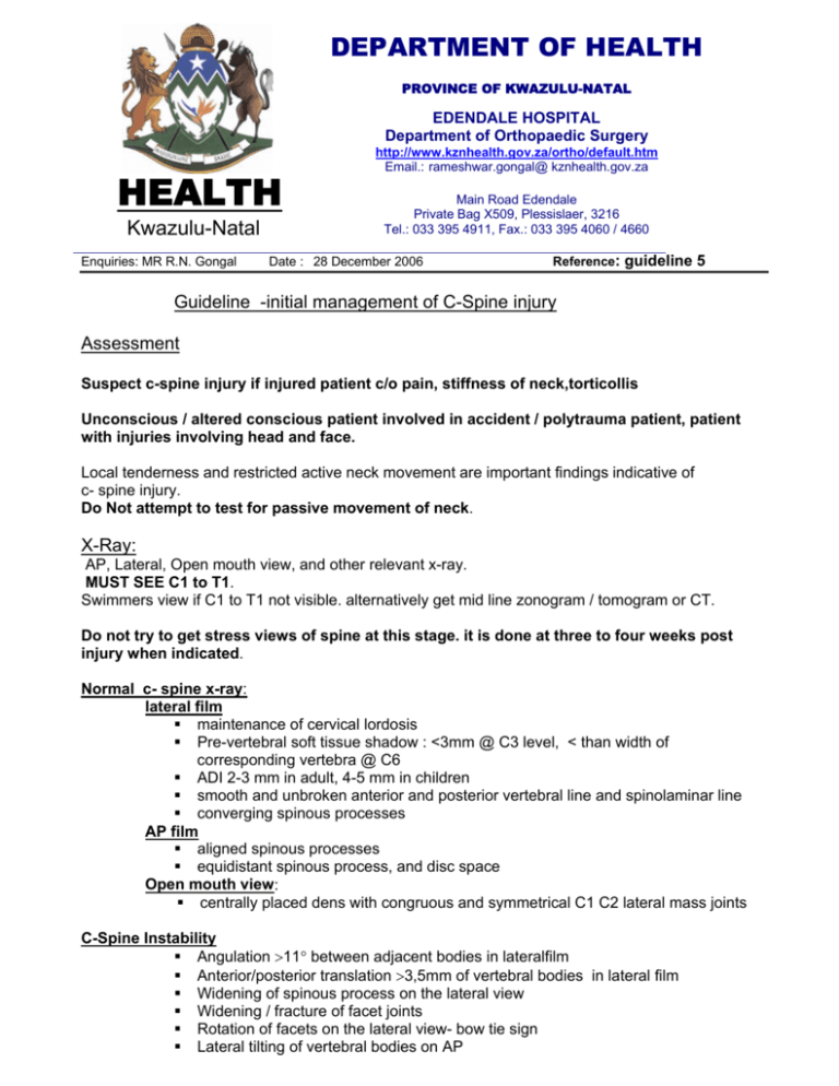
DEPARTMENT OF HEALTH PROVINCE OF KWAZULU-NATAL EDENDALE HOSPITAL Department of Orthopaedic Surgery HEALTH Kwazulu-Natal Enquiries: MR R.N. Gongal http://www.kznhealth.gov.za/ortho/default.htm Email.: rameshwar.gongal@ kznhealth.gov.za Main Road Edendale Private Bag X509, Plessislaer, 3216 Tel.: 033 395 4911, Fax.: 033 395 4060 / 4660 Date : 28 December 2006 Reference: guideline 5 Guideline -initial management of C-Spine injury Assessment Suspect c-spine injury if injured patient c/o pain, stiffness of neck,torticollis Unconscious / altered conscious patient involved in accident / polytrauma patient, patient with injuries involving head and face. Local tenderness and restricted active neck movement are important findings indicative of c- spine injury. Do Not attempt to test for passive movement of neck. X-Ray: AP, Lateral, Open mouth view, and other relevant x-ray. MUST SEE C1 to T1. Swimmers view if C1 to T1 not visible. alternatively get mid line zonogram / tomogram or CT. Do not try to get stress views of spine at this stage. it is done at three to four weeks post injury when indicated. Normal c- spine x-ray: lateral film maintenance of cervical lordosis Pre-vertebral soft tissue shadow : <3mm @ C3 level, < than width of corresponding vertebra @ C6 ADI 2-3 mm in adult, 4-5 mm in children smooth and unbroken anterior and posterior vertebral line and spinolaminar line converging spinous processes AP film aligned spinous processes equidistant spinous process, and disc space Open mouth view: centrally placed dens with congruous and symmetrical C1 C2 lateral mass joints C-Spine Instability Angulation >11° between adjacent bodies in lateralfilm Anterior/posterior translation >3,5mm of vertebral bodies in lateral film Widening of spinous process on the lateral view Widening / fracture of facet joints Rotation of facets on the lateral view- bow tie sign Lateral tilting of vertebral bodies on AP Abnormal disc narrowing / or increased disc space. neurological deficit severe damage/ fracture of anterior or posterior element of spine fracture of dens, Management: - ATLS principles, Neurogenic shock, spinal shock Documentation of neurology, vital sign ,breathing Do not turn and twist or flex the neck, - keep it extended. in the presence of neurology • NPO • pressure care • bowel and bladder care - Cone’s calliper traction for displaced cervical fracture or fracture dislocation with or without neurology ie. unstable spine ( weight not to exceed 2 kg for head and C1 and plus one kg add for each vertebral level. – upper level when two level injury; decrease the weight to half once reduction achieved) get CT scan of skull in small children before putting a cone’s calliper to assess the thickness of skull. continuous monitoring (neurology and vital signs) during reduction Split mattress to be used to keep the neck extended. tortocollis with normal looking x-ray– Halter neck traction undisplaced hangman’s fracture – Halter neck traction. normal looking x-ray with increased pre-vertebral soft tissue shadow – Hard Collar normal looking x-ray but with local neck pain - Soft Collar. Do not discharge the patient without consultant’s opinion. - Mr.R.N.Gongal FCS(SA)Ortho Principal and HOD Review January 2008
