Why Does My Heart Have Valves?
advertisement
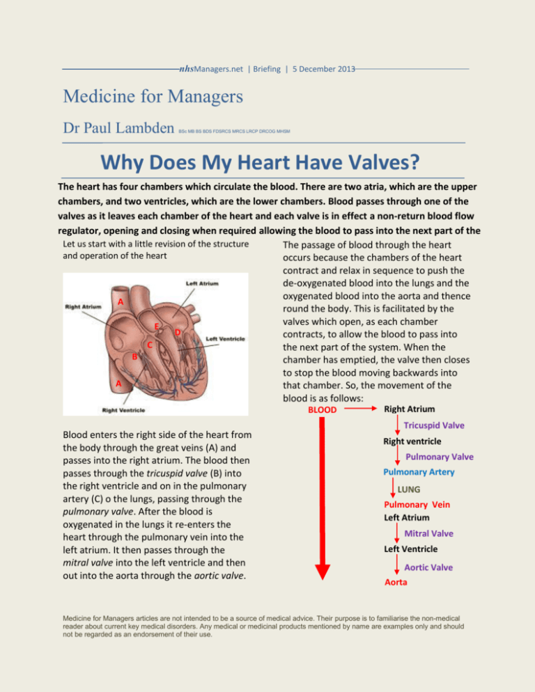
nhsManagers.net | Briefing | 5 December 2013 Medicine for Managers Dr Paul Lambden BSc MB BS BDS FDSRCS MRCS LRCP DRCOG MHSM Why Does My Heart Have Valves? The heart has four chambers which circulate the blood. There are two atria, which are the upper chambers, and two ventricles, which are the lower chambers. Blood passes through one of the valves as it leaves each chamber of the heart and each valve is in effect a non-return blood flow regulator, opening and closing when required allowing the blood to pass into the next part of the Let us start with a little revision of the structure The passage of blood through the heart system. and operation of the heart occurs because the chambers of the heart contract and relax in sequence to push the de-oxygenated blood into the lungs and the oxygenated blood into the aorta and thence A round the body. This is facilitated by the valves which open, as each chamber E D contracts, to allow the blood to pass into C the next part of the system. When the B chamber has emptied, the valve then closes to stop the blood moving backwards into A that chamber. So, the movement of the blood is as follows: BLOOD Blood enters the right side of the heart from the body through the great veins (A) and passes into the right atrium. The blood then passes through the tricuspid valve (B) into the right ventricle and on in the pulmonary artery (C) o the lungs, passing through the pulmonary valve. After the blood is oxygenated in the lungs it re-enters the heart through the pulmonary vein into the left atrium. It then passes through the mitral valve into the left ventricle and then out into the aorta through the aortic valve. Right Atrium Tricuspid Valve Right ventricle Pulmonary Valve Pulmonary Artery LUNG Pulmonary Vein Left Atrium Mitral Valve Left Ventricle Aortic Valve Aorta Medicine for Managers articles are not intended to be a source of medical advice. Their purpose is to familiarise the non-medical reader about current key medical disorders. Any medical or medicinal products mentioned by name are examples only and should not be regarded as an endorsement of their use. The valve action is co-ordinated in order to achieve the flow required. Therefore, for example, when the right ventricle is filled and contracts to pass blood into the pulmonary artery, the pulmonary valve opens to allow forward flow and the tricuspid valve closes to prevent backward flow. Similarly, when the left ventricle contracts to pass blood to the body via the aorta, the aortic valve opens and the mitral valve closes. Mitral Valve Tricuspid Valve Aortic Valve Pulmonary Valve All the valves have three cusps (except the mitral valve between the left atrium and the left ventricle which has two). The cusps are composed of tough sheets of connective tissue and act like flaps. They only open in one direction allowing blood to pass onwards through the valve but closing to prevent backflow. Heart valvular disease occurs when the valves do not work efficiently and allow blood to leak through when they should be closed. If a valve becomes diseased it may malfunction in one of two ways. It may fail to close properly causing regurgitation (leakage) of the valve resulting in blood flowing backwards from the ventricles to the atria (in the case of the mitral and tricuspid valves) and from to great blood vessels into the ventricles (in the case of the aortic and pulmonary valves). If the valves become damaged through infection or scarring the result is narrowing (stenosis) which impedes the passage of blood forwards out of the atria or ventricles. The result is that the blood must pump harder to push the blood onwards. Such conditions may affect one or more of the valves and both malfunctions may exist in any one valve at the same time. In mild valvular disorders there may be no symptoms. In more severe cases of valvular damage the patient may experience palpitations, fatigue or dizziness and may show signs of heart failure such as breathlessness or ankle swelling. In the particular case of bacterial endocarditis, which is an infection affecting the inner lining of the heart and the valves, the symptoms may be accompanied by a persistent fever. Heart valve damage may also be caused by a heart attack. Diagnosis may be made using a number of methods. Examination of the patient may demonstrate heart failure and applying a stethoscope to the chest will allow the doctor to hear the changes associated with valve malfunction. Such changes are called murmurs which may be easy or difficult to hear. The nature and timing of the murmur will allow a doctor (particularly a specialist cardiologist) to identify the valve affected and the type of abnormality. Other tests will include an electrocardiogram (recording of the heart’s electrical activity) and a chest X- Medicine for Managers articles are not intended to be a source of medical advice. Their purpose is to familiarise the non-medical reader about current key medical disorders. Any medical or medicinal products mentioned by name are examples only and should not be regarded as an endorsement of their use. ray. The patient may also be subject to a stress test where the patient is exercised on a treadmill and the electrical activity of the heart, heart rate and blood pressure are monitored. More definitive for valve disease is the echocardiogram. Ultrasound waves are bounced off the heart and are converted to images which show the heart size and shape but also display the movements of the valves and the heart generally. The degree of efficiency of the heart can be measured by the ejection fraction, which is the volume of blood as a proportion of the predicted amount which is pumped out of the heart when it contracts. The patient may also undergo cardiac catheterisation. In this simple procedure a catheter is passed via the blood vessels into the heart to measure pressure abnormalities or to identify backflow using radio-opaque dye which can demonstrate respectively stenosis or regurgitation. ball moves up when blood pushes it but seals the opening when blood presses on it from the other side. A number of other designs of prosthetic (artificial) valves are also available. Heart valves are for life, not just or Christmas, and the heart should be cared for by ensuring that blood pressure and cholesterol are controlled and exercise is taken regularly. It is said that between 10 and 15 percent of all heart surgery is valve surgery. It doesn’t look likely that valve surgeons will run out of business any time soon. paullambden@compuserve.com Treatment of valvular disease depends on the severity and the general condition of the patient. Sometimes the damaged valve can be repaired. For other patients it is necessary to replace the defective valve and either the valve of a pig or an artificial valve may be employed. Positioned where valve was located Ball moves up and down with blood flow The illustration is of a Starr Edwards valve which consists of a ball in a chamber. The circular ring is placed in the heart where the damaged valve was formerly located. The Medicine for Managers articles are not intended to be a source of medical advice. Their purpose is to familiarise the non-medical reader about current key medical disorders. Any medical or medicinal products mentioned by name are examples only and should not be regarded as an endorsement of their use.


