TNF-α Antibody - Cell Signaling Technology, Inc.
advertisement
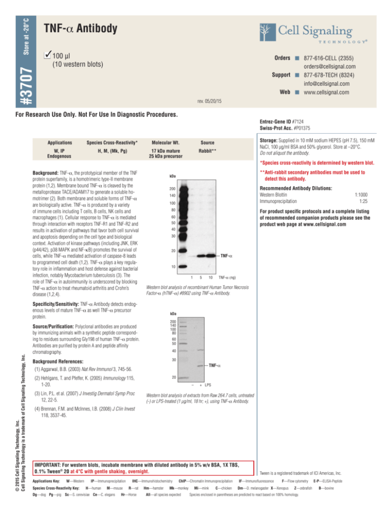
Store at -20°C TNF-α Antibody #3707 3 100 µl n Orders n 877-616-CELL (2355) orders@cellsignal.com Support n 877-678-TECH (8324) info@cellsignal.com Web n www.cellsignal.com (10 western blots) rev. 05/20/15 For Research Use Only. Not For Use In Diagnostic Procedures. Entrez-Gene ID #7124 Swiss-Prot Acc. #P01375 Applications Species Cross-Reactivity* Molecular Wt. Source W, IP Endogenous H, M, (Mk, Pg) 17 kDa mature 25 kDa precursor Rabbit** Storage: Supplied in 10 mM sodium HEPES (pH 7.5), 150 mM NaCl, 100 µg/ml BSA and 50% glycerol. Store at –20°C. Do not aliquot the antibody. *Species cross-reactivity is determined by western blot. Background: TNF-α, the prototypical member of the TNF protein superfamily, is a homotrimeric type-II membrane protein (1,2). Membrane bound TNF-α is cleaved by the metalloprotease TACE/ADAM17 to generate a soluble homotrimer (2). Both membrane and soluble forms of TNF-α are biologically active. TNF-α is produced by a variety of immune cells including T cells, B cells, NK cells and macrophages (1). Cellular response to TNF-α is mediated through interaction with receptors TNF-R1 and TNF-R2 and results in activation of pathways that favor both cell survival and apoptosis depending on the cell type and biological context. Activation of kinase pathways (including JNK, ERK (p44/42), p38 MAPK and NF-κB) promotes the survival of cells, while TNF-α mediated activation of caspase-8 leads to programmed cell death (1,2). TNF-α plays a key regulatory role in inflammation and host defense against bacterial infection, notably Mycobacterium tuberculosis (3). The role of TNF-α in autoimmunity is underscored by blocking TNF-α action to treat rheumatoid arthritis and Crohn’s disease (1,2,4). Recommended Antibody Dilutions: Western Blottin 1:1000 Immunoprecipitation1:25 200 140 100 80 For product specific protocols and a complete listing of recommended companion products please see the product web page at www.cellsignal.com 60 50 40 30 20 TNF-α 10 1 5 10 TNF-α (ng) Western blot analysis of recombinant Human Tumor Necrosis Factor-α (hTNF-α) #8902 using TNF-α Antibody. Specificity/Sensitivity: TNF-a Antibody detects endogenous levels of mature TNF-a as well TNF-a precursor protein. © 2015 Cell Signaling Technology, Inc. Cell Signaling Technology is a trademark of Cell Signaling Technology, Inc. **Anti-rabbit secondary antibodies must be used to detect this antibody. kDa kDa 200 140 100 80 Source/Purification: Polyclonal antibodies are produced by immunizing animals with a synthetic peptide corresponding to residues surrounding Gly198 of human TNF-α protein. Antibodies are purified by protein A and peptide affinity chromatography. 60 50 40 Background References: (1) Aggarwal, B.B. (2003) Nat Rev Immunol 3, 745-56. 30 (2) Hehlgans, T. and Pfeffer, K. (2005) Immunology 115, 1-20. 20 TNF-α (3) Lin, P.L. et al. (2007) J Investig Dermatol Symp Proc 12, 22-5. – + LPS Western blot analysis of extracts from Raw 264.7 cells, untreated (-) or LPS-treated (1 µg/ml, 18 hr; +), using TNF-α Antibody. (4) Brennan, F.M. and McInnes, I.B. (2008) J Clin Invest 118, 3537-45. IMPORTANT: For western blots, incubate membrane with diluted antibody in 5% w/v BSA, 1X TBS, 0.1% Tween® 20 at 4°C with gentle shaking, overnight. Applications Key: W—Western Species Cross-Reactivity Key: IP—Immunoprecipitation H—human M—mouse Dg—dog Pg—pig Sc—S. cerevisiae Ce—C. elegans IHC—Immunohistochemistry R—rat Hr—Horse Hm—hamster ChIP—Chromatin Immunoprecipitation Mk—monkey All—all species expected Mi—mink C—chicken Tween is a registered trademark of ICI Americas, Inc. IF—Immunofluorescence F—Flow cytometry Dm—D. melanogaster X—Xenopus Z—zebrafish Species enclosed in parentheses are predicted to react based on 100% homology. E-P—ELISA-Peptide B—bovine
