1 MICROBIOLOGY: Survey of Bacteria, Protists & Fungi
advertisement
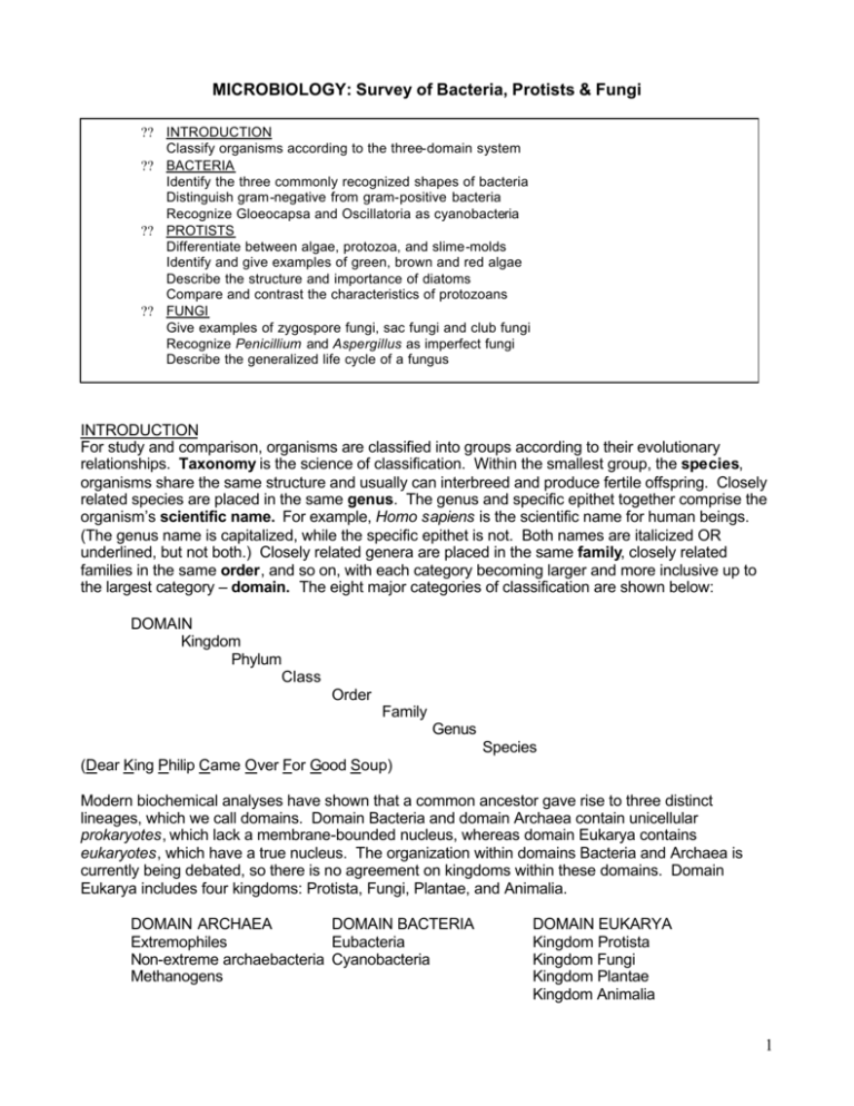
MICROBIOLOGY: Survey of Bacteria, Protists & Fungi ?? INTRODUCTION Classify organisms according to the three-domain system ?? BACTERIA Identify the three commonly recognized shapes of bacteria Distinguish gram-negative from gram-positive bacteria Recognize Gloeocapsa and Oscillatoria as cyanobacteria ?? PROTISTS Differentiate between algae, protozoa, and slime-molds Identify and give examples of green, brown and red algae Describe the structure and importance of diatoms Compare and contrast the characteristics of protozoans ?? FUNGI Give examples of zygospore fungi, sac fungi and club fungi Recognize Penicillium and Aspergillus as imperfect fungi Describe the generalized life cycle of a fungus INTRODUCTION For study and comparison, organisms are classified into groups according to their evolutionary relationships. Taxonomy is the science of classification. Within the smallest group, the species, organisms share the same structure and usually can interbreed and produce fertile offspring. Closely related species are placed in the same genus. The genus and specific epithet together comprise the organism’s scientific name. For example, Homo sapiens is the scientific name for human beings. (The genus name is capitalized, while the specific epithet is not. Both names are italicized OR underlined, but not both.) Closely related genera are placed in the same family, closely related families in the same order, and so on, with each category becoming larger and more inclusive up to the largest category – domain. The eight major categories of classification are shown below: DOMAIN Kingdom Phylum Class Order Family Genus Species (Dear King Philip Came Over For Good Soup) Modern biochemical analyses have shown that a common ancestor gave rise to three distinct lineages, which we call domains. Domain Bacteria and domain Archaea contain unicellular prokaryotes, which lack a membrane-bounded nucleus, whereas domain Eukarya contains eukaryotes, which have a true nucleus. The organization within domains Bacteria and Archaea is currently being debated, so there is no agreement on kingdoms within these domains. Domain Eukarya includes four kingdoms: Protista, Fungi, Plantae, and Animalia. DOMAIN ARCHAEA DOMAIN BACTERIA Extremophiles Eubacteria Non-extreme archaebacteria Cyanobacteria Methanogens DOMAIN EUKARYA Kingdom Protista Kingdom Fungi Kingdom Plantae Kingdom Animalia 1 DOMAINS ARCHAEA & BACTERIA All members of the Domains Archaea and Bacteria (formerly Kingdoms Archaebacteria & Eubacteria) are prokaryotic. Prokaryotes share the following features: ?? Unicellular ?? Lack organized nucleus and membrane-bounded organelles ?? Usually less than 1 um in diameter ?? Reproduction is primarily asexual via fission ?? May be autotrophic, chemotrophic, saprophytic or heterotrophic Because the archaebacteria are poorly understood, they will be eliminated from this microbiological survey. I. BACTERIA A. Classification Bacteria are the smallest and most numerous organisms. Bacteria exhibit great diversity. They are responsible for many diseases and also play a major role in modern medicine and agriculture. Most bacteria are found in three basic shapes: bacillus (rod-shaped), coccus (round or spherical), and spirillum (spiral or helical). Cocci may form clusters or chains, and rods may form long filaments. Some bacteria form endospores. An endospore contains a copy of the genetic material encased by heavy protective spore coats. Spores survive unfavorable conditions and germinate the form vegetative cells when conditions improve. B. In addition to being differentiated by shape, bacteria can be separated according to how they react to a staining procedure called Gram stain, named in honor of a nineteenth-century microbiologist, Hans Gram. Gram-positive bacteria are purple after being stained by the Gram stain procedure, while Gram-negative bacteria appear pink. The Gram stain reaction is important to bacteriologists because it is one of the first steps in identifying an unknown bacterium. Furthermore, the Gram stain reaction indicates a bacterium’s susceptibility or resistance to certain antibiotics, substances that inhibit the growth of bacteria. Procedures 1. Bacterial Shapes Study a “bacteria type” slide illustrating the three shapes of bacteria. You’ll need to use the high dry and oil immersion objectives on your compound microscope. (Don’t forget to use immersion oil with the 100X lens!) Draw and name the shapes of the bacteria you are observing below: _________________ Figure 1 __________________ _________________ Drawings of the three bacterial shapes (________X) 2 2. Gram stain reaction of various bacteria Examine the pre-stained slides indicated in the table below. Gram-positive bacteria are susceptible to penicillin, while Gram-negative bacteria are not. State the staining characteristics for the bacteria listed: Bacterial species Bacillus megaterium Rhodospirillum rubrum Escherichia coli Staphylococcus epidermidis 3. II. Gram reactions (+ or -) Culture common bacteria (per pair of students) a. Obtain two sterile cotton swabs and one closed petri dish containing sterile nutrient agar. b. Divide the plate into two sections by drawing a line down the center of the bottom of the plate. (Do not open the plate.) c. Drag the tip of the cotton swab over a surface, such as the fish tank, door handle, your tooth, etc. d. Open the petri plate and gently drag the exposed swab over half of the surface of the agar (as demonstrated by your TA). e. Find another surface and repeat steps “c” and “d” on the second half of the plate using a new swab. f. Close the lid and label the bottom edge of the dish with your initials and the surfaces that you swabbed. g. Place the plate upside down in the drawer at your lab bench. h. Examine the agar for bacterial growth in one week. CYANOBACTERIA Cyanobacteria, formerly called blue-green algae, are the photosynthetic members of the Domain Bacteria. Although cyanobacteria do not possess chloroplasts, they do have thylakoid membranes, where photosynthesis occurs. Procedure: Examine cyanobacteria Prepare a wet mount of the following organisms, or examine prepared slides, using the high dry and/or oil immersion objectives on your compound microscope. Draw what you see. A. Oscillatoria - This is a filamentous cyanobacterium with individual cells that resemble a stack of pennies. Oscillatoria takes its name from the characteristic oscillations that you may be able to see if your sample is alive. Observe this living culture, are oscillations visible? ___________ Oscillatoria (________X) 3 B. Gloeocapsa - Gloeocapsa is surrounded by a thick, gelatinous sheath. How does Gloeocapsa differ in appearance from Oscillatoria? In what ways are they similar? Gloeocapsa (_______X) III. NITROGEN FIXATION BY BACTERIA Atmospheric nitrogen is unavailable to plants. However, plants require nitrogen in relatively large quantities. Fortunately, certain bacteria and cyanobacteria are able to convert atmospheric nitrogen into usable form for plants. This process is called nitrogen fixation. Many of these “nitrogen-fixing” bacteria inhabit the roots of certain plants in “nodules”, while others reside in the soil. Demo – Root nodules 1. Observe the root nodules on display. The nodules containing nitrogen-fixing bacteria. 2. Examine the demonstration “root tubercle” slide. Can you locate the bacteria within the nodule? Sketch the roots and label the nodules containing nitrogen-fixing bacteria: ============================================================================= DOMAIN EUKARYA All members of the Domain Eukarya (Kingdoms Protista, Fungi, Plantae & Animalia) are eukaryotic. Eukaryotes share the following features: ?? Uni- or multicellular ?? Possess an organized nucleus and membrane-bounded organelles ?? Eukaryotic cells are generally larger than prokaryotic cells ?? Eukaryotic cellular division requires the formation of a spindle apparatus ?? May be autotrophic, saprotrophic or heterotrophic I. KINGDOM PROTISTA Representatives of the Kingdom Protista include all eukaryotes that lack the distinguishing characteristics of fungi, plants and animals = algae, protozoans, slime molds, and water molds. Its members are primarily unicellular, with few multicellular forms. The complexity and diversity of protists make it difficult to classify them. The following classification is used to study the protists: 4 CLASSIFICATION: THE PROTISTS Domain Eukarya Kingdom Protista Algae* Phylum Chlorophyta = green algae Phylum Phaeophyta = brown algae Phylum Rhodophyta = red algae Phylum Euglenophyta = euglenoids Phylum Bacillariophyta = diatoms Phylum Dinoflagellata = dinoflagellates Protozoans* Phylum Rhizopoda = amoebas and allies Phylum Ciliophora = ciliates Phylum Zoomastigophora = zooflagellates Phylum Apicomplexa = sporozoans Slime Molds* Phylum Gymnomycota = slime molds Water Molds* Phylum Oomycota = water molds *Categories not used in the classification of organisms, but added here for clarity. A. ALGAE – “plant-like” protists The algae make an enormous impact on the biosphere – both positively and negatively. Among their greatest contribution is the production of oxygen, because these are the photosynthetic protistans. Pond scum, frog spittle, seaweed, the stuff that clogs your aquarium if it’s not cleaned routinely, the debris on an ocean beach after a storm at sea, the nuisance organisms of a lake – these are the many images that pop into our mind when we first think about the organisms called algae. But many algae are also phytoplankton, the weakly swimming or floating algae that are at the base of the aquatic food chain. 1. Observation: Green Algae The green algae (Phylum Chlorophyta) may be ancestral to the first plants because both of these groups possess chlorophylls a and b, both store reserve food as starch, and both have cell walls that contain cellulose. You will examine a filamentous form (Spirogyra) and a colonial form (Volvox). a. Spirogyra Spirogyra is a filamentous alga that lives in fresh water and is often seen as a green scum on the surface of ponds and lakes. The most prominent feature of the cells is the spiral, ribbonlike chloroplast. ? Make a wet mount of live Spirogyra or observe a prepared slide. Draw what you see under the microscope: Spirogyra (_______X) 5 b. Volvox Volvox is a green algal colony. It is motile (capable of locomotion) because the thousands of cells that make up the colony have flagella. These cells are connected by delicate cytoplasmic extensions. Volvox is capable of both asexual and sexual reproduction. Certain cells of the adult colony can divide to produce daughter colonies that reside for some time in the parental colony. A daughter colony escapes the parental colony by releasing an enzyme that dissolves away a portion of the matrix of the parental colony. During sexual reproduction, some colonies of Volvox have cells that produce sperm, and others have cells that produce eggs. ? Using a depression slide, make a wet mount of live Volvox or observe a prepared slide. Draw what you see under the microscope and label the daughter colonies, if any. Volvox (________X) 2. Observation: Brown Algae The vast majority of the brown algae (Phylum Phaeophyta) are found in cold, marine environments. All members are multicellular, and most are macroscopic. Their color is due to the accessory pigment fucoxanthin, which is so abundant that it masks the green chlorophylls. Some species are used as food, while others are harvested for fertilizers. Of primary economic importance is algin, a cell wall component of brown algae that makes ice cream smooth, cosmetics soft, and paint uniform in consistency, among other uses. Kelps are large (up to 100 m long), complex brown algae. They are common along the seashores in cold waters. Fucus is a common brown alga of the coastal shore, especially abundant attached to rocks where the plants are periodically wetted by splashing waves and the tides. ? Examine the demonstration specimen of Fucus, noting the branching nature of the body. 3. Observation: Red Algae Like most brown algae, the red algae (Phylum Rhodophyta) are multicellular, but they occur chiefly in warmer seawater, both in shallow waters and as deep as light penetrates. Some forms of red algae are filamentous, but more often, they are complexly branched with a feathery, flat, and expanded or ribbonlike appearance. Red algae are the source of agar, a substance extracted from their cell walls. Agar is the solidifying agent in media on which some microorganisms are cultured. 6 4. Observation: Diatoms Diatoms possess a yellow-brown pigment in addition to chlorophyll. The diatom cell wall is in two sections, with the larger one fitting over the smaller as a lid fits over a box. Since the cell wall is impregnated with silica, diatoms are said to “live in glass houses”. The glass cell walls of diatoms do not decompose, so they accumulate in thick layers that are subsequently mined as diatomaceous earth and used in filters and as a natural insecticide. Diatoms, being photosynthetic and extremely abundant, are important food sources for the small heterotrophs in both marine and freshwater environments. ? View a prepared slide of diatoms. Draw the shapes that you identify under the microscope. Diatoms (________X) B. PROTOZOANS – “animal-like” protists The term protozoan refers to unicellular heterotrophs that ingest food via formation of a food vacuole. Protozoans are classified according to their mode of locomotion and food capture. 1. Observation: Amoeboid protozoans The best-known amoeboid protozoans (Phylum Sarcodina) are the amoebas, organisms that continually change shape by forming projections called pseudopodia (singular, pseudopodium, “false foot”). Organisms that locomote by forming pseudopodia are said to exhibit “amoeboid movement”. Food capture is by way of phagocytosis – the organism surrounds and engulfs a food organism with its pseudopodia, forming a food vacuole. One notorious human pathogen is Entamoeba histolytica, the cause of amoebic dysentery. ? Examine the prepared slide of Amoeba. Draw what you observe under the microscope. Amoeba (_________X) 7 2. Observation: Ciliated protozoans Most members of the Phylum Ciliophora are covered with numerous short locomotory structures called cilia. The cilia beat in synchrony to move the organism. One of the larges ciliates is the predatory Paramecium caudatum. Beating cilia drive food particles into the oral groove of the Paramecium. Food is then transported to the gullet. The particles become enclosed by a food vacuole, where digestion will take place. ? Prepare a wet mount of Paramecium caudatum or examine a prepared slide. Draw what you observe under the microscope. Paramecium caudatum (_____X) 3. Observation: Flagellated protozoans Members of the Phylum Mastigophora have one or more flagella to provide motility. The group includes some fairly notorious human parasites that cause disease, including giardiasis (from drinking contaminated water), some sexually transmitted diseases (via Trichomonas vaginalis), and African sleeping sickness, caused by Trypanosoma brucei. ? Examine a prepared slide of human blood that contains the parasitic flagellate Trypanosoma. Also view the image under the Protist microslide viewer. Draw what you see. Trypanosoma brucei (_____X) 4. Observation: Sporozoans All members of the Phylum Apicomplexa are parasites, infecting a wide range of animals, including humans. Plasmodium vivax causes one type of malaria in humans. P. vivax is transmitted to humans through the bite of an infected female Anopheles mosquito. The mosquito serves as a vector, a means of transmitting the organism from one host to another. Male mosquitoes cannot serve as vectors, because they lack the mouthparts for piercing skin and sucking blood. See Figure 25.9 on page 262 in Vodopich & Moore’s Biology Laboratory Manual, 6th edition. ? Examine the image on the Protist microslide under the microslide viewer. Draw what you see. 8 Plasmodium vivax (_____X) List two ways that you could distinguish between P. vivax (Phylum Apicomplexa) and T. brucei (Phylum Mastigophora) when observing a blood slide? o __________________________________________________ o C. __________________________________________________ SLIME MOLD – “fungus-like” protists The slime molds have both plantlike and animal-like characteristics. Because they engulf their food and lack a cell wall in their vegetative (nonreproductive) state, they are placed in the Kingdom Protista. However, when the reproduce, they produce spores with a rigid cell wall, similar to plants. Demo: Observe the demonstration plate of Physarum, a plasmodial slime mold. (Physarum is the yellow substance growing on an oatmeal food source.) D. II. WATER MOLD – “fungus-like” protists Some of the most notorious and historically important plant pathogens known to humans are water molds. Included in this group is Phytophthora infestans, the fungus that causes the disease known as late blight of potato. This disease spread through the potato fields of Ireland between 1845 and 1847. Most of the potato plants died, and 1 million Irish working-class citizens who had depended on potatoes as their primary food source starved to death. Another 2 million emigrated, many to the United States. Perhaps no other plant pathogen so poignantly illustrates the importance of environmental factors in causing disease. While Phytophthora infestans had been present previous to 1845 in the potato-growing fields of Ireland, it was not until the region experienced several consecutive years of wet and especially cool growing seasons that the late blight became a major problem. KINGDOM FUNGI Members of the kingdom fungi are primarily multicellular eukaryotes. Fungi are heterotrophic by absorption. Fungi are important decomposers, parasites, and foodstuffs. Structure: A fungal body, called a mycelium, is composed of many strands, called hyphae. Sometimes, the nuclei within a hypha are separated by walls, and sometimes they are not. Reproduction: Fungi produce windblown spores (small, haploid bodies with a protective covering) when they reproduce sexually or asexually. Following sexual union, a collection of specialized hyphae, called a fruiting body, is found in some groups. Spores are produced and released by the fruiting bodies. Classification: Fungi are classified according to differences in their life cycle and the type of structure that produces spores during sexual reproduction. 9 DOMAIN EUKARYA KINGDOM FUNGI Phylum Zygomycota – zygospore fungi Phylum Ascomycota – sac fungi Phylum Basidiomycota – club fungi Phylum Deuteromycota – imperfect fungi Draw the generalized life cycle of a fungus (refer to Figure 26.2 on page 268 in Ex. 26 of the Vodopich & Moore Biology Lab Manual): Generalized life cycle of a fungus A. PHYLUM ZYGOMYCOTA: zygospore fungi Representatives: Soil and dung molds, black bread molds (Rhizopus) Commonly called “zygomycetes”, all members of this phylum produce a thick-walled zygote called a zygosporangium. Most zygomycetes are saprobes. The common black bread mold, Rhizopus, is a representative zygomycete. Before the introduction of chemical preservatives into bread, Rhizopus was an almost certain invader, especially in high humidity. Observation: Rhizopus demo plate 1. Observe the demo plate of Rhizopus. Note many hyphae (individual strands) that make up the mycelium. 2. Now note that the culture contains numerous black “dots”. These are the sporangia (singular, sporangium). Sporangia are containers of spores by which Rhizopus reproduces asexually. During sexual reproduction in Rhizopus, two different mycelia must grow in close proximity. (The difference in the mycelia is genetic rather than structural. Because they are impossible to distinguish, the mycelia are simply referred to as + and – mating types.) As the hyphae from each mating type grow close, gametangia are produced at their tips. Each gametangium contains many haploid nuclei of a single mating type. When the gametangia make contact, the wall between the two dissolves and the cytoplasms of each mix. The many haploid nuclei fuse. The resulting cell containing the diploid nuclei is called a zygospore. Eventually a thick wall forms around the zygospore forming the zygosporangium. Observation: Prepared slide of Rhizopus 1. Obtain a prepared slide of Rhizopus. Examine it with the medium- and highpower objective of your compound microscope. 2. Find the stages of sexual reproduction in Rhizopus, including gametangia and zygosporangia. 10 ? B. Draw what you see and label the structures. Rhizopus (______X) PHYLUM ASCOMYCOTA: sac fungi Representatives: Many small, wood-decaying fungi, yeasts (Saccharomyces), molds (Neurospora), morels, cup fungi, truffles; plant parasites: powdery mildews, ergots Members of the ascomycetes produce spores (ascospores) in a sac, the ascus (plural, asci), which develops as a result of sexual reproduction. Asexual reproduction takes place when the fungus produces asexual spores called conidia (singular, conidium). The phylum includes organisms of considerable importance, such as the yeasts crucial to the baking and brewing industries, as well as numerous plant pathogens. A few are highly prized as food, including morels and truffles. Truffles cost in excess of $400 per pound! Yeasts differ from most other fungi because they are not composed of hyphae. Even though yeasts are unicellular, they form an ascus during sexual reproduction. Usually, however, yeasts reproduce asexually, either my mitosis and cell division or by budding. Observation: Saccharomyces demo plate and prepared slide Saccharomyces is a type of yeast commonly used in the brewing and baking industry. 1. Observe the demonstration plate on display. Note the absence of a mycelium and individual hyphae. Why is this? _______________________________ 2. Examine a prepared slide of yeast cells. The cup fungi are commonly found on soil during cool early spring and fall weather. Their sexual spore-containing structures are cup shaped, hence their common name. Actually, the structure we identify as the cup fungus is the fruiting body, produced as a result of sexual reproduction by the fungus. Most of the organism is present within the soil as an extensive mycelium. Specifically, the fruiting body is called an ascocarp. Observation: Peziza, a cup fungus 1. Observe a preserved or dried specimen of a cup fungus. 2. Obtain a prepared slide of the ascocarp of Peziza. Examine the slide with the medium and high power objectives of your compound microscope. 3. Identify the elongate fingerlike asci, which contain dark-colored, spherical ascospores. ? Sketch an ascus and the ascospores. Peziza (_____X) 11 C. PHYLUM BASIDIOMYCOTA: club fungi Representatives: Mushrooms, stinkhorns, puffballs, bracket and shelf fungi, coral fungi; plant parasites: rusts, smuts Members of this group of fungi are probably what first come to mind when we think of fungi, because this phylum contains those organisms called mushrooms. Actually the mushroom is only a portion of the fungus – it’s the fruiting body, specifically a basidiocarp, containing the sexually produced haploid basidiospores. These basidiospores are produced by a club-shaped basidium for which the group is named. Much (if not most) of the fungal mycelium grows out of sight, within the substrate on which the basidiocarp is found. Mushrooms called gill fungi have their sexual spores produced on sheets of hyphae that look like the gills of fish. Pore mushrooms have pores (tubes) instead of gills. Observation: Mushrooms 1. Obtain an edible mushroom and identify as many of the following structures as possible, based on the description given: a. Stalk: The upright portion that supports the cap. b. Annulus: A membrane surrounding the stalk where the immature (button-shaped) mushroom was attached. c. Cap: The umbrella-shaped portion of the mushroom. d. Gills: On the underside of the cap, radiating lamellae on which the basidia are located. e. Basidia: On the gills, club-shaped structures where basidiospores are located. f. Basidiospores: Spores produced by basidia. 2. Sketch your mushroom and label all of the parts. Mushroom 3. 4. 5. Return the mushroom to its original location in the room. View a prepared slide of a cross section of Coprinus. Using the three dry microscope objectives, look for the gills, basidia, and basidiospores. Sketch what you see below. Label the parts. Coprinus (_____X) 12 Observation: Schizophyllum A common plant pathogen, compare the structure of Schizophyllum, a basidiomycete, to that of the mushroom. How are they different? How are they similar? D. PHYLUM DEUTEROMYCOTA: imperfect fungi (i.e., no known means of sexual reproduction) Representatives: Athlete’s foot, ringworm, candidiasis, Penicillium, Aspergillus Deuteromycetes reproduce asexually by forming spores called conidia on upright hyphae known as conidiophores. They are “imperfect” in the sense that no sexual stage has yet been observed (leading to the use of deutero, meaning “second”) and may not exist. Cell structure and biochemistry indicate that imperfect fungi are sac fungi (ascomycetes) that have lost the ability to reproduce sexually. Imperfect fungi include the blue mold Penicillium species, which are famous for the production of the antibiotic penicillin, and the green mold Aspergillus species, which are used to manufacture products ranging from soy sauce to chewing gum. These molds grow on a variety of materials, such as fruits, cheese, leather, paper, cloth, etc. Candida albicans is a yeastlike imperfect fungus that causes vaginal infection in females. Observation: Penicillium and Aspergillus 1. Observe the demonstration plate of Penicillium. Note, especially, the color of the organism. 2. Examine a prepared slide of Penicillium. 3. Examine a prepared slide of Aspergillus. Compare the conidiophores and conidia to that of Penicillium. How do they differ? Sketch the conidiophores and conidia of Penicillium and Aspergillus below: Penicillium (____X) E. Aspergillus (____X) MUTUALISTIC FUNGI: LICHENS Fungi team up with plants or members of the Domain Bacteria or Kingdom Protista to produce some remarkable mutualistic relationships. The best way to envision such relationships is to think of them as good interpersonal relationships, in which both members benefit and are made richer than would be possible for each individual on its own. Lichens are organisms made up of fungi and either green algae (Kingdom Protista) or cyanobacteria (Domain Bacteria). The algal or cyanobacterial cells photosynthesize, and the fungus absorbs a portion of the carbohydrates produced. The fungal mycelium provides a protective, moist shelter for the photosynthesizing cells. Hence, both partners benefit from the relationship. 13 Observation: Lichens 1. Examine the demonstration specimen of a crustose lichen. What is it growing on? _________________________ Describe the appearance of the crustose lichen: 2. Examine the demonstration specimen of a foliose lichen. What is it growing on? _________________________ Describe the appearance of the foliose lichen: 3. Examine the demonstration specimen of a fruticose lichen. What is it growing on? _________________________ Describe the appearance of the fruticose lichen: 4. Examine a cross section of a lichen using all three dry objectives of the compound microscope. This particular lichen is composed of fungal and green algae cells. Locate the fungal mycelium and then the algal cells. (Draw and label below:) Lichen (____X) It’s likely that this slide has a cup-shaped fruiting body. To which fungal phylum does this fungus belong? _________________________________ For more information regarding bacteria, protists or fungi, refer to Exercises 23, 24, 25 and 26 of Vodopich & Moore’s Biology Lab Manual, 6th edition. Information contained in this handout was adapted from the following sources: Sylvia S. Mader, Laboratory Manual: Inquiry Into Life, 10th edition, McGraw-Hill Higher Education, 2003, pp.323-344. Perry, Morton & Perry, Laboratory Manual for Starr and Taggart’s “Biology: The Unity and Diversity of Life” and Starr’s “Biology: Concepts and Applications”, Wadsworth Group, Thomson Learning, 2002, pp.245-320. Vodopich & Moore, Biology Laboratory Manual, 6th edition, McGraw-Hill Higher Education, 2002, pp.229-278. 14
