Annotated Bibliography
advertisement
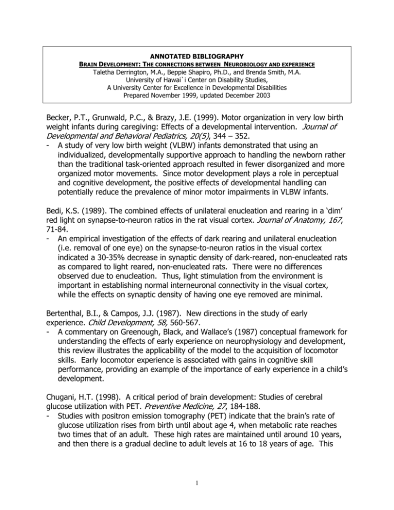
ANNOTATED BIBLIOGRAPHY BRAIN DEVELOPMENT: THE CONNECTIONS BETWEEN NEUROBIOLOGY AND EXPERIENCE Taletha Derrington, M.A., Beppie Shapiro, Ph.D., and Brenda Smith, M.A. University of Hawai`i Center on Disability Studies, A University Center for Excellence in Developmental Disabilities Prepared November 1999, updated December 2003 Becker, P.T., Grunwald, P.C., & Brazy, J.E. (1999). Motor organization in very low birth weight infants during caregiving: Effects of a developmental intervention. Journal of Developmental and Behavioral Pediatrics, 20(5), 344 – 352. - A study of very low birth weight (VLBW) infants demonstrated that using an individualized, developmentally supportive approach to handling the newborn rather than the traditional task-oriented approach resulted in fewer disorganized and more organized motor movements. Since motor development plays a role in perceptual and cognitive development, the positive effects of developmental handling can potentially reduce the prevalence of minor motor impairments in VLBW infants. Bedi, K.S. (1989). The combined effects of unilateral enucleation and rearing in a ‘dim’ red light on synapse-to-neuron ratios in the rat visual cortex. Journal of Anatomy, 167, 71-84. - An empirical investigation of the effects of dark rearing and unilateral enucleation (i.e. removal of one eye) on the synapse-to-neuron ratios in the visual cortex indicated a 30-35% decrease in synaptic density of dark-reared, non-enucleated rats as compared to light reared, non-enucleated rats. There were no differences observed due to enucleation. Thus, light stimulation from the environment is important in establishing normal interneuronal connectivity in the visual cortex, while the effects on synaptic density of having one eye removed are minimal. Bertenthal, B.I., & Campos, J.J. (1987). New directions in the study of early experience. Child Development, 58, 560-567. - A commentary on Greenough, Black, and Wallace’s (1987) conceptual framework for understanding the effects of early experience on neurophysiology and development, this review illustrates the applicability of the model to the acquisition of locomotor skills. Early locomotor experience is associated with gains in cognitive skill performance, providing an example of the importance of early experience in a child’s development. Chugani, H.T. (1998). A critical period of brain development: Studies of cerebral glucose utilization with PET. Preventive Medicine, 27, 184-188. - Studies with positron emission tomography (PET) indicate that the brain’s rate of glucose utilization rises from birth until about age 4, when metabolic rate reaches two times that of an adult. These high rates are maintained until around 10 years, and then there is a gradual decline to adult levels at 16 to 18 years of age. This 1 pattern correlates with rates of synapse formation and supports the idea of critical periods for maximal learning capacity. Chugani, H.T., Behen, M.E., Muzik, O., Juhasz, C., Nagy, F. & Chugani, D.C. (2001). Local brain functional activity following early deprivation: A study of postinstitutionalized Romanian orphans. NeuroImage, 14, 1290-1301. - Positron emission tomography (PET) was used to identify brain regions involved in the long-term deficits exhibited by U.S.-adopted Romanian orphans who experienced early global deprivation. Compared to 2 control groups, ten children aged 7 to 11 exhibited significantly decreased glucose metabolism in medial temporal structures, the brain stem, and in the orbital frontal, prefrontal limbic, and lateral temporal cortices. These areas are strongly interconnected and are damaged as a result of prolonged stress. The authors suggest that the early chronic stress endured by these Romanian orphans resulted in pathological development of these circuits, which may be the mechanism underlying their persistent behavior disorders. Chugani, H.T., Phelps, M.E., & Mazziotta, J.C. (1987). Positron emission tomography study of human brain functional development. Annals of Neurology, 22, 487-497. - Positron emission tomography (PET) was used to study glucose utilization rates in different brain regions of 29 children aged 5 days to 15 years. Data indicate a different time course in various brain regions for an increase in glucose utilization to twice the adult rate by 3 - 4 years. This time course parallels the time course for initial overproduction and subsequent elimination of excessive neurons and synaptic connections in the developing brain, and it matches the developmental timeline of behavioral acquisition. The authors conclude that PET is a potentially powerful approach to studying the mechanisms of normal and abnormal brain development. Dehaene-Lambertz, G., & Dehaene, S. (1994). Speed and cerebral correlates of syllable discrimination in infants. Nature, 370, 292-294. - Behavioral inhibition research indicates that young infants possess remarkable language capabilities, for example, the ability to discriminate more phonemes than are present in their native language, which regresses during their first year. In order to determine what brain mechanisms are involved and how fast infants can detect phonetic change, this study recorded the event-related potentials in the brains of 16 infants aged 2-3 months who were listening to different syllables. Processing was localized to the temporal lobes, and phonemic discrimination occurred in less than 400 ms. Eimas, P. (1985). The perception of speech in early infancy. Scientific American, 252(1), 46-52. - Behavioral research with infants suggests than infants are born with many of the capacities involved in later speech perception and comprehension, as illustrated by their ability to perceive linguistic contrasts in foreign languages, which declines rapidly by 1 year of age. Authors postulate that innate perceptual capacities evolved 2 in speech centers to biologically equip infants to acquire their parents’ language as quickly as possible. Fischer, K.W. (1987). Relations between brain and cognitive development. Child Development, 58, 623-632. - Reviewing research on brain and cognitive development, the author reports that the developmental pattern of synaptogenesis, head growth spurts, and changes in electroencephalogram readings show synchronized developmental discontinuities at the same time as the emergence of behavioral skills which signify the attainment of developmental stages. Gauthier, I., Tarr, J.J., Anderson, A.W., Skudlarski, P. & Gore, J.C. (1999) Activation of the middle fusiform ‘face area’ increases with expertise in recognizing novel objects. Nature Neuroscience, 2(6), 568-573. - Functional magnetic resonance imaging (fMRI) shows changes associated with increasing expertise in the fusiform gyrus (FG), an area known to have an activation “preference” for faces. Acquisition of expertise with novel objects (greebles) led to increased activation in the right FG for matching of upright greebles as compared to matching inverted greebles. Expertise seems to be one factor that leads to facial specialization of the FG. Goldman-Rakic, P.S. (1987). Development of cortical circuitry and cognitive function. Child Development, 58, 601-622. - Electron microscopic studies of areas of the cerebral cortex of non-human primates indicate that the number and density of synapses increases rapidly to higher than adult levels between 2 and 4 months of age, and then slowly declines to adult levels. The ability to perform delayed response tasks emerges around 4 months, suggesting that a critical mass of synapses is necessary for the emergence of behavior, and the elimination of excess synapses during childhood and adolescence is necessary for fully mature abilities. Greenough, W.T., Black, J.E., & Wallace, C. (1987). Experience and brain development. Child Development, 58, 539-559. - Reviewing neurological and psychological research, the authors advance the idea that experience effects neurological structure through two mechanisms: experienceexpectant, which can be described as a biologically driven overproduction of synapses which prepares the brain to receive information from the environment, followed by the elimination of synapses that are not stimulated by environmental experience; and experience-dependent, which involves the active formation of synapses in response to the experience which is to be stored. Although these mechanisms are probably not mutually exclusive, they provide a better description of the neural processes involved in learning and can explain both the accelerated learning ability of infants and children, and the ability for adults to learn and for victims of brain injury to recover. 3 Gunnar, M.R. (1996, December). Quality of care and the buffering of stress physiology: Its potential role in protecting the developing brain. Paper presented at the National Center for Clinical Infant Programs Conference, Washington, D.C. - This paper discusses an example of the brain’s ability to change with experience in the functioning of the hypothalamic-pituitary-adrenocortical (HPA) system. Stressful experiences stimulate this system, which produces cortisol, a hormone which facilitates neural death and synapse elimination; chronic stimulation can cause shrinkage of brain areas like the hippocampus (memory), impair selective attention, and ultimately cause the HPA to remain overactive, resulting in hypervigilant, anxious behavior. The HPA system is buffered through secure interpersonal relationships with caregivers, suggesting a method of dampening the deleterious effects of stress on neurological development. Haxby, J.V., Ungerleider, L.G., Clark, V.P., Schouten, J.L., Hoffman, E.A., & Martin, A. The effect of face inversion on activity in human neural systems for face and object perception. Neuron, 22, 189-199. - This study used functional magnetic resonance imaging (fMRI) to study the effect of inversion on face and object perception. Upright facial processing consistently occurred in the fusiform gyrus. Inversion elicited an additional, increased response in regions normally responsive to objects; however, inversion of objects did not result in a reciprocal activation of the fusiform gyrus. Huttenlocher, P.R. (1994). Synaptogenesis, synapse elimination, and neural plasticity in human cerebral cortex. In C.A. Nelson (Ed.), Threats to optimal development: integrating biological, psychological, and social risk factors. The Minnesota symposia in child psychology, (27, pp.35-54). New Jersey: Lawrence Erlbaum. - Evidence from several lines of research on brain development indicates that genes signal for the creation and migration of most cortical neurons before the child is born. After birth, genes code for the proliferation of connections between neurons, peaking at twice adult levels between 1 and 3 years of age, depending on the cortical region, before slowly declining to adult levels in adolescence. Which circuits will persist depends upon sensory experience from the environment, and synapse formation is directly linked to the emergence of skills and behaviors. The increased functional plasticity of infants’ and young children’s brains may result in increased efficacy of education on cognitive development during this period, and also account for the remarkable recovery of ability after brain injuries sustained early in life. Jessell, T.M. (1991). Neuronal survival and synapse formation. In Kandel, E.R., Schwartz, J.H., & Jessell, T.M. (Eds.) Principles of Neural Science, (3rd ed., pp. 930944). New York, NY: Elsevier. - An outline of empirical evidence on the widespread phenomenon of neuronal death, indicating that a neuron’s survival depends on having connections to other neurons. The mechanisms that are responsible for the formation and maintenance of 4 synapses are discussed, and evidence indicates that these processes are dependent on active communication between two neurons. Julesz, B. (1991). Early vision and focal attention. Reviews of Modern Physics, 63(3), 735-772. - This review of brain research and the field of psychobiology focuses on vision and focal attention. The author concludes that binocularity develops from the bottom up, i.e. through visual input from the retina which then travels to the visual cortex for processing, without input from higher cognitive centers (e.g. attentional and reasoning processes, termed top-down). Although genes create strong neurological predispositions for an organism to be able to see at birth, there is a maturational window during the first months of life where visual experience is crucial, and, if missed, will result in permanent stereo-blindness. Kandel, E.R. (1991). Brain and behavior. In Kandel, E.R., Schwartz, J.H., Jessell, T.M. (Eds.) Principles of Neural Science, (3rd ed., pp. 5-17). New York, NY: Elsevier. - This chapter provides a survey of neurological evidence and theories on how the human brain controls human behavior through the action of neurons and supporting glial cells. Research has revealed that specific areas of the brain control specific behavioral skills, and that a person’s environment, including other people’s behavior, can affect the functioning of individual neurons. Kanwisher, N., McDermott, J. & Chun, M.M. (1997). The fusiform face area: A module in human extrastriate cortex specialized for face perception. The Journal of Neuroscience, 17(11), 4302-4311. - This study used functional magnetic resonance imaging (fMRI) to locate occipitotemporal areas specialized for face perception. Fifteen subjects passively viewed photographs of faces and assorted common objects, and strength of cortical specialization was measured by the responsiveness of the same region of cortex to different stimuli. In 12 subjects, the right fusiform gyrus was selectively activated by faces compared to various control stimuli. Kermoian, R., & Campos, J. (1988). Locomotor experience: A facilitator of spatial cognitive development. Child Development, 59, 908-917. - Two studies were designed to test the influence of locomotor experience on performance in tests on object permanence in spatial search. Data indicate that the longer an infant had been moving him/herself, the better (s)he did in spatial search. A third study indicated that more organized and less effort-intensive locomotion, e.g. hands-and-knees vs. belly crawling, also predicted better performance. Kirkwood, A., Lee. H., & Bear, M. (1995). Co-regulation of long-term potentiation and experience-dependent synaptic plasticity in visual cortex by age and experience. Nature, 375(6529), 328-331. 5 - The creation of synapses that mediate binocular vision requires simultaneous input from both eyes during a postnatal critical period in rats. The visual cortex of rats is most susceptible to long-term potentiation (LTP), which is a lasting enhancement of activity in a neuron following specific stimulation, during the postnatal critical period. Data support the idea that LTP is a mechanism which mediates experiencedependent synaptic modifications in the developing mammalian brain. Kupfermann, I. (1991). Hypothalamus and limbic system: Peptidergic neurons, homeostasis, and emotional behavior. In Kandel, E.R., Schwartz, J.H., Jessell, T.M. (Eds.) Principles of Neural Science, (3rd ed., pp. 735-749). New York, NY: Elsevier. - An examination of the anatomy of the hypothalamus and limbic system which discusses the role of this system in regulating endocrine and autonomic systems and emotional behavior. Studies indicate that the limbic system undergoes structural and biochemical changes in response to behavioral demands, for example, neuronal degeneration occurs in response to chronically circulating hormones which are released in response to stress. Locke, J.L. (1992). Thirty years of research on developmental neurolinguistics. Pediatric Neurology, 8(4), 245-250. - The study of neurolinguistics in the last 30 years has established a genetic, neuroanatomic, and functional basis for a variety of disorders, such as dyslexia, autism, and delayed language. Research in several areas indicates that it is possible to detect early signs of developmental language delay during the first year of life, as the ability to take turns, to gesture, and to babble with complexity are important precursors to language ability and may reflect a state of neural maturation necessary for spoken language. Mayeux, R. & Kandel, E.R. (1991). Disorders of language: The aphasias. In Kandel, E.R., Schwartz, J.H., Jessell, T.M. (Eds.) Principles of Neural Science (3rd ed., pp. 839851). New York, NY: Elsevier. - Language is characterized from the neurobiological point of view. Functional control of language is localized to one of the two cerebral hemispheres, usually the left, and the different capabilities involved in language are further localized to different regions within the hemisphere. Language capability is present at birth, but environment has a profound impact on the development of language skills. McCarthy, G. (1997) Face specific processing in the human fusiform gyrus. Journal of Cognitive Neuroscience; 9(5), 1-9. - The researchers employed functional magnetic resonance imaging (fMRI) to identify face-specific cortical areas. Subjects viewed faces and then flowers presented in continuously amongst common objects or nonobjects. The posterior fusiform gyrus was bilaterally activated by faces and by objects when viewed among nonobjects. Only a focal region in the right lateral fusiform gyrus was activated by faces viewed among objects; flowers viewed among objects did not activate this region. 6 Nelson, C.A. (2000). The neurobiological bases of early intervention. In J.R. Shokoff & S.J. Meisels (Eds.), Handbook of Early Childhood Intervention (2nd ed., 204-227). New York, NY: Cambridge University Press. - A review of neurobiological development and research with humans and animals indicates the neurobiological mechanisms by which early intervention can help children with disabilities. Nicholls, J.G., Martin, A.R., & Wallace, B.G. (1992). From Neuron to Brain (3rd ed.). Sunderland, Massachusetts: Sinauer Associates, Inc. - Experiments on single cells or simple assemblies of neurons in a wide range of species and how their functioning translates into behavior are presented. The data indicate that the brain is constructed according to a highly-ordered design made up of relatively simple components and widespread mechanisms, such as the death of neurons and the elimination of synapses that result from neuronal inactivity. Perry, B., & Azad, I. (1999). Posttraumatic stress disorder in children and adolescents. Current Opinion in Pediatrics, 11, 310-316. - Over 30% of children exposed to traumatic experiences develop posttraumatic stress disorder, which can lead to depression, suicidal behavior, alcoholism, organic disease, and poor health as an adult, to name but a few. Trauma alters the structure and functioning of the limbic system, causing damage to memory and circuits linking brain to body, and decreased emotional regulatory capacity. A selfpromoting negative cycle can ensue leading to additional physical, cognitive, and emotional pathology, which is not likely to remit without treatment. Four treatment approaches are described, as is the idea of resiliency to the noxious effects of trauma on the nervous system afforded by a secure relationship with a caregiver. Pierce, D., & Courchesne, E. (2000). Exploring the neurofunctional organization of face processing in Autism. Archives of General Psychiatry, 57(4), 344-346. - Social dysfunction in autism is severe and likely involves abnormalities in multiple neural regions. Neurodevelopmental evidence is suggestive of the potential to increase activation in the fusiform gyrus (FG) area of the brain in order to develop “expertise” with faces. Future research on the neurological bases of social information processing will bring the field closer to identifying etiologies and effective treatments for autism. Porter, F.L., Grunau, R.E., & Anand, K.J. (1999). Long-term effects of pain in infants. Journal of Developmental Behavioral Pediatrics, 20(4), 253-261. - The growing body of evidence that early pain and stress have long-term effects on human neural development is examined. The explosion of synapse formation in the early postnatal period makes the brain most plastic and provides a critical period that maximizes environmental influence on the brain, and therefore on subsequent behavior. Repetitive exposure to stress early in life causes structural and functional 7 change in the developing nervous system which can lead to anxiety, depression, hyper-responsivity to pain and stress, cognitive deficits, morbidity, and early mortality. The provision of pain-management could be an important buffer for the deleterious effects of early pain and stress on neural development. Reilly, J.S., Bates, E.A., & Marchman, V.A. (1998). Narrative discourse in children with early focal brain injury. Brain and Language, 61, 335-375. - The linguistic proficiency and narrative competence of 30 children with pre- or perinatal brain damage was examined when the children were 3 yr., 7 mo. to 10 yr., 10 mo. old. Comparing their performance to age-matched controls, the data indicate that brain-damaged children performed lower on these indices. Although children with early brain damage go on to develop nearly normal language ability as compared to adult victims of brain injury, deficits in ability are apparent when children face more complex linguistic challenges. Ruben, R.J. (1997). A time frame of critical/sensitive periods of language development. Acta Otolaryngol,117(2), 202-205. - The author presents a summary of the evidence for critical/sensitive periods in language development. The critical period for phonology is from 24 weeks gestational age to 1 year of age. The critical period for syntax runs through 4 years, and for semantics the period lasts through age 15 to 16. Although these elements of language have different critical/sensitive periods, they are developmentally interdependent; for example, insufficient auditory input (e.g. due to hearing loss from otitis media) decreases the amount of phonological input, which results in decreased semantic and syntactic capacities. Abnormal sensory input during critical periods is described as a disease vector; conversely, early, linguistically enriched and organized sensory input will lay the foundations for sophisticated language abilities. Sengpiel, F., Stawinski, P., and Bonhoeffer, T. (1999). Influence of experience on orientation maps in cat visual cortex. Nature Neuroscience, 2(8), 727-732. - This study considers the effects of rearing kittens in a striped environment on the development of ocular dominance columns in the visual cortex. Data indicate that experience plays an instructive role whereby some neurons shift their genetically predisposed orientation preference to a preference for the orientations experienced by the young kitten, supporting the idea that genetically predetermined functioning of neurons can be altered by experience. Schultz, R.T., Gauthier, I., Klin, A., Fulbright, R.K., Andersen, A.W., Volkmar, F., et al. (2000). Abnormal ventral temporal cortical activity during face discrimination among individuals with Autism and Asperger Syndrome. Archives of General Psychiatry, 57(4), 331-340. - Functional magnetic resonance imaging (fMRI) of face and object perception in adults with autism was compared to 2 age- and IQ-matched control groups of typically developing individuals. During face processing, individuals with autism 8 exhibited significantly greater activation in the inferior temporal gyrus and less activation of the fusiform gyrus (FG). This pattern of activation suggests a featurebased strategy more typical of nonface object perception. The authors hypothesize about the developmental course and consequence of this difference, suggesting that inadequate attention to faces characteristic of children with autism means the FG cannot “practice” and become “expert” at facial processing, which is a requisite for good social skills. Siegel, D.J. (1999). The Developing Mind. New York: Guilford Press. - This book provides an in-depth look at the neurodevelopmental processes of the brain, including the biology of memory, attachment, emotion, modes of processing, states of mind, self-regulation, interpersonal connections, and integration. Since the brain is undergoing rapid, experience-dependent development for the first several years of life, this suggests that intervention will be most influential on the neuroanatomy of the brain if provided early. Yim, G. (1998, November). Early brain development and windows of opportunity. Presented at the meeting of the Hawai‘i Association for the Education of Young Children (HAEYC) Conference. Honolulu, Hawai‘i. - Basic principals of brain development, neuroanatomy, functional control of specific brain regions, and the existence of windows of opportunity for promoting optimal development are presented. The author discusses the ages where these windows are “wide-open” to allow experience to influence development of different developmental skills, as well as activities that a caregiver can provide for a child to promote development by taking advantage of these windows. Also discussed is a specific application of these ideas to attention deficit disorder. 9


