Fetal Pig Review Cont'd KEY - OG
advertisement
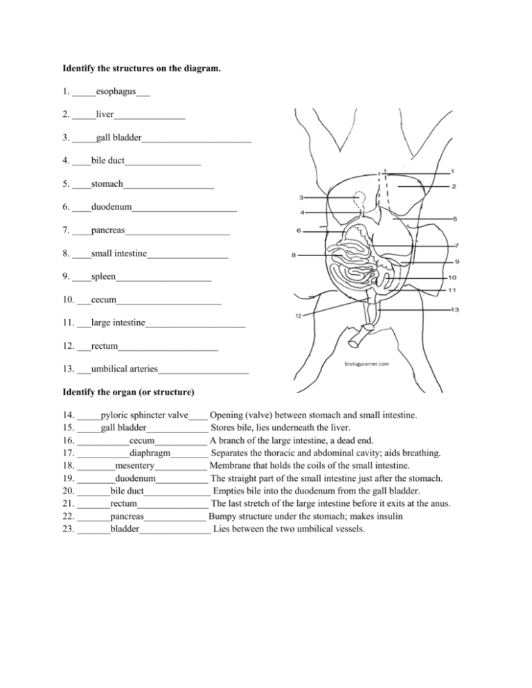
Identify the structures on the diagram. 1. _____esophagus___ 2. _____liver_______________ 3. _____gall bladder_______________________ 4. ____bile duct________________ 5. ____stomach___________________ 6. ____duodenum______________________ 7. ____pancreas______________________ 8. ____small intestine_________________ 9. ____spleen____________________ 10. ___cecum______________________ 11. ___large intestine_____________________ 12. ___rectum_____________________ 13. ___umbilical arteries___________________ Identify the organ (or structure) 14. _____pyloric sphincter valve____ Opening (valve) between stomach and small intestine. 15. _____gall bladder_____________ Stores bile, lies underneath the liver. 16. ___________cecum___________ A branch of the large intestine, a dead end. 17. ___________diaphragm________ Separates the thoracic and abdominal cavity; aids breathing. 18. ________mesentery___________ Membrane that holds the coils of the small intestine. 19. ________duodenum___________ The straight part of the small intestine just after the stomach. 20. _______bile duct______________ Empties bile into the duodenum from the gall bladder. 21. _______rectum_______________ The last stretch of the large intestine before it exits at the anus. 22. _______pancreas_____________ Bumpy structure under the stomach; makes insulin 23. _______bladder_______________ Lies between the two umbilical vessels. Identify by number: Aorta __2__ Dorsal Aorta _9__Pulmonary Trunk _1_ Common carotid _4__ Left & Right Carotid _7,8__ Coronary vessels _6__ Left Subclavian__5__ Right Subclavian __10__ Right Brachiocephalic _3___ Right Atrium __12__ Left Atrium _13__ Intercostal __11___ Ventricle __14_ Identify the structure. 1. _______pericardium________ Membrane over the heart. 2. _____trachea________ Airway from mouth to lungs 3. ____carotids_______ Blood supply to head 4. ____ventricles_________ Lower heart chambers 5. ____dorsal aorta_______ Blood supply to lower body 6. ____diaphragm_______ Muscle to aid breathing 7. ____vena cava_______ Returns blood to heart 8. ____aorta (or pulmonary)____ Large vessel at top of heart 9. ____larynx______ Used to make noises 10. ___coronary______ Arteries on heart surface. Label the structures: http://www.biologycorner.com/pig/fetal.html - Students look at picture, hover over ‘hot spots’ to see what organ it is, and what its function is http://www.biologycorner.com/myimages/fetal-pig-dissection/ -Pictures unlabelled/labeled of fetal pig, dissected http://faculty.clintoncc.suny.edu/faculty/michael.gregory/files/bio%20102/bio%20102%20laboratory/fe tal%20pig/fetal%20pig.htm – Goes thru dissection with instructions and labeled pictures http://biology.uco.edu/animalbiology/pigweb/pig.html - Categorized by system; students look at numbered photos and identify structures; key is given at end http://www.biologycorner.com/pig/quiz2.html - Practice Pig Practical – Print the answer sheet to go with it - http://www.biologycorner.com/worksheets/lab_practical_blanks.html

