Pulmonology Review
advertisement
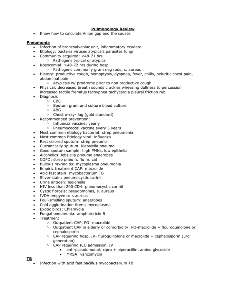
Pulmonology Review Know how to calculate Anion gap and the causes Pneumonia Infection of broncoalveolar unit, inflammatory ecudate Etiology: bacteria viruses atypicals parasites fungi Community acquired; <48-71 hrs o Pathogens typical or atypical Nosocomial: >48-72 hrs during hosp o Pathogens commonly gram neg rods, s. aureus History: productive cough, hemoptysis, dyspnea, fever, chills, peluritic chest pain, abdominal pain o Atypicals w/ prodrome prior to non productive cough Physical: decreased breath sounds crackles wheezing dullness to percussion increased tactile fremitus tachypnea tachycardia pleural friction rub Diagnosis o CBC o Sputum gram and culture blood culture o ABG o Chest x-ray- lag (gold standard) Recommended prevention: o Influenza vaccine; yearly o Pneumococcal vaccine every 5 years Most common etiology bacterial: strep pneumonia Most common Etiology viral: influenza Rest colored sputum: strep pneumo Currant jelly sputum: klebsiella pneumo Good sputum sample: high PMNs, low epithelial Alcoholics: lebsiella pneumo anaerobes COPD: strep pneu h. flu m. cat Bullous myringitis: mycoplasma pneumonia Empiric treatment CAP: macrolide Acid fast stain: mycobacterium TB Silver stain: pneumocystic carinii Urine antigen: legionella HIV less than 200 CD4: pneumocystic carinii Cystic fibrosis: pseudomonas, s. aureus IVDA empyema: s aureus Foul-smelling sputum: anaerobes Cold agglutination titers; mycoplasma Exotic birds: Chlamydia Fungal pneumonia: amphotericin B Treatment o Outpatient CAP, PO: macrolide o Outpatient CAP in elderly or comorbidity: PO-macrolide + flouroquinolone or cephalosporin o CAP requiring hosp, IV- fluroquinolone or macrolide + cephalosporin (3rd generation) o CAP requiring ICU admission, IV anti-pseudomonal: cipro + piperacillin, amino glycoside MRSA: vancomycin TB Infection with acid fast bacillus mycobacterium TB Transmission: aerosolized droplets o Only pts with active TB are contagious Primary TB: bacilli inhaled and deposited in lung o Ingested by alveolar macrophages o Multiply and disseminate via blood and lymph o Granulomas form wall off bacilli in oxygen rich areas o Organisms remain dormant in granulomas o Insult to immune system reactivates infection (5-10%) o Usually asymptomatic Secondary TB: reactivation o Usually manifests in most oxygenated lung areas o Produces clinical manifestations of TB o Complicated by ciliary or extra pulmonary TB History: fever, cough, night sweats, weight loss, hemoptysis, dyspnea Diagnosis: o CXR: upper lobe infiltrates with cavitations o Sputum: acid fast stain o PPD: 0.1mL (antigen from dead TB cells), intradermal, read 48-72 hours, measure induration Ghon's complex: calcified primary focus Ranke's complex: calcified primary focus plus hilar LAD PPD measured in: 48-72 hours Positive if induration is: more than 15mm Positive in high risk: more than 10 Positive in HIV: more than 5 Treatment of primary TB: INH x 6-9 months Treatment of active TB: PERIS (must give 4 drugs at the same time) Hypersensitivity type: PPD: type IV, delayed, cell mediated Prevent INH neuropathy: vitamin b6, pyridoxine Pleural Effusion Abnormal accumulation of fluid in pleural space Etiology: transudative or exudative History: asymptomatic dyspnea pleuritic chest pain Physical: dullness to percussion decreased tactile fremitus and breath sounds Diagnosis o CXR: blunting of costophernic angles o Thoracentesis and pleural fluid analysis Treatment: o Treatment underlying condition, observe o Therapeutic thoracentesis and chest tube for drainage Intact capillaries: transudative Protein rich: exudative MCC of exudative: pneumonia, malignancy MCC of transudative: CHF, cirrhosis Both transudate and exudate: pulmonary embolus Elevated capillary pressure: transudative Decreased oncotic pressure: transudative Decreased lymphatic drainage: exudative pH of fluid<7.2: empyema Chest pain: exudative Pneumothorax Air accumulation in pleural space Etiology: spontaneous or traumatic History: asymptomatic, dyspnea, pleuritic chest pain, cough Physical decreased breath sounds, hyperresonance, decreased tactile fremitis o Tension pneumothorax: one way valve with increase in pressure causing contralateral mediastinal shift and hemodynamic compromise Diagnosis o CXR: demonstration of visceral pleural line Treatment: o Observation, catheter aspiration, chest tube Hypotension JVD tracheal shift: tension pneumothorax, treatment- immediate decompression Tall, thin boys: primary spontaneous s/p IJV catheter placement: iatrogenic Pericordial crunch: hamman sign Chest lag on affected side: hoover sign Tx of recurrences: pleurodesis Less than 15% hemothorax: observation Radiolucent outline of heart: MEDIASTINAL EMPHYSEMA Interstitial lung disease Inflammatory process involving alveoli resulting in generalized fibrosis that leads to distortion of lung architecture and impaired gas Etiology o Environmental lung disease o Coal miners lung o Silicosis o Asbestosis o Granulomatous disease: sarcoidosis History: asymptomatic dyspnea fatigue cough Physical: bibasilar rales, digital clubbing, signs of pulmonary hypertension Diagnosis o CXR: diffuse changes, ground glass, honey comb o PFTs: restrictive pattern increased FEV1/FVC o All lung volumes low, hypoxia o Tissue biopsy treatment o Observation, catheter aspiration, Mining milling pipes insulation ship yards: asbestos Quarry tunneling glass pottery Sandblasting: silicosis Non caseating granulomas: sarcoidosis Bilateral hilar LAD: sarcoidosis Elevated ACE: sarcoidosis Mesothelioma: asbestos Egg shell calcifcation: silicosis Erythema nodosum: sarcoidosis Diffuse pleural thickening: asbestosis COPD Chronic bronchitis: chronic productive cough for at least 3 months per year for 2 consec years Emphysema: permanent enlargement of airspaces distal to terminal bronchioles destruction of alveolar walls Etiology: smoking alpha 1 antitrypsin deficiency History: cough sputum dyspnea Physical: wheezes, decreased breath sound, AP increase, tachypnea, tachycardia, cyanosis, hyperresonance Diagnosis o PFT: decrease FEV1, decrease FEV1/FVC ratio, increase TLC, residual volume o CXR: hyperinflation, flat diaphragm, hyperlucency o Measure alpha1 antitrypsin MC form emphysema in smokers: centriacinar MC form emphysema decrease AAT: panacinar Overweight: Chronic Bronchitis Quiet chest: emphysema Elevated hemoglobin: Chronic Bronchitis Freq chest infections: Chronic bronchitis Most important life change: smoking cessation Treatment prolongs life: oxygen therapy Superior bronchodilator: anticholinergic Cor pulmonale Pulmonary htn: elevation of intravascular pressure within the pulmonary circulation causing resistance to blood flow Etiology: pathological changes to pulmonary vessels Cor pulmonale: RVH and eventual right sided heart failure due to pulm disease in absence of left heart disease Etiology: MC, COPD, hypoxemia History: exertional dyspnea chest pain weakness Diagnosis o Ekg: RVH RAD tall peaked P waves o CXR: RVH prominent proximal pulmonary arteries o Echo: RVH absence of left sided dysfunction o Cardiac catheritizaiton level of pulmonary HTN Treatment o Oxygen salt fluid restriction digoxia vasodilators Pulmonary embolus Thrombus originating in the venous circulation or right side of the heart Risks: cancer COPD smoking immobility surgery History: tachypnea, dyspnea, pleuritic chest pain, hemoptysis diaphoresis cough syncope fever o Virchow's triad: venous stasis hypercoagulabitly endothelial vessel injury Physical nonspecific ABG: respiratory alkalosis Ekg tachycardia non-specific, st-t wave changes Treatment: heparin then warfarin, thrombolytics Tx recurent PE: IVC filter- greenfield filter Prophylaxis in surgical patients: ambulation, stockings Mc origin of clot: DVT Screening lab test: D-dimer VQ scan finding: perfusion mismatch Recurrent subacute PE may cause: primary pulmonary HTN Recurrent subacute DVT may cause: venous insufficiency Dx gold standard for PE: pulmonary angiogram Lung cancer Major cell types contributing to lung cancer o Small cell cancer: central o Squamous cell cancer: central o Adenocarcinoma: periphery o Large cell carcinoma Risks: smoking air pollution, radiation asbestos History: Cough weight loss dyspnea chest pain hemoptysis diagnosis o cxr or ct: routine screening not recommended o Biopsy Staging: non small cell (TNM), small limited vs. extensive Know the risks and history Asthma Pathophysiology triad: airway inflammation, hyperresponsivness, and reversible airflow obstruction o Extrinsic: atopic IgE eczema hay fever o Intrinsic: not related to atopy or environmental triggers Triggers: viral URI (number one cause) , pollen dust mold cockroaches cats dogs air temp smoking medication exercise History: intermittent dyspnea wheezing chest tightness cough worse at night Diagnosis o PFTs: decrease FEV1, decrease FEV1/FVC o CBC: eosinophilia o CXR: normal or hyperinflation Initial dx test: peak flow Bronchoprovocation test: methacholine (induce an asthma attacks) Tx for acute attacks: B2-agonist Normal peak flow value:>350-400 Medication for mod-severe disease: inhaled steroids Prophylactic medication: montelukast-leukotriene inhibitor Severe airflow obstruction (peak flow): <100 Daily symptoms, freq exacerbations: moderate persistent Inspiratory/expiratory ratio: prolonged expiration ARDS Diffuse bilateral inflammatory process due to primary disease Neutrophil activation in systemic or pulmonic circulation is the primary mechanism Massive intrapulmonary shunting of blood due to atelectasis and surfactant dysfunction Sever hypoxemia with no improvement with 100 percent oxygen Interstitial and alveolar edema Stiff, non-compliant lungs Risks: sepsis aspiration trauma CABG History: tachypnea dyspnea tachycardia fever Physical :crackels, bronchi CXR: bilateral pulmonary infiltrates ABG: respiratory alkalosis with hypoxemia Ph- 7.2, pco2-50, hco3- 24: acute resp acidosis Ph-7.2, pco2-30, hco3- 14: comp met acidosis Ph-7.5, pco2-50, hco3-30: comp met alkalosis pH-7.2, pco2-50, hco3-30: comp resp acidosis Anion gap must be adjusted for: albumin level Causes of normal anion gap metabolic acidosis: diarrhea or RTA Name the acid base disorder: 1. The following ABG results are from an 11 yo girl who presented to ER with flu like symptoms o Ph-7.48 pco2- 26, hc03-21 o Respiratory alkylosis 2. A 14 yr old girl with cystic fibrosis has complained of an increased cough productive of green sputum over the last week o Ph-7.30, pco2-50 hco3-24 o2 saturation 65% o Respiratory acidosis 3. Cardiac arrest for 3-4 min before EMS arrived. CMS and defibrillation were required to start his heart beating again o Ph-7.22 pco2-30 hco3-10 o2 sat- 80% o Metabolic acidosis o Man probably also initially experienced resp acidosis o Hyperventilate. When blood stops flowing tissues work in the absence of oxygen 4. An elderly gentleman is in a coma after suffering a stroke he has been placed in ICU on ventilator pH-7.5 pco2- 30 hco3-24 Respiratory alkalosis Cause: his ventilation settings are too high 5. Ph-7.50, pco2-40 hco3-32 Metabolic alkalosis Cause vomiting acid gastric secretion Compensation decrease respiratory Grossly purulent fluid: emphysema ph more than 7.30 Pleural fluid protein more than .5: exudative effusion A wbc more than 10,000 with hx breast cancer: malignancy Abdominal pain w/ increase in amylase: pancreatitis
