Anatomy Workshop #2
advertisement

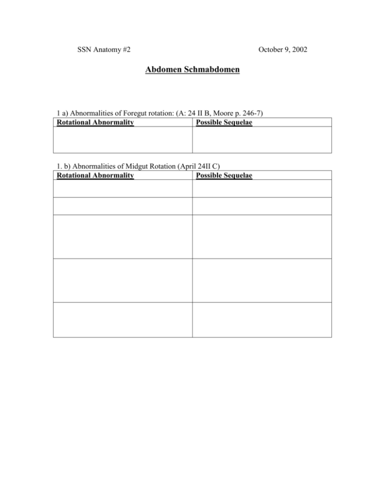
SSN Anatomy #2 October 9, 2002 Abdomen Schmabdomen 1 a) Abnormalities of Foregut rotation: (A: 24 II B, Moore p. 246-7) Rotational Abnormality Possible Sequelae 1. b) Abnormalities of Midgut Rotation (April 24II C) Rotational Abnormality Possible Sequelae 2. Subdivisions of GI Tract: (A:25, Moore p. 233)) Region Foregut Midgut Structures Arterial Supply Innervation 1) 2) 3) 4) 5) 6) 7) 1) 2) Parasympathetic: 3) Sympathetic: Sympathetic: Parasympathetic: 4) 5) 6) 1) Hindgut Parasympathetic: 2) Sympathetic: 3) Spinal Level Region of Referred Pain 3. Somatic Nerves of Posterior Abdomen (A: p. 397-398) Nerve Motor Sensory Functions Spinal Functions Level Iliohypogastric Ilioinguinal Genitofemoral -Genital -Femoral Lateral Femoral Cutaneous Femoral Obturator Lumbosacral Trunk Associated Reflex 4.Access to the peritoneal cavity: (A: 23 IV A-B; Moore p. 189-191) Incision Vertical: Rectus sheath Horizontal: Rectus Sheath Paramedian: Rectus Sheath Vertical: Linea Alba Transverse: Ventrolateral abdominal wall Vertical: Lateral Abdominal Wall Nerve supply cut Arterial supply cut Other pros & cons 5. Potential sites of herniation: (A: 23 VII D-F, Moore p. 205-207)) Weakness in Boundaries Hernia type anterior wall Triangle of 1). Hasselbach 2) Site of emergence 3) 1) Deep Inguinal Ring 2) 3) 1) Femoral Ring 2) 3) 4) 6.a) How is the arcuate line formed? (A: 28 III C, Moore p.184, Netter p. 235) b) What is its clinical significance? 7. Primary derivatives of ventral and dorsal mesenteries. (A: 24 II A, ) Ventral Mesentery Dorsal Mesentery 8. Peritoneal subdivisions. (A: 338 Table 24-1) Explain what each term means and give an example of each: Retroperitoneal Peritoneal Secondarily retroperitoneal 9. Peritoneal landmarks (A: 24 III B) Peritoneal Structure Boundaries Clinical Significance Foramen of Winslow Subphrenic recess Pouch of Morrison Right colic gutter 10. a)Which vessels contribute to the marginal artery of the colon? (A p.377, Netter plate 287) b) What are three clinical significance points of the marginal artery and why are they important? 11. Liver vasculature and biliary drainage. (A: 25 II D) Arterial Supply Biliary Drainage Liver lobe that is supplied or drained Right hepatic artery Left hepatic duct 12. Trace the sympathetic and afferent innervation of the kidney: (A: p.388, Moore p. 290) Sympathetic: Afferent (Sensory): 13. Ureter innervation (A: p. 390) Part of Ureter Innervation Upper abdominal ureter Refers pain to: Area of narrowing Lower abdominal ureter Pelvic ureter 14. From where does the adrenal vasculature supply arise and to where does the venous drainage empty? ( A p. 392) Arterial: 1) 2) 3) Venous: 1) 2) Clinical Cases 1. One day, a 48-year-old nurse practitioner comes to your office, complaining of a “colicky” pain in the epigastric region. She notices that eating foods that are high in fat exacerbates the pain. When you examine her, you find that she is jaundiced. Upon taking her history, you also find that she has two children and that she had been slightly obese until she started her “Deal-a-Meal” program a couple of months ago. (A: p.359) a. What do her symptoms and history indicate as a diagnosis? b. Upon further testing, you decide that a gallbladder removal is indicated. After entering the peritoneum, what should you locate before clamping or severing any structures? c. What are the boundaries of this structure? d. What is its significance? 2. In the ER, you are presented with a 13 y/o girl who complains of diffuse, colicky pain in the umbilical region. Her dad says she is just faking a stomachache because she wants to avoid going to a family reunion. You feel her abdomen and it shows no guarding (i.e. no muscle contraction to protect peritoneum upon touch). (A; p.370) a. What is her differential diagnosis? Due to several trauma cases that take you away, the girl and her father end up waiting for three hours in the ER. The next time you come in, the girl is doubled over, her abdomen displays guarding, and she shrieks when you press the lower-right quadrant. a. Explain the guarding and the localized pain, and how this affects your diagnosis. 3. A 48 y/o male alcoholic visits his physician asking for treatment of painful hemorrhoids. The patient’s liver is found to be enlarged, and a diagnosis of portal hypertension is made. (A: p.379) a. What causes portal hypertension? b. Name three other manifestations of portal hypertension. c. What surgical means are used to circumvent portal hypertension? 4. You’re a third-year medical student in the ER and a 50 y/o male comes in complaining of lower back pain. You suspect kidney stones. (A: p.389) a. Given the location of his pain, where do you think the stone has lodged? b. What is the best way to access the kidney at the renal pelvis? c. What complication is associated with this structure? d. Upon surgical access to the kidney it is found that there is no stone, but rather an acute kidney infection. How can this infection spread to other parts of the body? “Quickies” (April page #) What’s the difference between the falx inguinalis and the conjoined tendon? (324) Falx inguinalis: Conjoined tendon: What’s the “rule of 3’s” regarding a Meckel’s diverticulum? (365) What are the “lineas” that surround the rectus abdominis? Lateral: Medial: What three muscles comprise the posterior abdominal wall? (393) What’s the significance of the “bloodless line” at the gastroduodenal junction? (348) Kidney: Which kidney lies lower in the abdominal wall and why? (385) Is the kidney supported by mesentary? (385) Name the three structures at the renal hilus. (386) Where do the left and right gonadal and phrenic veins empty? (388) Right: Left: Where is the kidney vascular and parenchymal pain principally referred? (388)h The vascular supply of the ureters comes from: (390)


