printer-friendly version
advertisement
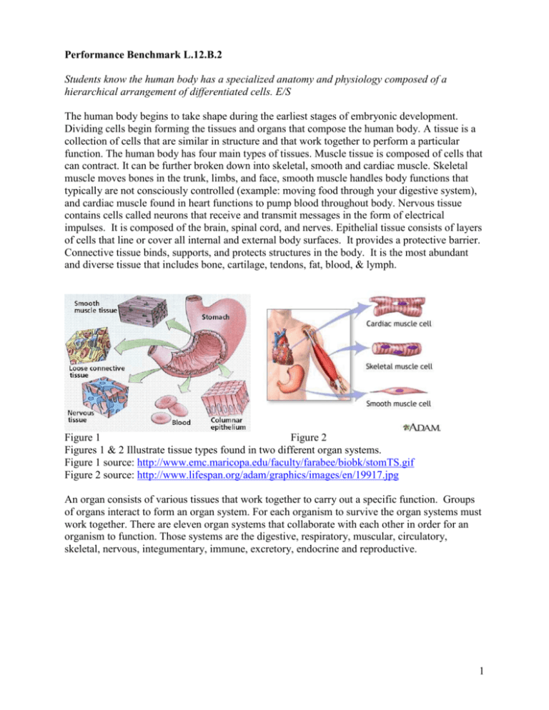
Performance Benchmark L.12.B.2 Students know the human body has a specialized anatomy and physiology composed of a hierarchical arrangement of differentiated cells. E/S The human body begins to take shape during the earliest stages of embryonic development. Dividing cells begin forming the tissues and organs that compose the human body. A tissue is a collection of cells that are similar in structure and that work together to perform a particular function. The human body has four main types of tissues. Muscle tissue is composed of cells that can contract. It can be further broken down into skeletal, smooth and cardiac muscle. Skeletal muscle moves bones in the trunk, limbs, and face, smooth muscle handles body functions that typically are not consciously controlled (example: moving food through your digestive system), and cardiac muscle found in heart functions to pump blood throughout body. Nervous tissue contains cells called neurons that receive and transmit messages in the form of electrical impulses. It is composed of the brain, spinal cord, and nerves. Epithelial tissue consists of layers of cells that line or cover all internal and external body surfaces. It provides a protective barrier. Connective tissue binds, supports, and protects structures in the body. It is the most abundant and diverse tissue that includes bone, cartilage, tendons, fat, blood, & lymph. Figure 1 Figure 2 Figures 1 & 2 Illustrate tissue types found in two different organ systems. Figure 1 source: http://www.emc.maricopa.edu/faculty/farabee/biobk/stomTS.gif Figure 2 source: http://www.lifespan.org/adam/graphics/images/en/19917.jpg An organ consists of various tissues that work together to carry out a specific function. Groups of organs interact to form an organ system. For each organism to survive the organ systems must work together. There are eleven organ systems that collaborate with each other in order for an organism to function. Those systems are the digestive, respiratory, muscular, circulatory, skeletal, nervous, integumentary, immune, excretory, endocrine and reproductive. 1 System Major Structures Functions Digestive Mouth, throat, esophagus stomach, liver, pancreas small and large intestines Extracts and absorbs nutrients from food; removes wastes; maintains water and chemical balances Respiratory Lungs, nose, mouth, trachea Moves air into and out of lungs; controls gas exchange between blood and lungs Muscular Skeletal, smooth, and cardiac muscle tissues Moves limbs and trunk; moves substances through body; provides structure and support Circulatory Heart, blood vessels, blood (cardiovascular) Transports nutrients, wastes, hormones, and gases lymph nodes and vessels, lymph (lymphatic) Skeletal Bones and joints Protects and supports the body and organs; interacts with skeletal muscles, produces red blood cells, white blood cells, and platelets Nervous Brain, spinal cord, and sense organs Regulates behavior; maintains homeostasis; regulates other organ systems; controls sensory and motor functions Table 1: The structures and functions of the six major body systems (http://www.sirinet.net/~jgjohnso/intro.html) Digestive System Before your body can use the nutrients in the food you consume, the nutrients must be broken down physically and chemically. The process of breaking down food into molecules the body can use is called digestion. In humans, digestion begins in the oral cavity where food is chewed (mastication) with the teeth. The food enters the stomach upon passage through the esophageal sphincter. In the stomach, food is further broken apart through a process of churning and is thoroughly mixed with a digestive fluid, composed chiefly of hydrochloric acid. After being processed in the stomach, food is passed to the small intestine. This is where most of the digestive process occurs. After going through the small intestine, the food then goes to the large intestine. The food that cannot be broken down is called feces. Feces are stored in the rectum until they are expelled through the anus. 2 Figure 3 and 4 Illustrate the human digestive system Figure 3: http://www.tuberose.com/Graphics/Intestinal_Health.jpg Figure 4: http://medicalimages.allrefer.com/large/digestive-system-organs.jpg Respiratory System It is the function of the respiratory system to transport gases to and from the circulatory system. The respiratory system involves both external respiration and internal respiration. External respiration is the exchange of gases between the atmosphere and the blood. Internal respiration is the exchange of gases between the blood and the cells of the body. Figures 5 and 6: http://www.spiderspun.net/images/lungs.gif http://www.umm.edu/respiratory/images/respiratory_anatomy.jpg If you are interested in the respiratory system and would like to know more about it go to http://www.emc.maricopa.edu/faculty/farabee/BIOBK/BioBookRESPSYS.html 3 Muscular System Muscles make up the bulk of the body and account for about one-third of its weight. Their ability to contract not only enables the body to move, but also provides the force that pushes substances, such as blood and food, through the body. Without the muscular system, none of the other organ systems would be able to function. Figure 7 and 8 Illustrate the muscle types and the parts of a muscle Figure 7: http://www.medic101.com/EMP_Lessons/cardiovascular/images/actin_myosin.gif Figure 8: http://www.nides.bc.ca/Assignments/Body/Bundles_files/musc.gif To learn more about the muscular system go to http://webschoolsolutions.com/patts/systems/muscles.htm Circulatory System Most of the cells in the human body are not in direct contact with the external environment. The circulatory system acts as a transport service for these cells. Two fluids move through the circulatory system: blood and lymph. The blood, heart, and blood vessels form the cardiovascular system. The lymph, lymph nodes, and lymph vessels form the lymphatic system. The cardiovascular system and lymphatic system collectively make up the circulatory system. Blood transports oxygen from the lungs to cells and carries carbon dioxide from the cells to the lungs. To learn more about the heart go to http://sln.fi.edu/biosci/heart.html To learn more in general about the circulatory system go to http://www.emc.maricopa.edu/faculty/farabee/BIOBK/BioBookcircSYS.html 4 Figure 9 and 10 Illustrate the circulatory system Figure 9: http://en.wikipedia.org/wiki/Image:3DScience_cardiovascular_system.jpg Figure 10: http://www.drugdevelopmenttechnology.com/projects/lipitor/images/1_Labelled_Human_Heart.jpg Skeletal System The adult human body consists of approximately 206 bones, which are organized into an internal framework called the skeleton. Because the human skeleton is an internal structure, biologists refer to it as an endoskeleton. The variation in size and shape among the bones that make up the skeleton reflects their different roles in the body. Figure 11 Illustrate the human skeleton http://www.contmediausa.com/shop/app/products/Human3D/Images/BS000A.jpg 5 Figure 12 Illustrate the human bone anatomy and structure http://www.sirinet.net/~jgjohnso/bonestruct4.jpg To learn more about the skeletal system go to http://www.bio.psu.edu/people/faculty/strauss/anatomy/skel/skeletal.htm and for additional information go to http://www.mnsu.edu/emuseum/biology/humananatomy/skeletal/skeletalsystem.html Nervous System Mental and physical activity and many aspects of homeostasis are controlled by the nervous system, a complex network of cells that communicate with one another. Within this communications network, a carefully organized division of labor exists so that each component of the nervous system operates effectively. As a result, a football player can weave through opposing tacklers, an architect can create an original design, and a student can understand this information. To learn more about the organ systems go to http://en.wikipedia.org/wiki/Human_biology http://biology.about.com/od/organsystems/a/aa031706a.htm http://www.innerbody.com/htm/body.html http://www.sirinet.net/~jgjohnso/biologyII.html 6 Performance Benchmark L.12.B.2 Students know the human body has a specialized anatomy and physiology composed of a hierarchical arrangement of differentiated cells. E/S Common misconceptions associated with this benchmark 1. Students incorrectly believe that the only gas humans breathe out during respiration is carbon dioxide. External respiration is the exchange of gases between the atmosphere and the blood and involves more than just the exchange of oxygen in and carbon dioxide out. Actually when a person breathes the majority of gas that is exhaled is carbon dioxide, but it is not the only gas that escapes from the mouth or nose. In addition to carbon dioxide, a small amount of oxygen and water vapor is released back into the atmosphere. Although most students are aware that oxygen is the gas that we inhale and rely on to breath, they quickly forget about the water vapor that is exhaled with every breath. The water vapor is clearly seen in cold weather and it is at that very moment that people exclaim that they “can see their breath.” It can also be easily seen by exhaling onto a mirror or window. For further information regarding the respiratory system visit http://www.sirinet.net/~jgjohnso/respiratory.html or http://www.42explore.com/respsyst.htm 2. Students incorrectly think the body systems operate in isolation from each other. The levels of structural organization starts at the cellular level, then continues to the tissue level, organ level, organ systems, and ends at the organism. The principle organ systems of the human body in fact do not work alone at all. Each system carries out a specific function in the body, but in order for an organism to survive the systems must work together. This system collaboration is known as integration of organ systems. It is not until these systems working together do they constitute the “total” organism and a completely living individual. To learn more about why the body systems need to work together visit http://www.sirinet.net/~jgjohnso/intro.html or http://www.kidinfo.com/Health/Human_Body.html 3.Students incorrectly think blood leaves the vessels and enters parts of the body. Students that have developed an understanding about blood leaving the vessels in the human body are unknowingly confusing the concept with less complex organisms. Many invertebrates do not have a circulatory system at all. Their cells are close enough to their environment for oxygen, other gases, nutrients, and waste products to simply diffuse out of and into their cells. Open circulatory systems (evolved in crustaceans, insects, mollusks and other invertebrates) pump blood into a hemocoel with the blood diffusing back to the circulatory system between cells. Blood is pumped by a heart into the body cavities, where tissues are surrounded by the blood. In animals with multiple layers of cells, especially large land animals, this will not work, as their cells are too far from the external environment for simple osmosis and diffusion to function quickly enough in exchanging cellular wastes and needed material with the environment. In this case a closed circulatory system is utilized. Closed circulatory systems have the blood closed at all times within vessels of different size and wall thickness. In this type of system, blood is pumped by a heart through vessels, and does not normally fill body cavities. For 7 further information regarding the human circulatory system visit http://www.sirinet.net/~jgjohnso/circulation.html or http://www.howstuffworks.com/heart.htm 4. Students incorrectly think blood vessels end in a dead end. They do not reconnect. Blood flows backward or travels through the body to another vessel going back. The circulatory systems of all vertebrates are closed, meaning that the blood never leaves the system of blood vessels consisting of arteries, capillaries and veins. Arteries bring oxygenated blood to the tissues (except pulmonary arteries), and veins bring deoxygenated blood back to the heart (except pulmonary veins). Blood passes from arteries to capillaries then to veins where it returns to the heart. Capillaries are the thinnest and most numerous of blood vessels and are responsible for exchanging gasses and nutrients to the cells in exchange for their waste products. In the closed circulatory system of mammals, there are two subdivisions—the systemic circulation and the pulmonary circulation. The pulmonary circulation involves circulation of deoxygenated blood from the heart to the lungs, so that it may be properly oxygenated. Systemic circulation takes care of sending blood to the rest of the body. Once the blood flows through the system of capillaries at the body’s tissues, it returns through the venous system. If all the vessels of this network in an adult human body were laid end-to-end, they would extend for about 60,000 miles (more than 96,500 kilometers); far enough to circle the Earth more than twice. For further information on the closed circulatory system of humans refer to http://www.sirinet.net/~jgjohnso/circulation.html or http://www.medtropolis.com/VBody.asp 5. Students mistakenly believe muscle cells can push and pull. Skeletal muscle is voluntary, striated, and attached to the skeleton. Each skeletal muscle is an organ of 100’s or 1000’s of muscle fibers, as well as nerves and connective tissue. When muscle cells contract, they pull on the connective tissue (tendons), which transfers the force elsewhere. The connective tissue also gives the muscle elasticity. Skeletal muscle, unlike other muscles, requires conscious effort that originates in the brain in order to contract. Because of the large amounts of energy used in contraction and the large quantity of wastes generated, a rich blood supply is needed by skeletal muscle. When muscles contract, they pull on the bones, which move bones at the joints. Joints are set up as lever systems: the fulcrum is where the two bones meet, one force is produced by the muscle, and the other by a load on the bone. Muscles can not push bones, they can only pull. Each bone is usually controlled by at least two muscles. One muscle pulls the bone one direction, the other pulls it back to its original position. To learn more about the process by which muscles provide movement to the human body visit http://www.sirinet.net/~jgjohnso/muscle.html or http://users.rcn.com/jkimball.ma.ultranet/BiologyPages/M/Muscles.html 6. Students erroneously believe the small intestine is short; the large intestine is long. The intestine is the portion of the digestive tract between the stomach and the anus. It is divided into two major sections: small intestine and large intestine. The small intestine is about 6 meters (20 feet) long. The large intestine has a larger width but is only 1.5 meters (5 feet) long. To learn more about the intestines function in the human body visit http://www.sirinet.net/~jgjohnso/nutrition.html or http://kidshealth.org/kid/body/digest_noSW.html 8 Performance Benchmark L.12.B.2 Students know the human body has a specialized anatomy and physiology composed of a hierarchical arrangement of differentiated cells. E/S Sample Test Questions 1. A muscle can a. push a bone. b. pull a bone. c. both push and pull a bone simultaneously. d. sometimes push and sometimes pull a bone. 2. The advantage of a closed circulatory system over an open circulatory system is that a. blood moves more efficiently through the tubes of a closed circulatory system. b. a closed circulatory system prevents blood from leaking out of the body. c. blood is able to be pumped by a muscular heart in a closed circulatory system. d. lungs are able to function in animals with a closed circulatory system. 3. The human body releases gases during the process of respiration. What gases escape from the mouth or nose during exhalation? a. CO2 b. CO2 and water vapor c. CO2, nitrogen, water vapor and oxygen d. CO and water vapor 4. From the smallest functional units to the largest, the body is organized as follows: a. Cell, organ system, organ, tissue, organism b. Organ, cell, tissue, organ system, organism c. Organism, organ system, organ, tissue, cell d. Cell, tissue, organ, organ system, organism 5. The heart and the blood vessels are separate organs that form the a. Skeletal system. b. Cardiovascular system. c. Digestive system. d. Circulatory system. 6. Systemic circulation takes care of sending blood to the all of the following except the a. Rest of the body. b. Heart. c. Lungs. d. Brain. 9 7. Refer to the illustration below. Most of the end products of digestion are absorbed into the circulatory system from which structure? a. Structure 1 b. Structure 2 c. Structure 3 d. Structure 4 8. The structure responsible for churning the food during the digestion process is a. Structure 1 b. Structure 3 c. Structure 4 d. Structure 5 10 Performance Benchmark L.12.B.2 Students know the human body has a specialized anatomy and physiology composed of a hierarchical arrangement of differentiated cells. E/S Answers to the Sample Questions 1. (b) 2. (a) 3. (c) 4. (d) 5. (b) 6. (c) 7. (d) 8. (d) 11 Performance Benchmark L.12.B.2 Students know the human body has a specialized anatomy and physiology composed of a hierarchical arrangement of differentiated cells. E/S Intervention Strategies and Resources The following list of intervention and resources that will facilitate student understanding of this benchmark. 1. Introduction to Organ Systems The Partnership for Environmental Education and Rural Health (PEER) through Texas A&M University has produced “Welcome to the Organ Systems.” Curriculum content and classroom lessons present a perspective on the human body as an integrated group of systems working together in order to maintain homeostasis and organism survival. It can be found at http://peer.tamu.edu/curriculum_modules/OrganSystems/index.htm. The unit has five modules that relate to the organ systems, but only three of them specifically relate to this benchmark. The direct links to these three lessons are: Organ Systems – Digestion http://peer.tamu.edu/curriculum_modules/OrganSystems/Module_2/index.htm Organ Systems – Respiration and Circulation http://peer.tamu.edu/curriculum_modules/OrganSystems/Module_4/index.htm Organ Systems – Nervous System and Brain http://peer.tamu.edu/curriculum_modules/OrganSystems/Module_5/index.htm See L12B3 for additional activities associated with the nervous system and the brain. 2. Biology – Anatomy and Physiology A biology website at Frederick High School in Frederick, Oklahoma provides additional information on the organ systems. The site is maintained by Jerry Johnson, a biology teacher at the high school. The website covers all of the necessary organ systems and provides study guides and testing materials for each system. The link to the site is http://www.sirinet.net/~jgjohnso/biologyII.html 3. The Interactive Body – Source of Interactive Anatomy Activities The British Broadcasting Company has developed a web page devoted to the human body and mind. The site contains a section focused on the body and covers the organs, muscles, skeletal system, nervous system and senses. Each area allows the student to work through an interactive that tests their knowledge of that area. It also provides a fact file that offers student access to a “Did you know” segment. The site can be found at http://www.bbc.co.uk/science/humanbody/body/interactives/3djigsaw_02/index.shtml?organs 12
