From www.bloodjournal.org by guest on March 5, 2016. For personal use only.
Plenary paper
Dynamic molecular modeling of pathogenic mutations in the spectrin
self-association domain
Zhushan Zhang, Scott A. Weed, Patrick G. Gallagher, and Jon S. Morrow
Disruption of spectrin self-association underlies many inherited hemolytic disorders. Using dynamic modeling and energy minimization, the 3-dimensional
structure of the self-association domain
has been estimated in human erythrocyte
spectrin and the structural consequences
of 17 elliptogenic mutations determined.
The predicted structure of the normal
self-association domain was remarkably
similar to the crystal structure of the
Drosophila ␣-spectrin 14th repeat unit,
despite replacement in the human sequence of over 70% of the amino acids
relative to fly spectrin, including 2 prolines in the human sequence that appear
in helical regions of the fly structure. The
predicted structure placed all hydrophilic
residues at the surface and identified 4
salt bridges, 9 hydrophobic interactions,
and 4 H-bonds that stabilize the native
self-association unit. Remarkably, every
pathologic point mutation, including
seemingly conservative substitutions
such as G for A, A for V, or K for R
(single-letter amino acid codes), led to
conformational rearrangements in the predicted structure. The degree of structural
disruption, as measured by root-meansquare deviation of the predicted backbone structure from the Drosophila structure, correlated strongly with the severity
of clinical disease associated with each
mutation. This approach thus enables an
accurate prediction, from the primary sequence, of the clinical consequences of
specific point mutations in spectrin. The
3-dimensional structure of the self-association domain derived here is likely to be
accurate. It provides a powerful heuristic
model for understanding how point mutations disrupt cytoskeletal function in a variety of hemolytic disorders. (Blood. 2001;98:
1645-1653)
© 2001 by The American Society of Hematology
Introduction
The shape and stability of the human erythrocyte derives from a
delicately balanced surface area–volume ratio and the lateral distribution
and abundance of its integral membrane proteins, as modified by their
attachment to an underlying spectrin-actin skeleton.1-4 Although the
energetics of the membrane may be dominated by interactions of
integral proteins within the bilayer,5 the homogeneous spectrin skeleton
modifies cell stability by modifying bilayer elasticity, by tethering or
trapping membrane proteins so as to limit their lateral diffusability and
ensure their quasi-random distribution, and by dampening the rate of
protein rearrangement during cyclic cell deformation (as occurs during
capillary passage).
One pathway by which the energetics of elasticity and membrane protein-distribution control can be altered is by failure of the
spectrin skeleton to adequately form an anastamosing meshwork
beneath the membrane, usually due to failures in self-association.
Such disorders typically lead to asymmetrical distortions in the
shape and deformability of the red blood cell (RBC), as in various
forms of hereditary elliptocytosis (HE) (reviewed in Morrow et al,2
Tse and Lux,3 Delaunay and Dhermy,6 and Gallagher and Jarolim7).
Spectrin itself is composed of a heterodimer of 2 structurally
similar proteins, ␣- and -spectrin,2,8 encoded by separate genes.9,10
Spectrin heterodimers self-associate in an end-on fashion to form
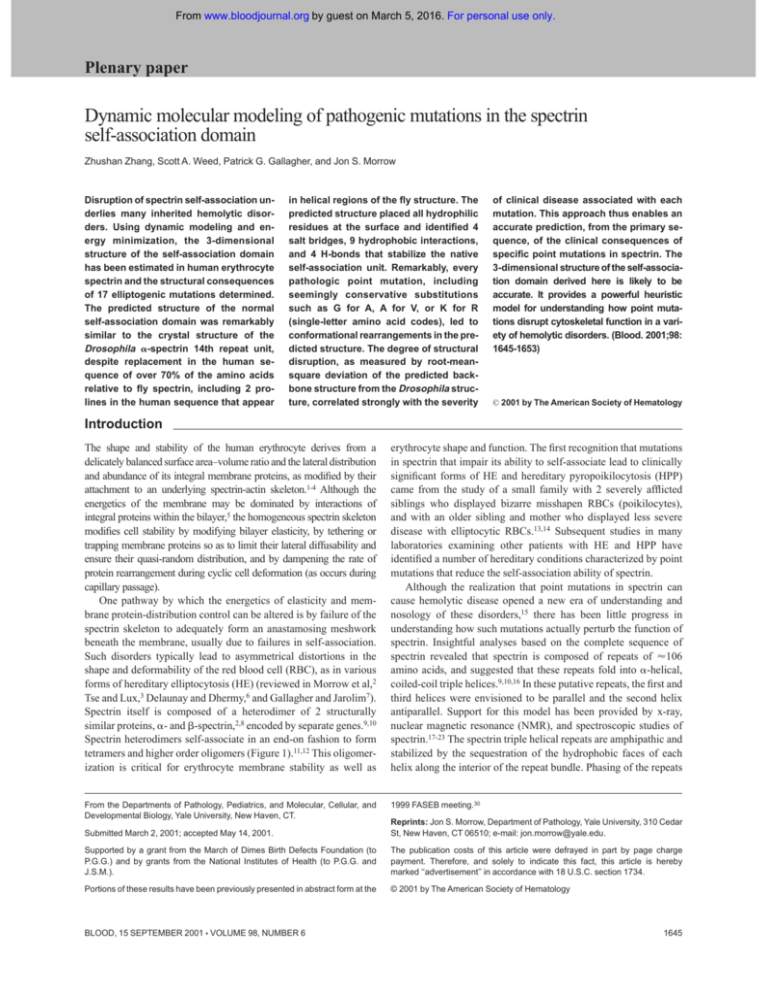
tetramers and higher order oligomers (Figure 1).11,12 This oligomerization is critical for erythrocyte membrane stability as well as
erythrocyte shape and function. The first recognition that mutations
in spectrin that impair its ability to self-associate lead to clinically
significant forms of HE and hereditary pyropoikilocytosis (HPP)
came from the study of a small family with 2 severely afflicted
siblings who displayed bizarre misshapen RBCs (poikilocytes),
and with an older sibling and mother who displayed less severe
disease with elliptocytic RBCs.13,14 Subsequent studies in many
laboratories examining other patients with HE and HPP have
identified a number of hereditary conditions characterized by point
mutations that reduce the self-association ability of spectrin.
Although the realization that point mutations in spectrin can
cause hemolytic disease opened a new era of understanding and
nosology of these disorders,15 there has been little progress in
understanding how such mutations actually perturb the function of
spectrin. Insightful analyses based on the complete sequence of
spectrin revealed that spectrin is composed of repeats of ⬇106
amino acids, and suggested that these repeats fold into ␣-helical,
coiled-coil triple helices.9,10,16 In these putative repeats, the first and
third helices were envisioned to be parallel and the second helix
antiparallel. Support for this model has been provided by x-ray,
nuclear magnetic resonance (NMR), and spectroscopic studies of
spectrin.17-23 The spectrin triple helical repeats are amphipathic and
stabilized by the sequestration of the hydrophobic faces of each
helix along the interior of the repeat bundle. Phasing of the repeats
From the Departments of Pathology, Pediatrics, and Molecular, Cellular, and
Developmental Biology, Yale University, New Haven, CT.
1999 FASEB meeting.30
Submitted March 2, 2001; accepted May 14, 2001.
Reprints: Jon S. Morrow, Department of Pathology, Yale University, 310 Cedar
St, New Haven, CT 06510; e-mail: jon.morrow@yale.edu.
Supported by a grant from the March of Dimes Birth Defects Foundation (to
P.G.G.) and by grants from the National Institutes of Health (to P.G.G. and
J.S.M.).
The publication costs of this article were defrayed in part by page charge
payment. Therefore, and solely to indicate this fact, this article is hereby
marked ‘‘advertisement’’ in accordance with 18 U.S.C. section 1734.
Portions of these results have been previously presented in abstract form at the
© 2001 by The American Society of Hematology
BLOOD, 15 SEPTEMBER 2001 䡠 VOLUME 98, NUMBER 6
1645
From www.bloodjournal.org by guest on March 5, 2016. For personal use only.
1646
BLOOD, 15 SEPTEMBER 2001 䡠 VOLUME 98, NUMBER 6
ZHANG et al
Figure 1. Cartoon of the self-association site of spectrin. The
self-association site is formed by a contribution of one helix from
the amino-terminus of ␣-spectrin, and 2 helices from the incomplete 17th repeat unit near the COOH-terminus of -spectrin.
revealed that there are incomplete repeats at the NH2-terminus of
␣-spectrin and the COOH-terminus of -spectrin (by 1 and 2 helices,
respectively). When a point mutation was identified in the COOHterminal partial repeat of -spectrin from an HE/HPP kindred with
severe anemia and impaired spectrin self-association, it was postulated
that the self-association site in spectrin must be formed from an atypical
triple helical unit involving the interaction of the COOH-terminus of
-spectrin (helices A and B) and the NH2-terminus of ␣-spectrin (helix
C) (Figure 1, bottom).24 Additional studies have identified over 20
mutations associated with HE or HPP in this atypical triple helical unit
that are defective in spectrin self-association (Table 1). Other work
detected further correlations between the clinical HE/HPP phenotype
and the presence of mutations in this putative triple helical unit.6,25
Studies examining directly the self-association activity of spectrin using
recombinant peptides also came to a similar conclusion,26-29 and have
demonstrated that this atypical repeat forms the critical region of the
spectrin self-association site.
Despite these advances, the challenge of understanding in detail the
structure of the spectrin self-association domain remains. Given the
lability and low-affinity of this noncovalent interaction (Ka ⬇10 M),
the direct resolution of its structure by either multidimensional NMR or
crystallography is a daunting challenge. As an alternative, we have
explored the power of computational strategies to develop plausible
models of this dynamic structure. These efforts involve the conceptual
folding of 2 peptide sequences representing the self-association domains
of ␣- and -spectrin.26,27 This modeling seeks to minimize the total
Table 1. Positions of point mutations in the spectrin self-association domain
Position in 106
residue repeat
Amino
acid
Mutant
Comment
RMS⌬*
(Å)
Clinical
severity
2018
11
A
2019
12
S
G
Spectrin Cagliari
4.5729
2
43
P
Spectrin Providence
4.4747
3
2023
16
A
37
V
4.9564
3
2024
17
44
W
R
3.3006
2
2025
44
18
L
R
4.9359
3
42
2053
46
A
P
4.3999
3
24
2061
54
W
R
ND
2
25, 45
2064
57
R
P
ND
2
46
24
79
I
S
28
83
R
H
28
83
R
S
6.2351
4
48, 50
28
83
R
L
10.0354
4
48, 50
28
83
R
C
5.1409
3
48, 51
34
89
R
W
Spectrin Genova
3.7954
2
52
Spectrin Tunis
3.1034
1
53
3.1557
2
54
ND
1
55
4.7368
3
56
2.4822
0
50
3.1990
1
56
Codon
Reference
-Spectrin
Spectrin Buffalo
Spectrin Cosenza
␣-Spectrin
Spectrin Tunisia
41
96
R
W
45
100
R
S
45
100
R
T
Spectrin Anastasia
46
101
G
V
Spectrin Culoz
48
103
K
R
49
104
L
F
Spectrin Lyon
2.8271
1
44
5.3170
3
38, 47-49
*Root-mean square deviation of the predicted backbone structure compared to the Drosophila 14th repeat crystal structure, as reported by Yan and colleagues.17 The fit for
the wild-type Drosophila sequence ⫽ 1.9897 Å; wild-type (normal) human ⫽ 2.8457 Å.
From www.bloodjournal.org by guest on March 5, 2016. For personal use only.
BLOOD, 15 SEPTEMBER 2001 䡠 VOLUME 98, NUMBER 6
system energy derived from all backbone and side-chain interactions,
including interactions with a surrounding hydration shell. Dynamic
modeling is used to facilitate the refinement of the structure and to
relieve forbidden steric or angular dependencies. The resultant structures
are triple helical, and for wild-type sequences are closely similar to the
crystal structures of a single spectrin repeat unit. Interestingly, we find
that even ostensibly conservative single residue replacements can
significantly destabilize the predicted structure, with the degree of
destabilization closely paralleling the clinical severity associated with
each mutation.
Materials and methods
Molecular modeling
Programs used to calculate the total system energy of different conformations, and to present the resultant structures as 3-dimensional representations, included X-plor, version 3.1m,31 and the program Insight32; both were
operated on a Silicon Graphics Indigo2 Extreme workstation. The starting
structure for the modeling was that portion of the crystal structure of
Drosophila spectrin that represents a single repeat unit.17 The 2-repeat triple
helical crystal structure of chicken ␣-spectrin23 was also considered as a
starting point for the modeling determinations, but was deemed to be less
suitable because it shared a backbone structure that was almost indistinguishable from the fly (root-mean-square deviation [RMS⌬] ⫽ 1.98 Å); was less
well resolved relative to the fly structure (2.0 Å versus 1.8 Å); and
surprisingly, shared less primary sequence homology with the putative
human self-association domain than did the Drosophila sequence (25%30% homology for avian spectrin versus 33% for the fly 14th repeat). No
attempts were made to model a structure based on the alternatively phased
conformation of the avian 2-repeat structure,23 because this conformation
was deemed to be an unlikely candidate given the absence of covalent
continuity between the putative B and C helices of the self-association site
(see below), continuity that would a priori appear to be a prerequisite to
extend the B helix at the expense of the C helix in this conformation. To
establish the validity of the modeling simulations and to explore the
parameters needed to achieve a creditable approximation, the structure of
the Drosophila ␣-spectrin 14th structural repeat itself was modeled and
compared with its crystal structure. The molecular dynamics at 300°K of
the Drosophila ␣-spectrin 14th repeat were initially modeled in a vacuum,
using the Verlet method.33 This approach yielded helices at the NH2- and
COOH-termini that unwound excessively, and the orientation of the amino
acid side chains were dissimilar to the crystal structure. This same result
Figure 2. The self-association domain of spectrin can
be modeled as a triple helix. (Top) Alignment of the
primary sequence of Drosophila ␣-spectrin 14th unit with
the partial repeat units from the amino-terminus of human
␣⌱-spectrin (residues 19-51) and from the 17th repeat unit
of ⌱-spectrin (residues 2008-2080), using the program
Bestfit.35 The position of the 3 helices as determined from
the crystal structure of the Drosophila spectrin are shown.
Continuity was not assumed where the 2 subunits join in
the concatenated sequence. This concatenated sequence, with or without the mutations described in the
text, was then treated as a pseudo-106 residue repeat
unit for the dynamic modeling studies. Dros indicates
Drosophila; term, terminus. (Bottom) Comparison of the
calculated structure of Drosophila ␣-spectrin 14th repeat
(blue) or the concatenated human ␣⌱⌱ self-association
domain (green) with the crystal structure of the Drosophila 14th repeat (red). Two views are shown, longitudinal
on the left, and an end-on view on the right. The 6-Å
hydration shell involving 1100 water molecules is not
shown. This hydration shell is required for the predicted
structure to fold to a triple-helical unit. Note the close
correspondence of the fitted structures with the crystal,
even though in the case of the self-association domain,
70% of the residues differ from those in the Drosophila
protein.
SPECTRIN MUTATIONS
1647
was obtained when a simulated annealing starting at high temperature was
used. These results suggested that simulation in a vacuum was inappropriate
for this super helix of the 3 helices, and suggested that interactions between
spectrin and the aqueous environment were critical to its structure. To solve
this problem, and given practical limitations on the computational complexity that could be managed, the effects of modeling this structure in a 6-Å
water shell using both molecular dynamics at 300°K and simulated
annealing from high temperatures were evaluated. The starting structure
was “placed” in a 6-Å-thick water shell that contained about 1100 water
molecules. Five hundred steps of Powell conjugate gradient energy
minimization34 were first performed to minimize the system energy. Then
Verlet molecular dynamics was used to accomplish the simulated annealing.
The simulated temperature was raised to 1000°K, and then cooled to 110°K
in steps of 10°K. At each temperature, 400 time-steps of dynamic
calculation were performed. Time-step increments (TSk) were determined
by the following formula:
TS k ⫽
{
}
18
fs
100 ⫺ k
k ⫽ 0, 1, . . . 89
The total simulation time was 16.5 picoseconds (ps). The initial velocity
was randomly set according the Maxwell velocity distribution. The
frictional coefficient was set to 100 ps⫺1 for all atoms. A structure was
sampled after each temperature step. The 90 sampled structures were
energy minimized by the Powell conjugate gradient energy minimization
with 2000 steps, and the 10 structures with lowest energy minima were
averaged. Energy minimization was then again performed on the averaged
structure with 2000 steps. The tolerance for the norm of the gradient of Etotal
was 0.01 during all of the energy minimization procedures. This approach
yielded better agreement with the known crystal structure (described below
and Figure 2).
Alignment of the structural unit with the
self-association domain
For purposes of molecular modeling, the self-association domain of ␣⌱⌱
spectrin was evaluated as a single concatenated sequence. To approximate
the positioning of the putative triple helical self-association unit relative to
the Drosophila structural repeat, the sequence of the self-association unit of
␣⌱⌱-spectrin was aligned with the 14th repeat unit of Drosophila
␣-spectrin using the program BestFit35 (Figure 2, top). The corresponding
residues of human spectrin were then graphically incorporated into the
published crystallographic coordinates of fly spectrin, thereby creating a
starting structure for the dynamic modeling computations. Residues 1 to 73
of the ␣ chain in the Drosophila crystal structure were replaced with the
From www.bloodjournal.org by guest on March 5, 2016. For personal use only.
1648
ZHANG et al
partial 17th structural unit of ⌱-spectrin. Residues 74 to 106 of the crystal
structure were replaced with the extended amino-terminus of ␣⌱-spectrin. A
pseudo repeat unit with a continuous peptide chain was thus generated for
use in the modeling calculations (although to faithfully reflect the native
state, the calculations did not assume continuity between the ␣- and
-spectrin concatenated sequences). A total of 73 residues were substituted
relative to the Drosophila sequence for the wild-type self-association
domain, and 72 ⫹ 1 (mutant) residues for each mutant examined. Replaced
structures were used as the starting structure for the molecular modeling
algorithms. It is noteworthy that many of the replaced residues even in
the wild-type spectrin represent nonconservative substitutions relative
to the Drosophila sequence, including the inclusion of a proline residue
in the middle of helix C. Yet, and despite these many substitutions, the
modeling algorithms converged on a predicted structure for the normal
human self-association domain that was remarkably similar in its overall
structural features to the Drosophila structure unit (Figure 2, bottom).
Determination of side-chain interactions
Salt-bridge interactions and hydrophobic interactions between residues
were scored as positive if they fell within 5 Å of each other in the refined
structure. Hydrogen bonds were scored as present if the distance between
the electronegative atoms (N & O) within O-H-N or O-H-O groups were
⬇2.9 Å apart.
Evaluation of clinical severity
In evaluating the clinical impact of each spectrin mutation, consideration
was given to the degree of hemolysis or RBC cell fragility, steady-state
hemoglobin or hematocrit (in nontransfused subjects), the reticulocyte
count, transfusion dependency, whether splenectomy had been necessary,
the degree of morphologic abnormality, and the overall morbidity and
mortality. These parameters were extracted from clinical data described in
the original reports (Table 1). Patients with no abnormalities in erythrocyte
shape or stability were scored 0; shape abnormalities without evidence of
hemolysis or clinical disease were ⫹1; mild hemolysis but not requiring
splenectomy, ⫹2; severe hemolysis with or without splenectomy, possibly
lethal in homozygous state, ⫹3; and the most severe disease, transfusion
dependent, always lethal in homozygotes, ⫹4.
Results
Molecular modeling predicts a plausible model of the spectrin
self-association domain
The simulated structure of the 14th structural unit of Drosophila
␣-spectrin derived by this method was superimposed onto its
crystal structure (Figure 2, bottom). As noted in a previous study
using a similar approach,36 the predicted structure, including the
orientation of the side chains, agrees well with the crystal structure.
The RMS⌬ for the comparison between the crystal structure and
that estimated by the above modeling procedure for Drosophila
␣-spectrin was 1.99 Å for the backbone atoms and 2.42 Å for all
heavy atoms. Overall, the calculated structure is a bit less compact
compared to the crystal structure. It is not known if this small
difference represents errors in the computational approximation or
a real difference in compactness between the solution structure and
that of the crystal (which is prepared but not modeled in a high-salt
environment).
The predicted structure for the normal spectrin self-association
domain is also similar to the structure of the Drosophila repeat,
with a backbone RMS⌬ of just 2.846Å (Figure 2). This is a
satisfactory result, given that over 70% of the structure has been
replaced with different amino acids, including 2 proline residues at
positions 23 in helix A and 64 in helix B (-spectrin residues 2030
and 2071, respectively). It is also important to note that the
BLOOD, 15 SEPTEMBER 2001 䡠 VOLUME 98, NUMBER 6
similarity of the predicted normal human self-association unit to
the Drosophila structural repeat is unlikely to reflect an insensitivity of the modeling algorithms to the variant sequences, because as
demonstrated below, single residue replacements that are known to
affect the function of the self-association domain have a profound
effect on the predicted structure.
Interactions of helix C with helices A and B primarily stabilize
the self-association unit
Based on the plausibility of the predicted overall tertiary and
quaternary structure of the self-association unit, an analysis was
carried out to identify interactions predicted to maximally stabilize
the functional unit (as described in “Materials and methods”).
Three types of linkages were evaluated: hydrophobic interactions
involving nonpolar side chains; hydrogen bonds, and ionic charge
interactions (salt bridges). These results are summarized in Figure
3 and Table 2. Interestingly, most of the putative strong interactions
identified in the modeled structure involve helix C interacting with
helix A or B. Just 2 direct interactions were predicted between
helices A and B, and one of these is marginal (internuclear distance
⬎ 5 Å), suggesting that in the absence of a third helix (ie, the helix
provided by ␣-spectrin), the self-association domain probably
exists in a more open conformation with its 2 helices at least
partially extended. This prediction is consonant with the intermediate open state identified in studies examining the protease-resistant
structure of -spectrin.26
Mutations of ␣- and -spectrin destabilize the
self-association domain
Spectrin Providence was first identified in a family with recurrent
fatal erythroblastosis fetalis. The cause was traced to a P for S
(single-letter amino acid codes) mutation at codon 2019 of
-spectrin. This residue falls in the middle of helix A. In the
heterozygous state, these patients display elliptocytosis and marked
erythrocyte fragility. Biochemical studies have established a substantial loss of self-association ability in this spectrin mutation. The
predicted structural consequences of this mutation are profound
(Figure 4, top, and Figure 5). This single amino acid mutation leads
the modeling algorithms to predict a markedly disrupted tertiary
structure, with a splaying of helices A and B, and a loss of the
narrow pocket into which helix C (␣-spectrin) normally docks.
Based on this predicted structure, it is hard to imagine how an
effective self-association interaction could be achieved. These
results fit well the measured self-association constant for this
spectrin, which is more than 2 logs weaker than for normal
spectrin.37 The severe disruption of the self-association domain that
follows the Providence mutation can be better appreciated in the
stereo presentation (Figure 5).
Another site of mutation that commonly causes hemolytic
disease is at codon 28 of ␣⌱-spectrin (Table 1). The predicted
structure of spectrin with an R3S mutation at this locus is shown
(Figure 4, bottom). Given that both S and R are hydrophilic, it is
surprising that this substitution would have such dramatic structural consequences. Similarly, spectrin Corbeil, an R3H mutation
at this locus, is also associated with significant poikilocytic
hemolytic anemia and defective spectrin self-association.38 The
model of spectrin Corbeil (Figure 6) also predicts major conformational changes. Given that ␣28 is the codon most commonly
mutated in HE and HPP, it is gratifying that these modeling studies
reveal it to be critical for maintaining the secondary, tertiary, and
quaternary structure of the self-association unit. Indeed, even
From www.bloodjournal.org by guest on March 5, 2016. For personal use only.
BLOOD, 15 SEPTEMBER 2001 䡠 VOLUME 98, NUMBER 6
SPECTRIN MUTATIONS
1649
Figure 3. Helical wheel depiction of the self-association unit. The predicted structure of the normal spectrin
self-association unit was examined for the presence of
various types of noncovalent interactions, based on their
chemical characteristics and proximity. These interactions are depicted and are summarized in Table 2. Note
that nearly all of the interactions stabilizing the triple
helical self-association unit arise between helix A or B
with helix C. One interaction between helix A and B
involves a salt bridge with R2079 (labeled), which falls in
the loop sequence between helix B and C. Helices A and
B are derived from -spectrin (shaded), helix C from
␣-spectrin.
ostensibly conservative substitutions at this locus (such as R3H)
are disruptive. Presumably, this sensitivity reflects the requirement
for the 2 hydrogen bonds formed between R28 and residues 2018
and 2022 of -spectrin.
All pathogenic mutations disrupt the predicted tertiary
structure of the self-association domain
To establish the generality and predictive value of the modeling
algorithm, 17 different mutations involving either the ␣- or
-subunit of spectrin were modeled. All of these mutations have
been identified because they generate a detectable phenotype. The
severity of these phenotypes ranges from severe and often fatal
hemolytic anemias or erythroblastosis fetalis to minor disturbances
in erythrocyte shape without measurable disturbances in stability.
These mutations are summarized in Table 1, and the derived
structures are shown in Figure 6. It is apparent that for all
pathologic mutations, there is some disruption of the secondary,
tertiary, and quaternary structure. The RMS⌬ values of these
Table 2. Side-chain interactions in the putative spectrin self-association domain
Position in helix and residue
Position in
106–amino
acid motif
Codon
Interaction
distance
(Å)*†
Position in
helix and
residue
Position in
106 amino
acid motif
Codon
Interaction type
Interacting residues between helix B and helix C
B7-R
43
2055
3.04
C26-G
101
46
H-bond (⫾)
B22-F
58
2070
4.93
C11-V
86
31
Hydrophobic
B18-W
54
2066
3.52
C15-Y
90
35
Hydrophobic
B18-W
54
2066
3.50
C18-F
93
38
Hydrophobic
B11-F
47
2059
3.15
C22-V
97
42
Hydrophobic
B11-F
47
2059
4.57
C23-A
98
43
Hydrophobic
B4-L
40
2052
3.41
C29-L
104
49
Hydrophobic
A11-A
11
2018
2.76
C7-R
82
28
H-bond
A15-E
15
2022
3.04
C7-R
82
28
H-bond (⫾)
A22-E
22
2029
2.79
C21-R
96
41
H-bond
Interacting residues between helix A and helix C
A7-F
7
2014
4.07
C4-I
79
24
Hydrophobic
A25-L
25
2032
3.74
C18-F
93
38
Hydrophobic
Salt bridge
A15-E
15
2022
3.81
C7-R
82
27
A15-E
15
2022
4.94
C14-R
89
34
Salt bridge
A22-E
22
2029
4.53
C21-R
96
41
Salt bridge
A3-E
3
2010
4.68
B36-R
72
2079
Salt bridge
A7-F
7
2014
5.35
B25-L
61
2068
Hydrophobic (⫾)
Interacting residues between helix A and helix B
*Distances for hydrophobic interactions were calculated from the 2 closest atoms of the interacting pair.
†Distances for salt bridges were calculated from center-to-center of each salt charge in the pair.
From www.bloodjournal.org by guest on March 5, 2016. For personal use only.
1650
ZHANG et al
BLOOD, 15 SEPTEMBER 2001 䡠 VOLUME 98, NUMBER 6
and for predicting the structural consequences of point mutations
involving this domain in either ␣- or -spectrin. This conclusion is
supported by several observations: (1) modeling the Drosophila
␣-spectrin 14th repeat unit accurately reflects its structure as
measured by x-ray crystallography; (2) the predicted structure of
the self-association domain of normal spectrin is very similar to the
Drosophila structure, and valid in comparison to criteria set by
other studies.17,23 This is so despite the replacement of over 70% of
its residues, including 2 proline substitutions present in the human
but not the Drosophila sequence, a conclusion consonant with the
well-recognized conservation across broad evolutionary distances
of secondary and tertiary structural features within a given protein
family; and (3) the plausibility of the predicted structure itself, with
polar residues on the surface, hydrophobic residues buried, an
absence of steric conflicts, and a satisfactory number and variety of
stabilizing noncovalent interactions.
Given the presumed validity of this model, what does it tell us
about spectrin? Perhaps the most profound lesson, beyond refining
Figure 4. Mutations of ␣- and -spectrin disrupt the normal predicted structure
of the spectrin self-association domain. (Top pair) Structure of normal spectrin
versus spectrin Providence, showing the position of the S3P amino acid replacement at codon 2019 (position 12 in the A helix). Note the predicted severe distortion
of this helix, with loss of the -spectrin pocket into which ␣-spectrin (helix C) docks.
(Bottom pair) A similar analysis revealing the predicted effect of a mutation in codon
28 of ␣-spectrin (R28S), a mutation that leads to hereditary pyropoikilocytosis. Even
though the serine in this mutation is hydrophilic like the residue it replaces (R), the
loss of the 2 putative salt bridges formed by the lost arginine (Table 2) destabilizes the
self-association binding site. In each model, longitudinal and end-on views are
shown. The mutated residue is depicted in space-filled form.
structures relative to the crystal structure of Drosophila ␣-spectrin
range from a value equivalent to the native structure (K28R
␣⌱-spectrin) to the markedly deviant structure predicted for the
R28L mutation in ␣⌱-spectrin.
Molecular modeling correlates well with the clinical severity of
each mutation
When the degree of predicted disruption in the structure of the
self-association unit, as measured by the RMS⌬ of its predicted
backbone structure versus the Drosophila crystal structure, is
compared to the approximate clinical severity of patients carrying
the mutation, a striking correlation emerges (Figure 7). Although
this correlation must of necessity be only approximate, given the
clinical heterogeneity observed for any given mutation even within
the same family (eg, due to the presence of a low expression
spectrin allele in cis or trans),39 these findings strongly imply that
the structural changes anticipated by the modeling algorithms are
directly reflected in the functional integrity of the self-association domain.
Discussion
The results presented here establish a computational approach for
generating plausible models of the spectrin self-association domain
Figure 5. Stereo drawing of the spectrin Providence mutation. (A) Predicted
structure of the normal self-association unit. (B) Predicted structure of the selfassociation unit in spectrin Providence. This spectrin mutation leads to severe
hemolytic disease and erythroblastosis fetalis in the homozygous state.37
From www.bloodjournal.org by guest on March 5, 2016. For personal use only.
BLOOD, 15 SEPTEMBER 2001 䡠 VOLUME 98, NUMBER 6
SPECTRIN MUTATIONS
1651
Figure 6. Modeling of 17 pathogenic mutations in the spectrin self-association domain. (A) Mutations involving ␣⌱-spectrin. (B) Mutations involving ⌱-spectrin. Note the
predicted disruption, sometimes severe, that accompanies the substitution of even a single native residue. Longitudinal and end-on views are shown for each mutation. The
mutated residue is depicted in each case using a solid-filled representation. The coordinates for each of these modeled structures are available as .PDB files (downloadable as
.RTF files) on the Blood website as supplemental material; see the Supplemental Data Sets link at the top of the online article.
Figure 7. The degree of predicted structural disruption correlates well with the
clinical severity induced by the mutation. For each of the 17 mutations in the
self-association domain that were modeled (Table 1), the RMS⌬ of their backbone
structure versus that of the Drosophila crystal structure was plotted versus an
estimate of the clinical severity of the condition, on a scale of 0 to 4 (as described in
“Materials and methods”). Also included in this plot is the RMS⌬ value for the native
structure (2.846 Å, clinical severity ⫽ 0). Note that the deviation of the mutant
structures, as predicted by dynamic molecular modeling from a knowledge of the
primary sequence and the crystal structure of Drosophila spectrin, correlates well
with the clinical severity of each mutation. This behavior can be approximated by the
equation shown, derived by a nonlinear least squares fit to the data.
by more rigorous analysis earlier concepts of the structure of the
self-association domain, is that the tertiary and quaternary structure
of the spectrin repeat may be precariously balanced between
multiple conformational states. Several biophysical studies have
noted conformational transitions in spectrin at physiologic temperatures,20 that its protease resistance is extremely sensitive to
temperature and ionic strength,40 and that alternative folding states
exist that might account for spectrin’s unusual hydrodynamic
properties.23 Yet, none of these studies would have predicted such
profound sensitivity of the spectrin repeat unit and self-association
unit to even seemingly minor residue substitutions.
These results also extend our understanding of the many
hemolytic disorders caused by defective spectrin self-association.
The majority of patients with HE and HPP have mutations in the
region of ␣--spectrin self-association.7,41 Heterozygous point
mutations in the ␣-spectrin self-association site (helix C) are
among the most common mutations identified in patients with HE
or HPP (Table 1). Homozygous patients with such point mutations
have not been described, perhaps because the complete loss of
spectrin self-association may be incompatible with life. Point
mutations in the -spectrin self-association site are generally mild
in the heterozygous state and severe, even fatal, in the homozygous
state. Truncations that remove all or part of the COOH-terminal
region of -spectrin in the heterozygous state are associated with
symptomatic elliptocytosis with poikilocytes and fragmented erythrocytes. As with homozygous ␣-spectrin self-association site
mutants, homozygotes with truncated -spectrin have not been
From www.bloodjournal.org by guest on March 5, 2016. For personal use only.
1652
BLOOD, 15 SEPTEMBER 2001 䡠 VOLUME 98, NUMBER 6
ZHANG et al
observed.7 With the insights gained in this study into the forces that
stabilize the self-association unit (Table 2 and Figure 3), these
clinical observations can now be better understood.
As noted above, residue 28 of ␣-spectrin (Table 1) appears to be
a “hot spot” for point mutations associated with variable clinical
manifestations and is never found in the homozygous state. From
molecular modeling, we see that mutations at this locus are
predicted to disrupt 2 important hydrogen bonds between this
residue and residues 2018 and 2022 of -spectrin, resulting in a
failure of helix C (␣-spectrin) to properly dock in the -spectrin
pocket. Correspondingly, when the -spectrin 2018 residue is
mutated, as it is in spectrin Cagliari, a similar phenotype results.
An example of a mutation in the -spectrin self-association
domain that is either fatal or associated with a severe, transfusiondependent hemolytic anemia is spectrin Buffalo, with an R3L
substitution at residue 2025.42 Dynamic modeling of the spectrin
Buffalo mutation predicts that hydrophobic interactions in the
interior of the triple helical repeat are disrupted by the replacement
of the hydrophobic uncharged leucine with a hydrophilic positively
charged arginine (Figure 6; also Gallagher et al42). Another
example is spectrin Providence, also fatal in the homozygous state,
with a P3S substitution at residue 2019.37 This residue is adjacent
to the critical hydrogen bond that stabilizes the self-association
unit, noted above between ␣28 and 2018. Modeling demonstrates
that the spectrin Providence mutation significantly disrupts the
forces that stabilize the -spectrin pocket (helices A and B) into
which ␣-spectrin (helix C) docks (Figures 4 and 5).
In summary, these results provide important information on the
putative interactions between specific amino acid residues that
compose the self-association unit of human ␣⌱⌱-spectrin, and
highlight the extraordinary sensitivity of the self-association unit to
disruption by the single point mutations that are associated with
clinical disorders of erythrocyte shape or stability. These data also
allow a prediction of the specific residues whose disruption is likely
to lead to a clinical phenotype. For example, in ␣-spectrin, 11
residues likely to be critical to the structure of the self-association
unit have been identified by this computational approach. Mutations have been discovered in, or immediately adjacent to, 8 of
these 11 residues. On this basis and on the strength of our model,
we predict that clinically significant mutations involving the other
3 critical ␣-spectrin residues (␣31, ␣38, and ␣43), or residues
immediately adjacent, await discovery. A final lesson to come from
this work is the clear demonstration that a complex clinical
syndrome, and its severity, can be predicted essentially from first
principles (ie, only with knowledge of the primary peptide
sequence and a starting approximation of its structure). To our
understanding, this represents perhaps the first time that such a
precise correlation has been possible for these types of cytoskeletal
disorders, and suggests that this approach may be valuable for
predicting the consequences of mutations in other functional
domains of spectrin, and potentially for other proteins as well.
Acknowledgments
Drs Scott Kennedy, Carol Cianci, and Bernard Forget are thanked
for their many helpful insights and assistance with various aspects
of this project.
References
1. De Matteis MA, Morrow JS. Spectrin tethers and
mesh in the biosynthetic pathway. J Cell Sci.
2000;113:2331-2343.
12. Morrow JS, Marchesi VT. Self-assembly of spectrin oligomers in vitro: a basis for a dynamic cytoskeleton. J Cell Biol. 1981;88:463-468.
2. Morrow JS, Rimm DL, Kennedy SP, Cianci CD,
Sinard JH, Weed SA. Of membrane stability and
mosaics: the spectrin cytoskeleton. In: Hoffman J,
Jamieson J, eds. Handbook of Physiology. London, UK: Oxford; 1997:485-540.
13. Knowles WJ, Morrow JS, Speicher DW, et al.
Spectrin from patients with hereditary pyropoikilocytosis have self-association defects and an abnormal tryptic digestion map. ICN/UCLA Symposium on Differentiation and Function of
Hematopoietic Cell Surfaces, Keystone, CO,
February, 1981.
3. Tse WT, Lux SE. Red blood cell membrane disorders. Br J Haematol. 1999;104:2-13.
4. Corbett JD, Agre P, Palek J, Golan DE. Differential control of band 3 lateral and rotational mobility
in intact red cells. J Clin Invest. 1994;94:683-688.
5. Mohandas N, Evans E. Mechanical properties of
the red cell membrane in relation to molecular
structure and genetic defects. Annu Rev Biophys
Biomol Struct. 1994;23:787-818.
6. Delaunay J, Dhermy D. Mutations involving the
spectrin heterodimer contact site: clinical expression and alterations in specific function. Semin
Hematol. 1993;30:21-33.
7. Gallagher PG, Jarolim P. Red cell membrane disorders. In: Hoffman R, Benz EJ Jr, Shattil SJ, et
al, eds. Hematology: Basic Principles and Practice. 3rd ed. New York, NY: Churchill Livingstone;
2000:576-610.
8. Winkelmann JC, Forget BG. Erythroid and nonerythroid spectrins. Blood. 1993;81:3173-3185.
9. Sahr KE, Laurila P, Kotula L, et al. The complete
cDNA and polypeptide sequences of human erythroid ␣-spectrin. J Biol Chem. 1990;265:44344443.
10. Winkelmann JC, Chang JG, Tse WT, Scarpa AL,
Marchesi VT, Forget BG. Full-length sequence of
the cDNA for human erythroid beta-spectrin.
J Biol Chem. 1990;265:11827-11832.
11. Morrow JS, Speicher DW, Knowles WJ, Hsu CJ,
Marchesi VT. Identification of functional domains
of human erythrocyte spectrin. Proc Nat Acad Sci
U S A. 1980;77:6592-6596.
14. Knowles WJ, Morrow JS, Speicher DW, et al. Molecular and functional changes in spectrin from
patients with hereditary pyropoikilocytosis. J Clin
Invest. 1983;71:1867-1877.
15. Lux SE, Palek J. Disorders of the red cell membrane. In: Handin RI, Lux SE, Stossel TP, eds.
Blood: Principles and Practice of Hematology.
Philadelphia, PA: Lippincott; 1995:1701-1816.
16. Speicher DW, Marchesi VT. Erythrocyte spectrin
is comprised of many homologous triple helical
segments. Nature (London). 1984;311:177-180.
17. Yan Y, Winograd E, Viel A, Cronin T, Harrison SC,
Branton D. Crystal structure of the repetitive segments of spectrin. Science. 1993;262:2027-2030.
18. Pascual J, Pfuhl M, Rivas G, Pastore A, Saraste
M. The spectrin repeat folds into a three-helix
bundle in solution. FEBS Lett. 1996;383:201-207.
19. Pascual J, Pfuhl M, Walther D, Saraste M, Nilges
M. Solution structure of the spectrin repeat: a lefthanded antiparallel triple-helical coiled-coil. J Mol
Biol. 1997;273:740-751.
20. Ralston GB, Dunbar JC. Salt and temperaturedependent conformation changes in spectrin from
human erythrocyte membranes. Biochim Biophys
Acta. 1979;579:20-30.
21. Calvert R, Ungewickell E, Gratzer W. A conformational study of human spectrin. Eur J Biochem.
1980;107:363-367.
22. LaBrake CC, Wang L, Keiderling TA, Fung LW.
Fourier transform infrared spectroscopic studies
of the secondary structure of spectrin under different ionic strengths. Biochemistry. 1993;32:
10296-10302.
23. Grum VL, Li D, MacDonald RI, Mondragon A.
Structures of two repeats of spectrin suggest
models of flexibility. Cell. 1999;98:523-535.
24. Tse WT, Lecomte MC, Costa FF, et al. Point mutation in the beta-spectrin gene associated with
alpha I/74 hereditary elliptocytosis: implications
for the mechanism of spectrin dimer self-association. J Clin Invest. 1990;86:909-916.
25. Nicolas G, Pedroni S, Fournier C, et al. Spectrin
self-association site: characterization and study
of beta-spectrin mutations associated with hereditary elliptocytosis. Biochem J. 1998;332:8189.
26. Speicher DW, DeSilva TM, Speicher KD, Ursitti
JA, Hembach P, Weglarz L. Location of the human red cell spectrin tetramer binding site and
detection of a related “closed” hairpin loop dimer
using proteolytic footprinting. J Biol Chem. 1993;
268:4227-4235.
27. Kennedy SP, Weed SA, Forget BG, Morrow JS. A
partial structural repeat forms the heterodimer
self-association site of all -spectrins. J Biol
Chem. 1994;269:11400-11408.
28. Cherry L, Menhart N, Fung LW. Interactions of the
alpha-spectrin N-terminal region with beta-spectrin: implications for the spectrin tetramerization
reaction. J Biol Chem. 1999;274:2077-2084.
29. Lecomte MC, Nicolas G, Dhermy D, Pinder JC,
Gratzer WB. Properties of normal and mutant
polypeptide fragments from the dimer self-association sites of human red cell spectrin. Eur Biophys J. 1999;28:208-215.
30. Zhang Z, Weed SA, Morrow JS. Dynamic molecular modeling of pathogenic mutations in the
spectrin self-association domain [abstract].
FASEB J. 1999;13:A1394.
From www.bloodjournal.org by guest on March 5, 2016. For personal use only.
BLOOD, 15 SEPTEMBER 2001 䡠 VOLUME 98, NUMBER 6
31. Brünger AT. X-Plor: A system for x-ray crystallography and NMR (ed X-plor pre-release version
3.851, available via the Internet [1996]). New Haven, CT: Yale University Press; 1992.
32. Biosym Technologies. Insight II User guide (ed
2.3.0), 1993.
33. Verlet L. Computer experiments on classical fluid,
I: thermodynamical properties of Lennard-Jones
molecules. Physical Rev. 1967;159:98-103.
34. Powell MJD. Restart procedures for the conjugate gradient method. Mathematical Programming. 1977;12:141-254.
35. Devereux J, Haeberli P, Smithies O. A comprehensive set of sequence analysis programs for
the VAX. Nucleic Acids Res. 1984;12:387-395.
36. Stabach PR, Cianci CD, Glantz SB, Zhang Z,
Morrow JS. Site-directed mutagenesis of ␣⌱⌱
spectrin (fodrin) reveals determinants of its -calpain susceptibility. Biochemistry. 1997;36:57-65.
37. Gallagher PG, Weed SA, Tse WT, et al. Recurrent
fatal hydrops fetalis associated with a nucleotide
substitution in the erythrocyte beta-spectrin gene
[see comments]. J Clin Invest. 1995;95:11741182.
38. Garbarz M, Lecomte MC, Feo C, et al. Hereditary
pyropoikilocytosis and elliptocytosis in a white
French family with the spectrin alpha I/74 variant
related to a CGT to CAT codon change (Arg to
His) at position 22 of the spectrin alpha I domain.
Blood. 1990;75:1691-1698.
39. Wilmotte R, Marechal J, Morle L, et al. Low expression allele alpha LELY of red cell spectrin is
associated with mutations in exon 40 (alpha V/41
polymorphism) and intron 45 and with partial skipping of exon 46. J Clin Invest. 1993;91:20912096.
40. Speicher DW, Morrow JS, Knowles WJ, Marchesi
SPECTRIN MUTATIONS
VT. Identification of proteolytically resistant domains of human erythrocyte spectrin. Proc Natl
Acad Sci U S A. 1980;77:5673-5677.
41. Gallagher PG, Forget BG. Hematologically important mutations: spectrin variants in hereditary elliptocytosis and hereditary pyropoikilocytosis.
Blood Cells Mol Dis. 1996;22:254-258.
42. Gallagher PG, Petruzzi MJ, Weed SA, Zhang Z,
Marchesi SL, Mohandas N, Morrow JS, Forget
BG. Mutation of a highly conserved residue of
betaI spectrin associated with fatal and near-fatal
neonatal hemolytic anemia. J Clin Invest. 1997;
99:267-277.
43. Sahr KE, Coetzer TL, Moy LS, et al. Spectin Cagliari: an Ala3Gly substitution in helix 1 of beta
spectrin repeat 17 that severely disrupts the
structure and self-association of the erythrocyte
spectrin heterodimer. J Biol Chem. 1993;268:
22656-22662.
44. Parquet N, Devaux I, Boulanger L, et al. Identification of three novel spectrin alpha I/74 mutations in hereditary elliptocytosis: further support
for a triple-stranded folding unit model of the
spectrin heterodimer contact site. Blood. 1994;
84:303-308.
45. Glele-Kakai C, Garbarz M, Lecomte MC, et al.
Epidemiological studies of spectrin mutations related to hereditary elliptocytosis and spectrin
polymorphisms in Benin. Br J Haematol. 1996;95:
57-66.
46. Qualtieri A, Pasqua A, Bisconte MG, Le Pera M,
Brancati C. Spectrin Cosenza: a novel beta chain
variant associated with Sp alphaI/74 hereditary
elliptocytosis. Br J Haematol. 1997;97:273-278.
47. Baklouti F, Marechal J, Morle L, et al. Occurrence
of the alpha I 22 Arg3His (CGT3CAT) spectrin
mutation in Tunisia: potential association with severe elliptocytosis. Br J Haematol. 1991;78:108113.
48. Coetzer TL, Sahr K, Prchal J, et al. Four different
1653
mutations in codon 28 alpha spectrin are associated with structurally and functionally abnormal
spectrin alphaI/74 in hereditary elliptocytosis.
J Clin Invest. 1991;88:743-749.
49. Randon J, Boulanger L, Marechal J, et al. A variant of spectrin low-expression allele alpha LELY
carrying a hereditary elliptocytosis mutation in
codon 28. Br J Haematol. 1994;88:534-540.
50. Floyd PB, Gallagher PG, Valentino LA, Davis M,
Marchesi SL, Forget BG. Heterogeneity of the
molecular basis of hereditary pyropoikilocytosis
and hereditary elliptocytosis associated with increased levels of the spectrin alphaI/74-kilodalton
tryptic peptide. Blood. 1991;78:1365-1372.
51. Lorenzo F, Miraglia del Giudice E, Alloisio N, et al.
Severe poikilocytosis associated with a de novo
alpha 28 Arg3Cys mutation in spectrin. Br J
Haematol. 1993;83:152-157.
52. Perrotta S, Miraglia del Giudice E, Alloisio N, et
al. Mild elliptocytosis associated with the alpha 34
Arg3Trp mutation in Genova (alpha I/74). Blood.
1994;83:3346-3349.
53. Morle L, Morle F, Roux AF, et al. Spectrin Tunis
(Sp alpha I/78), an elliptocytogenic variant, is due
to the CGG3TGG condon change (Arg3Trp) at
position 35 of the alpha I domain. Blood. 1989;74:
828-832.
54. Lecomte MC, Garbarz M, Grandchamp B, et al.
Sp alpha I/78: a mutation of the alpha I spectrin
domain in a white kindred with HE and HPP phenotypes. Blood. 1989;74:1126-1133.
55. Perrotta S, Iolascon A, De Angelis F, et al. Spectrin Anastasia (alpha I/78): a new spectrin variant
(alpha 45 Arg3Thr) with moderate elliptocytogenic potential. Br J Haematol. 1995;89:933-936.
56. Morle L, Roux AF, Alloisio N, et al. Two elliptocytogenic alpha I/74 variants of the spectrin alpha I
domain: Spectrin Culoz (GGT3GTT; alpha I 40
Gly3Val) and spectrin Lyon (CTT3TTT; alpha I
43 Leu3Phe). J Clin Invest. 1990;86:548-554.
Erratum
In the article by Badros et al entitled “High response rate in refractory and
poor-risk multiple myeloma after allotransplantation using a nonmyeloablative conditioning regimen and donor lymphocyte infusions,” which appeared
in the May 1, 2001, issue of Blood (Volume 97:2574-2579), the name of Elias
Anaissie was misspelled in the byline.
From www.bloodjournal.org by guest on March 5, 2016. For personal use only.
2001 98: 1653
Updated information and services can be found at:
http://www.bloodjournal.org/content/98/6/1653.full.html
Articles on similar topics can be found in the following Blood collections
Information about reproducing this article in parts or in its entirety may be found online at:
http://www.bloodjournal.org/site/misc/rights.xhtml#repub_requests
Information about ordering reprints may be found online at:
http://www.bloodjournal.org/site/misc/rights.xhtml#reprints
Information about subscriptions and ASH membership may be found online at:
http://www.bloodjournal.org/site/subscriptions/index.xhtml
Blood (print ISSN 0006-4971, online ISSN 1528-0020), is published weekly by the American Society of
Hematology, 2021 L St, NW, Suite 900, Washington DC 20036.
Copyright 2011 by The American Society of Hematology; all rights reserved.

