Supplementary Material S5
advertisement
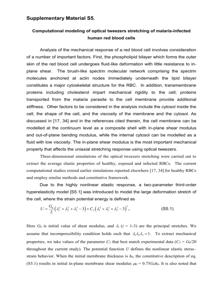
Supplementary Material S5. Computational modeling of optical tweezers stretching of malaria-infected human red blood cells Analysis of the mechanical response of a red blood cell involves consideration of a number of important factors. First, the phospholipid bilayer which forms the outer skin of the red blood cell undergoes fluid-like deformation with little resistance to inplane shear. The brush-like spectrin molecular network comprising the spectrin molecules anchored at actin nodes immediately underneath the lipid bilayer constitutes a major cytoskeletal structure for the RBC. In addition, transmembrane proteins including cholesterol impart mechanical rigidity to the cell; proteins transported from the malaria parasite to the cell membrane provide additional stiffness. Other factors to be considered in the analysis include the cytosol inside the cell, the shape of the cell, and the viscosity of the membrane and the cytosol. As discussed in [17, 34] and in the references cited therein, the cell membrane can be modelled at the continuum level as a composite shell with in-plane shear modulus and out-of-plane bending modulus, while the internal cytosol can be modelled as a fluid with low viscosity. The in-plane shear modulus is the most important mechanical property that affects the uniaxial stretching response using optical tweezers. Three-dimensional simulations of the optical tweezers stretching were carried out to extract the average elastic properties of healthy, exposed and infected RBCs. The current computational studies extend earlier simulations reported elsewhere [17, 34] for healthy RBCs and employ similar methods and constitutive framework. Due to the highly nonlinear elastic response, a two-parameter third-order hyperelasticity model [S5.1] was introduced to model the large deformation stretch of the cell, where the strain potential energy is defined as U 3 G0 2 1 22 32 3 C3 12 22 32 3 , 2 (S5.1) Here G0 is initial value of shear modulus, and i (i = 1-3) are the principal stretches. We assume that incompressibility condition holds such that 123 1 . To extract mechanical properties, we take values of the parameter C3 that best match experimental data (C3 = G0/20 throughout the current study). The potential function U defines the nonlinear elastic stress– strain behavior. When the initial membrane thickness is h0, the constitutive description of eq. (S5.1) results in initial in-plane membrane shear modulus 0 = 0.75G0h0. It is also noted that in-plane shear modulus of such a hyperelastic model characteristically stiffens when uniaxial stretching strain typically exceeds 50%. To perform the simulations that match the averaged response as well as the experimental scatter seen for RBC deformation (in terms of axial and transverse diameters at different infestation stages), the initial cell diameter was chosen to be approximately the averaged initial cell size; the initial large diameter of the cell was taken to be 7.0 ~ 7.8 m for the H-RBC, Pf-U-RBC, Pf-R-pRBC and Pf-T-pRBC cases so as to enforce consistency with experimental observations, RBC modeling assumes an initially biconcave shape [S5.2] and a contact diameter of 2 m, except for schizont stage (Pf-T-pRBC) cells, which have an initially spherical shape and a contact diameter of 1.5 m . As discussed in detail elsewhere [34], the bending modulus was chosen to be a fixed value of 2x10-19 Nּm, based on values available in the literature. Parametric studies at the continuum [17, 34] and spectrin [36] levels show that the large deformation stretch results are relatively insensitive to the specific choice of bending of phospholipid bilayer and the spectrin network, and a fixed volume of cytosol inside the red blood cell. Considering the symmetry of deformation during uniaxial stretching by optical tweezers, only one half of the three-dimensional cell was modeled using the general purpose finite element program, ABAQUS (ABAQUS Corporation, Pawtucket, Rhode Island, USA), and a total of 12,000 three-dimensional bi-linear shell elements are used in the finite element model. Since the transverse deformation of the RBCs cannot be accurately measured with the current experimental setup, the measured transverse diameters may be smaller than the actual values. By adjusting the input modulus parameters, the simulations that best match the experimental range of values are chosen to extract mechanical properties at different developmental stages of the parasite within the RBC. Supplementary Material S6 includes two video clips of the simulation for a healthy cell and a malaria infected cell (trophozoite stage), where much stiffer response can be seen for the latter case. Additional References for Supplementary Material S5 [S5.1] Yeoh OH. Characterization of elastic properties of carbon-black-filled rubber vulcanizers. Rubber Chem. Technol. 1990; 63:792-805. [S5.2] Evans EA, Fung YC. Improved measurements of erythrocyte geometry. Microvasc. Res. 1972; 4:335-347.
