Cholangiocarcinoma (Bile Duct Cancer)
The Bile Duct System (Biliary Tract)
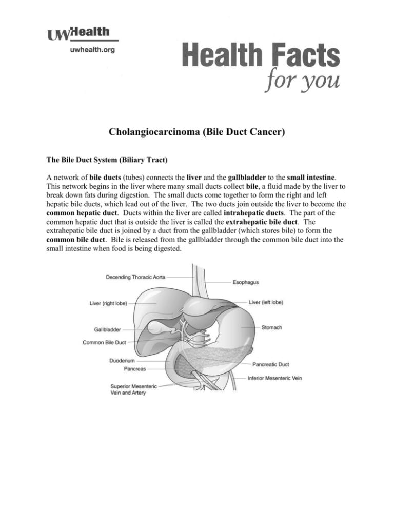
A network of bile ducts (tubes) connects the liver and the gallbladder to the small intestine.
This network begins in the liver where many small ducts collect bile, a fluid made by the liver to
break down fats during digestion. The small ducts come together to form the right and left
hepatic bile ducts, which lead out of the liver. The two ducts join outside the liver to become the
common hepatic duct. Ducts within the liver are called intrahepatic ducts. The part of the
common hepatic duct that is outside the liver is called the extrahepatic bile duct. The
extrahepatic bile duct is joined by a duct from the gallbladder (which stores bile) to form the
common bile duct. Bile is released from the gallbladder through the common bile duct into the
small intestine when food is being digested.
Bile Duct Cancer (Cholangiocarcinoma)
Bile duct cancer, a rare cancer in the United States, starts in the lining of the bile ducts
(epithelial cells). Bile duct cancers can start in the bile ducts within the liver. These are called
intrahepatic cholangiocarcinomas. Or they can start in the bile ducts outside of the liver.
These are called extrahepatic cholangiocarcinomas.
Risk Factors
The following disorders are risk factors for developing bile duct cancer:
Primary sclerosing cholangitis
Chronic ulcerative colitis
Choledochal (common bile duct) cysts
Infection with a Chinese liver fluke parasite
Symptoms
Bile duct cancers usually cause symptoms when the cancer blocks (obstructs) the biliary
drainage system causing yellow skin or whites of the eyes (jaundice). Itching (pruritis) often
occurs due to the deposits of bile salts in the skin. Other symptoms include pain, often in the
right upper abdomen, fever, clay-colored stools, and dark urine.
Prognosis
The prognosis (chance of recovery) and treatment options depend on:
The stage of the cancer (whether it affects only the bile duct or has spread to other
places in the body.)
Whether the cancer can be completely removed by surgery.
Whether the cancer is in the upper or lower part of the duct.
Whether the cancer is newly diagnosed or has recurred (come back).
Treatment options may also depend on the symptoms caused by the cancer. Bile duct cancer
found after it has spread may not be completely removed by surgery (unresectable). If the cancer
has spread, palliative treatment may improve the patient’s quality of life by controlling
symptoms and complications of the disease.
Diagnosis and Staging
These tests and procedures may be used to diagnose bile duct cancers and determine the stage of
disease (extent of the cancer). The stage of the disease is important to know in order to make a
treatment plan.
2
Physical exam and complete history of health habits, past illnesses, and treatments.
Ultrasound – a radiology procedure that bounces high-energy sound waves
(ultrasound) off tissues or organs to form a picture called a sonogram
CT scan (CAT scan) – detailed picture of the inside of the body taken by a special xray machine that is attached to a computer
MRI (magnetic resonance imaging) – a radiology procedure that uses a magnet, radio
waves, and a computer to make detailed pictures of the inside of the body
ERCP (endoscopic retrograde cholangiopancreatography) – a procedure performed
by a gastroenterologist (a doctor who specializes in digestive tract diseases) where a
small lighted tube (scope) is passed through the mouth, esophagus, stomach, and first
part of the small intestine. A smaller tube or catheter is passed into the ducts, a dye is
injected and x-rays taken. If the ducts are blocked, a small flexible tube (stent) may
be inserted into the duct to unblock it. Tissue samples (biopsies) may be taken.
PTC (percutaneous transhepatic cholangiography) – a procedure used to x-ray the
liver and bile ducts. A thin needle is inserted through the skin below the ribs and into
the liver. Dye is injected into the liver or bile ducts and x-rays are taken. If a
blockage is found, a flexible tube or stent is sometimes left in the liver to drain bile
into either the small intestine or to a collection bag outside the body. Tissue samples
or biopsies may also be taken.
Biopsy – the removal of cells or tissues to be examined under the microscope to
check for cancer. Tissue can be removed during an ERCP, a PTC, or surgery.
Liver function tests – blood tests that measure the amounts of certain substances
released into the blood by the liver. Higher than normal amounts can be a sign of
liver disease that may be caused by bile duct cancer.
Laparoscopy – surgery to look at the organs inside the abdomen to check for signs of
disease. A small lighted tube (scope) is inserted through a small incision in the
abdomen. Tissue samples (biopsies) may be taken through the scope. A laparoscopy
helps determine if the cancer can be removed surgically or if it has spread to other
organs in the abdomen.
Stages of Bile Duct Cancer
Stage 0 (Carcinoma in situ) – cancer is found only in the innermost layer of cells lining
the bile duct.
Stage I is divided into stage IA and stage IB.
Stage IA – cancer is found in the bile duct only
Stage IB – cancer has spread through the wall of the bile duct
Stage II is divided into stage IIA and stage IIB.
Stage IIA – cancer has spread to the liver, gallbladder, pancreas, and/or to either
the right or left branches of the hepatic artery or to the right or left branches of the
portal vein
Stage IIB – cancer has spread to nearby lymph nodes and:
o is found in the bile duct; or
o has spread through the wall of the bile duct; or
3
o has spread to the liver, gallbladder, pancreas, and/or the right or left
branches of the hepatic artery or portal vein
Stage III – cancer has spread:
to the portal vein or to both right and left branches of the portal vein; or
to the hepatic artery; or
to other nearby organs or tissues, such as the colon, stomach, small intestine, or
abdominal wall; or
perhaps to nearby lymph nodes.
Stage IV – cancer has spread to lymph nodes and/or organs far away from the bile duct.
Treatment Groups
Localized (and resectable) – the cancer is in a limited area and can be completely
removed by surgery.
Unresectable – the cancer has spread to nearby lymph nodes, blood vessels or other
organs and cannot be removed completely by surgery.
Methods of Treatment
Surgery
These types of surgery are used to treat bile duct cancer.
Removal of the bile duct – If-the tumor is small and only in the bile duct, the entire bile
duct may be removed. A new duct is made by connecting the duct openings in the liver
to the intestine. Lymph nodes are removed and examined under the microscope to see if
they contain cancer.
Partial hepatectomy – Removal of the part of the liver where cancer is found. The part
removed may be a wedge of tissue, an entire lobe, or a larger part of the liver, along with
some normal tissue (margin) around it.
Whipple procedure - A surgery in which the head of the pancreas, the gallbladder, part
of the stomach, part of the small intestine, and the bile duct are removed. Enough of the
pancreas is left to produce digestive juices and insulin.
Surgical biliary bypass – If the tumor cannot be removed but is blocking the small
intestine and causing bile to build up in the gallbladder, a biliary bypass may be done.
During this operation, the gallbladder or bile duct will be cut and sewn to the small
intestine to create a new pathway around the blocked area. This procedure helps to
relieve jaundice caused by the build-up of bile.
Stent placement – If the tumor is blocking the bile duct, a stent (thin tube) may be
placed in the duct to drain bile that has built up in the area. The stent may drain to the
outside of the body or it may go around the blocked area and drain the bile into the small
intestine. A stent may be place during an ERCP, a PTC, or surgery.
4
Radiation Therapy
Radiation therapy is a cancer treatment that uses high-energy x-rays or other types of radiation to
kill cancer cells. There are two types of radiation therapy. External beam radiation uses a
machine outside the body to send radiation toward the cancer. Internal beam radiation uses a
radioactive substance sealed in needles, seeds, wires, or catheters that are placed directly into or
near the cancer. The way the radiation therapy is given depends on the type and stage of the
cancer being treated.
Chemotherapy
Chemotherapy uses drugs to stop the growth of cancer cells, either by killing the cells or by
stopping the cells from dividing. Unlike surgery and radiation therapy, chemotherapy is a
systemic treatment that can reach cancer cells throughout the body. Chemotherapy is sometimes
used along with radiation therapy to make the radiation therapy more effective.
Clinical Trials
Clinical trials, exploring ways of improving local control may be available using chemotherapy
with or without radiation.
This information has been reproduced with permission of the National Cancer Institute. For
more information please visit their website at www.nci.nih.gov
HU
U
Your health care team may have given you this information as part of your care. If so, please use it and call if you
have any questions. If this information was not given to you as part of your care, please check with your doctor.
This is not medical advice. This is not to be used for diagnosis or treatment of any medical condition. Because
each person’s health needs are different, you should talk with your doctor or others on your health care team
when using this information. If you have an emergency, please call 911. Copyright © 2/2014 University of
Wisconsin Hospitals and Clinics Authority. All rights reserved. Produced by the Department of Nursing. HF#6702
5


