Envelope Technique
advertisement
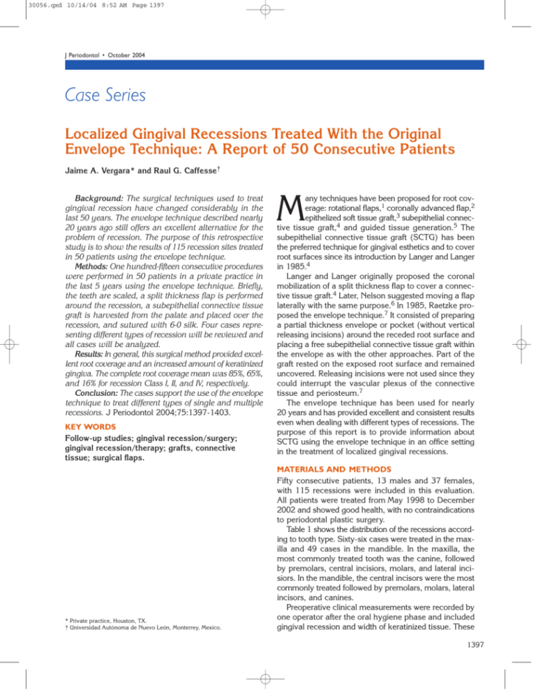
30056.qxd 10/14/04 8:52 AM Page 1397 J Periodontol • October 2004 Case Series Localized Gingival Recessions Treated With the Original Envelope Technique: A Report of 50 Consecutive Patients Jaime A. Vergara* and Raul G. Caffesse† Background: The surgical techniques used to treat gingival recession have changed considerably in the last 50 years. The envelope technique described nearly 20 years ago still offers an excellent alternative for the problem of recession. The purpose of this retrospective study is to show the results of 115 recession sites treated in 50 patients using the envelope technique. Methods: One hundred-fifteen consecutive procedures were performed in 50 patients in a private practice in the last 5 years using the envelope technique. Briefly, the teeth are scaled, a split thickness flap is performed around the recession, a subepithelial connective tissue graft is harvested from the palate and placed over the recession, and sutured with 6-0 silk. Four cases representing different types of recession will be reviewed and all cases will be analyzed. Results: In general, this surgical method provided excellent root coverage and an increased amount of keratinized gingiva. The complete root coverage mean was 85%, 65%, and 16% for recession Class I, II, and IV, respectively. Conclusion: The cases support the use of the envelope technique to treat different types of single and multiple recessions. J Periodontol 2004;75:1397-1403. KEY WORDS Follow-up studies; gingival recession/surgery; gingival recession/therapy; grafts, connective tissue; surgical flaps. * Private practice, Houston, TX. † Universidad Autónoma de Nuevo León, Monterrey, Mexico. M any techniques have been proposed for root coverage: rotational flaps,1 coronally advanced flap,2 epithelized soft tissue graft,3 subepithelial connective tissue graft,4 and guided tissue generation.5 The subepithelial connective tissue graft (SCTG) has been the preferred technique for gingival esthetics and to cover root surfaces since its introduction by Langer and Langer in 1985.4 Langer and Langer originally proposed the coronal mobilization of a split thickness flap to cover a connective tissue graft.4 Later, Nelson suggested moving a flap laterally with the same purpose.6 In 1985, Raetzke proposed the envelope technique.7 It consisted of preparing a partial thickness envelope or pocket (without vertical releasing incisions) around the receded root surface and placing a free subepithelial connective tissue graft within the envelope as with the other approaches. Part of the graft rested on the exposed root surface and remained uncovered. Releasing incisions were not used since they could interrupt the vascular plexus of the connective tissue and periosteum.7 The envelope technique has been used for nearly 20 years and has provided excellent and consistent results even when dealing with different types of recessions. The purpose of this report is to provide information about SCTG using the envelope technique in an office setting in the treatment of localized gingival recessions. MATERIALS AND METHODS Fifty consecutive patients, 13 males and 37 females, with 115 recessions were included in this evaluation. All patients were treated from May 1998 to December 2002 and showed good health, with no contraindications to periodontal plastic surgery. Table 1 shows the distribution of the recessions according to tooth type. Sixty-six cases were treated in the maxilla and 49 cases in the mandible. In the maxilla, the most commonly treated tooth was the canine, followed by premolars, central incisiors, molars, and lateral incisiors. In the mandible, the central incisors were the most commonly treated followed by premolars, molars, lateral incisors, and canines. Preoperative clinical measurements were recorded by one operator after the oral hygiene phase and included gingival recession and width of keratinized tissue. These 1397 30056.qxd 10/14/04 8:52 AM Page 1398 Gingival Recessions Treated With Envelope Technique Volume 75 • Number 10 Table 1. Table 2. Recessions by Tooth Type and Location Number of Recessions Treated per Patient N % 1 19 38 4 2 19 38 24 3 3 5 10 16 11 ≥4 7 14 Molar 8 7 50 100 Total 66 49 Type Maxilla Mandible Central 11 24 Lateral 7 Canine Premolar Recessions Total Table 3. measurements were recorded at the mid-facial aspect of each tooth with a Michigan manual probe. The recessions were further classified in either Class I, II, III, or IV according to Miller’s classification.8 Preoperative, surgical, and postoperative photographs were taken. All cases were anesthetized with septocaine 4% with 1:100,000 epinephrine. The exposed roots were thoroughly planed with hand and rotary instruments and a solution of 8% EDTA, pH7, was applied for 1 to 3 minutes with a cotton swab. An intrasulcular marginal incision was performed and a split thickness envelope flap was raised using Beaver blades of different sizes and shapes based on the tooth location and size. No vertical incisions were made. A conventional free gingival graft was obtained from the palate using a 15c Bard-Parker blade and its epithelium was partially or totally eliminated. The SCTG was placed within the envelope and both the graft and flap were sutured with 6-0 silk sutures internally and externally to eliminate any movement. An analgesic and chlorhexidine gluconate rinse were prescribed after surgery. The patients were seen one week after surgery for suture removal and oral hygiene instruction. The patients were instructed to refrain from brushing the surgical area for at least the first 2 weeks after surgery. All cases have been maintained and followed up for at least 6 months (longest follow-up is 48 months). The same measurements recorded at baseline were repeated by the same operator without knowledge of the initial recordings. All measurements and surgeries were performed by the same clinician (JV), and the post-treatment follow-up was recorded 6 months after surgery. No patients were lost to follow-up. Table 2 shows the number of sites treated per patient. Most of the patients had one or two areas treated. However, the range of recessions per patient treated varied from 1 to 9. When multiple grafts were performed, the maximum number of recessions treated in one surgery was six. Table 3 shows the age distribution by Miller classification (range: 14 to 62 years; mean: 38.6). Most of 1398 Age Distribution by Miller Classification Classification Age I II III IV ≤20 0 1 0 0 21-30 16 17 0 0 31-40 18 17 0 0 41-50 3 14 1 9 51-60 4 11 0 3 61-70 0 0 1 0 Total 41 60 2 12 the cases treated were in the 20 to 40 age group. While type I and II recessions were found in all age groups, type III and IV were only seen in patients over 41 years old. RESULTS Table 4 shows the distribution of different types of recession according to Miller. Most of the sites were Class II followed by Class I, IV, and III. Before treatment, Class I recessions ranged from 1 to 4 mm (mean: 2.65 ± 1.07); Class II recessions ranged from 1 to 7 mm (mean: 3.48 ± 1.25); Class III recessions ranged from 5 to 6 mm (mean: 5.5 ± 0.5); and Class IV ranged from 2 to 10 mm (mean 5.09 ± 2.07). After treatment, residual recessions for Class I ranged from 0 to 4 mm (mean 0.23 ± 0.71); Class II ranged from 1 to 7 mm (mean 0.49 ± 0.73); Class III ranged from 1 to 3 mm (mean 1.0 ± 1.5); and Class IV ranged from 2 to 10 mm (mean 1.86 ± 0.14). Table 5 shows the amount of mm of recession prior to treatment. Most of the sites had recessions ≥3 mm. Table 6 shows the residual recession after treatment when all types of recession were combined. Seventy- 30056.qxd 10/14/04 8:52 AM Page 1399 Vergara, Caffesse J Periodontol • October 2004 Table 4. Table 5. Clinical Measurements (mm) by Miller Classification Recession Depth Prior to Treatment Depth (mm) Pretreatment Post-Treatment N % 1 5 4 Class N Mean ± SD Range Mean ± SD Range 2 25 22 I 41 2.65 ± 1.07 1-4 0.23 ± 0.71 0-4 3 35 30 II 60 3.48 ± 1.25 1-7 0.49 ± 0.73 1-7 4 18 16 III 2 5.50 ± 0.50 5-6 1.00 ± 1.50 1-3 ≥5 32 28 IV 12 5.09 ± 2.07 2-10 1.86 ± 0.14 2-10 Total 115 100 Table 6. Residual Recession After Treatment Table 7. Residual Recession (mm) by Miller Class Recession (mm) N % 0 76 66 1 20 17 Class N 0 1 2 15 13 I 41 35 5 3 2 2 II 60 39 13 8 ≥4 2 2 III 2 0 1 0 1 0% 115 100 IV 12 2 1 7 2 16% Total Recession 2 >2 Complete Coverage 1 85% 65% Table 8. Number and Percentage of Complete Coverage of Class I and II Recessions by Age Table 9. Number and Percentage of Complete and Almost Complete Coverage in Single and Multiple Class I and II Recessions Age Group N Complete Coverage (N) Mean (%) 21-30 33 30 90 Recession 31-40 35 21 60 41-50 17 12 70 51-60 15 9 60 six sites (66%) had complete root coverage and 83% had very good esthetic results (<1 mm recession). Table 7 shows the amount of residual recession according to Miller’s classification of the original recession. In Class I recessions, complete coverage was achieved in 85% of the cases; 98% of this group of cases obtained very good esthetic results. In Class II cases, 65% had similar results. Table 8 shows the number and percentage of complete coverage of Class I and II recessions by age. Residual Recession N Recessions ≤1 mm 0 mm Single 15 (15 patients) 13 (87%) 12 (80%) Multiple 86 (26 patients) 75 (86%) 61 (70%) Table 9 shows the number of sites where complete or nearly complete coverage of recession was obtained when treating single or multiple Class I and II cases. Table 10 shows the response to therapy of Class I and II recessions by age. Patients <40 years old showed a 91% coverage and patients older than 40 years gained an 86% coverage. The number of cases achieving 100% coverage was 55 (79%) and 19 (59%), respectively. The healing in almost all grafts was uneventful. Five patients, four with single and one with multiple recessions, lost their grafts but only two recessions did not show any 1399 30056.qxd 10/14/04 8:52 AM Page 1400 Gingival Recessions Treated With Envelope Technique Volume 75 • Number 10 improvement in terms of root Table 10. coverage. Two grafted teeth had Response to Therapy of Class I and II Recessions by Age partial loss of grafted tissue in the first week. However, in both Pretreatment Post-Treatment cases, 2 mm out of 5 mm of root % N Complete % Complete surface were covered with the Age N Mean ± SD Range Mean ± SD Range Coverage Coverage Coverage grafts. One of the cases was a ≤40 68 3.09 ± 1.26 2-6 mm 0.30 ± 0.72 0-2 mm 91 55 79 smoker patient. All grafted teeth had an improvement in the ≥41 32 3.32 ± 1.19 1-7 mm 0.51 ± 0.71 0-2 mm 86 19 59 amount of keratinized gingiva. All the cases treated, irrespective of the results, were included in the data presented. Four cases are presented representing the four types of recessions according to Miller’s classification. The cases are representative of the results achieved in all those treated. CASE 1 (FIG. 1) A 42-year-old female patient presented for consultation regarding gingival recession on tooth #9. The recession represented an esthetic problem to the patient. Her medical history was non-contributory. Upon evaluation, a 2 mm recession was noticed in the area. Apical to the recession, there was a moderate amount of keratinized gingival tissue present. The distal interdental papillae was slightly blunted and tooth #10 presented a 1 mm recession. The recession on tooth #9 was classified as Class I and was treated with a connective tissue graft using a conventional envelope technique. Two years after surgery, 100% root coverage was observed, with improvement in the distal papilla and creeping attachment on the buccal aspect of tooth #10. The patient was satisfied with the esthetic results. CASE 2 (FIG. 2) A 33-year-old female patient presented with localized recession on teeth #24 and 25. The patient noticed that the recession had increased after orthodontic treatment had started. Her medical history was non-contributory. Upon evaluation, the gingival tissue around tooth #25 was inflamed and had recession to the mucogingival junction. There was a lack of attached gingiva. 1400 Figure 1. Class I recession. A) Presurgical photograph. B) Placement of SCTG over the root. C) Final results at 6 months post-surgery. Figure 2. Class II recession in tooth #25. A) Presurgical photograph. B) Placement of SCTG over the roots of #24 and 25. C) Final results at 6 months demonstrate complete root coverage on both teeth. 30056.qxd 10/14/04 8:52 AM Page 1401 Vergara, Caffesse J Periodontol • October 2004 Tooth #24 had a minor recession. No muscle pull affecting the area was present. The probing depths ranged from 2 to 3 mm on tooth #25, and clinical attachment level was 9 mm on the buccal aspect of the recession. The diagnosis included a Class II localized recession and a mucogingival problem in the area. An envelope technique was performed. After 6 months of healing, it was observed that complete root coverage was obtained. The amount of keratinized gingiva was increased by 8 mm. The new grafted tissue blended perfectly with the surrounding keratinized tissue. It was not necessary to perform gingivoplasty to the grafted area. CASE 3 (FIG. 3) A 42-year-old female patient consulted because of progressive gingival recession and presence of sensitivity to cold in the area of tooth #7. Her medical history was noncontributory. Upon evaluation, a 5 mm mid-facial recession was noticed. The tooth had extruded 2 mm due to periodontal disease. Scaling and root planing had been performed 24 months prior to consultation. Originally the recession was 2 mm in depth but after that root instrumentation, it progressed to 5 mm. Probing depths ranged from 2 to 3 mm in the area and the tooth had Class II mobility. The apical gingiva was thin with a minimal amount of keratinized but mobile tissue. Both interdental papillae had receded and the recession was classified as Class III according to Miller. A connective tissue graft was proposed and subsequently performed. The envelope technique was utilized. Six months later, it was noticed that most of the recession (75%) had been covered and the distal papilla had augmented after the procedure. There was no need to do a second procedure to eliminate any imperfection. Figure 3. Class III recession. A) Presurgical photograph. B) Radiograph; interproximal bone loss is observed on the mesial and distal of tooth #7. C) Placement of SCTG over the root. D) Final results at 6 months post-surgery. Figure 4. Class IV recession. A) Presurgical photograph. B) Radiograph; interproximal bone loss is observed between teeth #24 and 25. C) Placement of SCTG over the root. D) Final results at 6 months postsurgery. CASE 4 (FIG. 4) A 44-year-old female patient presented to the office complaining about severe sensitivy and longer teeth in the area of the mandibular anteriors. Her medical history was non-contributory. Upon evaluation, severe gingival recession was observed in the area of the lower 1401 30056.qxd 10/14/04 8:52 AM Page 1402 Gingival Recessions Treated With Envelope Technique central incisors caused by previous localized chronic periodontitis and the presence of a high frenum. Tooth #24 depicted complete lack of attached and keratinized gingiva on its facial aspect, and tooth #25 showed only a minimal amount. Gingival inflammation was observed involving the facial aspect of tooth #24 where plaque was present on the facial and interproximal tooth surfaces. Probing depths ranged from 2 to 4 mm around teeth #24 and 25. Clinical attachment loss ranged from 0 to 7 mm. Radiographically, there was horizontal bone loss reaching the middle third of the root surface. The defect was classified as Class IV according to Miller. A conventional envelope technique was utilized, internally severing the frenum attachment. While exposing the area, subgingival calculus tenaciously attached to the root surfaces was found. Three-month results showed excellent soft tissue augmentation in the area. The interdental papillae had been lengthened due to abundant amount of connective tissue placed during the surgery. Both teeth #24 and 25 showed great esthetic improvement. The amount of keratinized and attached gingiva increased considerably and the sensitivity subsided. The recessions on both areas were eliminated. DISCUSSION The present retrospective study evaluated the effectiveness of the original envelope technique for the treatment of single and multiple recessions. All four Miller class recessions were treated. The procedure is easy to perform, but is technique sensitive. The results showed improvement for all types of recession in terms of root coverage and increase in the amount of keratinized gingiva, supporting it as an effective method to obtain those objectives. It is evident that there were too few (two) Class III recessions to allow for specific conclusions about their response to therapy. The percentage for complete coverage was 85%, 65%, and 16% for Class I, II, and IV recessions, respectively. If Class I and II recessions are combined, 75% complete root coverage was achieved. These values are higher than those reported by Zabalegui et al.9 using the same envelope technique, who found complete root coverage in 66.7% of cases. However, it is lower than the values reported by Harris10 who obtained 85% and 84% root coverage in Class I and II recessions when treated with a coronally positioned flap technique or a double papilla technique, respectively. Zabalegui et al.9 concluded that this technique produced adequate early healing and highly predictable root coverage results even when treating Class III and IV recessions. The present study did not include enough Class III or IV recessions to confirm this observation. According to Miller,8 Class III and IV recessions are less prone to root coverage. However, the few cases reported here showed acceptable clinical results. Other studies using the envelope technique showed 1402 Volume 75 • Number 10 similar findings for Class I and II recessions. Cordioli et al. showed 89.7% of root coverage.11 Janke et al.12 obtained 80% of root coverage after 6 months, Bouchard et al.13 reported 69.2% also after six months of healing, and Müller et al.14 74% after 12 months of healing. The present findings agree with these results. Although the technique described here presents advantages such as early healing, good color matching, and no incision or suture marks, when treating multiple recessions a line at the junction between the original gingival margin and the grafted tissue is commonly observed (Fig. 4). Although not reported by Zabalegui et al., this was also noted in the clinical photographs that illustrated their publication. Some of the present cases needed slight gingivoplasty to remove the imperfections. This esthetic problem was mainly a concern for the periodontist and not for the patient. The thickness of the graft was not standardized and depended mainly on the thickness of the donor site. No clear evidence of the influence of the graft thickness on the results achieved could be drawn. In order to asses the effect that age could have on the results achieved, the Class I and II population was grouped in four cells according to their age from 21 to 60 years. The highest percentage of complete coverage was obtained in the 21 to 30 year group (Table 8). A trend toward less complete coverage in general followed. If patients are divided into two groups ≤40 and ≥41 years of age and recession is analyzed, it is evident that the response to therapy is similar irrespective of age (Table 10). The trend seems to indicate a more favorable result at an earlier age; however, the present findings seem to point toward the lack of effect of aging on the clinical results when mean values are considered. Nevertheless, if the percentage of cases achieving full coverage is taken into consideration, a better response was observed in the younger age group. Cases were treated where one isolated recession or multiple recessions were present. In order to evaluate the effect that single or multiple recessions could have on the results achieved, the percentage of complete and almost complete coverage was evaluated. Almost complete coverage was considered when the residual recession was ≤1 mm. While the percentages of almost complete coverage were practically the same for both groups (87% for single and 86% for multiple; Table 9), when complete coverage was evaluated, 80% success was achieved for Class I and II single recessions and 70% for multiple recessions (Table 9). Thus, a slightly better result could be expected when treating single recessions. In essence, this retrospective study reports very positive results in the treatment of Class I and II recessions using the envelope technique and an SCTG. The number of Class III and IV recessions was too few to arrive at definitive conclusions. Nonetheless, the clinical situation can be improved with this procedure. 30056.qxd 10/14/04 8:52 AM Page 1403 Vergara, Caffesse J Periodontol • October 2004 The envelope technique used in this report represents a simple approach to the treatment of gingival recessions. It favors the maintenance of tissue vascularization by avoiding the use of vertical releasing incisions and allows for excellent color blending. However, it is technique sensitive especially in those cases with lack of keratinized tissue and/or thin periodontal biotype. In the cases evaluated, the 6% failure rate reported was due mainly to a few patients in whom the grafts were lost. Despite the loss, only a few recessions did not show any change for the better from the original clinical situation. REFERENCES 1. Grupe J, Warren R. Repair of gingival defects by a sliding flap operation. J Periodontol 1956;27:290-295. 2. Harvey P. Management of advanced periodontitis. Part I. Preliminary report of a method of surgical reconstruction. N Z Dent J 1965;61:180-187. 3. Sullivan HC, Atkins JH. Free autogenous gingival grafts III. Utilization of grafts in the treatment of gingival recessions. Periodontics 1968;6:152-160. 4. Langer B, Langer L. Subepithelial connective tissue graft technique for root coverage. J Periodontol 1985;56:715-720. 5. Pini Prato GP, Tinti C, Vincenzi G, Magnani C, Cortellini P, Clauser C. Guided tissue generation versus mucogingival surgery in the treatment of human buccal gingival recession. J Periodontol 1992;63:919-928. 6. Nelson SW. The subpedicle connective tissue graft: A bilaminar reconstructive procedure for the coverage of denuded root surfaces. J Periodontol 1987;58:95-102. 7. Raetzke PB. Covering localized areas of root exposure employing the “envelope” technique. J Periodontol 1985; 56:397-402. 8. Miller PD. A classification of marginal tissue recession. Int J Periodontics Restorative Dent 1985;5(2):8-13. 9. Zabalegui I, Sicilia A, Cambra J, Gil J, Sanz M. Int J Periodontics Restorative Dent 1999;19:199-206. 10. Harris R. Connective tissue grafts combined with either double pedicle grafts or coronally positioned pedicle grafts: Results of 266 consecutively treated defects in 200 patients. Int J Periodontics Restorative Dent 2002;22: 463-471. 11. Cordioli G, Mortarino C, Chierico A, Grusovin MG, Majzoub Z. Comparison of 2 techniques of subepithelial connective tissue graft in the treatment of gingival recessions. J Periodontol 2001;72:1470-1476. 12. Jahnke PV, Sandifer JB, Gher ME, Gray JL, Richardson AC. Thick free gingival and connective tissue autographs for root coverage. J Periodontol 1993;64:315-322. 13. Bouchard P, Etienne D, Ouhayoun JP, Nilvéus R. Subepithelial connective tissue grafts in the treatment of gingival recessions. A comparative study of 2 procedures. J Periodontol 1994;65:929-936. 14. Müller HP, Eger T, Schorb A. Gingival dimensions after root coverage with free connective tissue grafts. J Clin Periodontol 1998;25:424-430. Correspondence: Dr. Jaime A. Vergara, 3730 Kirby Dr., Suite 601, Houston, TX 77098. Fax: 713/521-7347; e-mail: jvergara810@ pol.net. Accepted for publication January 7, 2004. 1403
