basics of pulmonary auscultation
BASICS OF PULMONARY AUSCULTATION
a Specialization in Cardiovascular and Respiratory Care
The clinical examination of the respiratory system includes patient history, inspection, palpation, percussion, and pulmonary auscultation. This particular article is limited to a description of the following segments of the auscultation process: vesicular murmur, certain adventitious sounds, auscultatory sites and findings.
When a patient breathes in during respiration, the air travels through the tracheobronchopulmonary tree. Breath sounds produced along the surface of the thoracic cage, amplified by the stethoscope, are called vesicular murmurs. They sound “soft” and can be heard during inspiration, fading at the beginning of expiration at a ratio of 3:1; these sounds are harsher in children and softer in individuals who are obese or who have limited lung capacity. Vesicular murmurs are longer during expiration if expiration is labored (as in bronchitis or pulmonary emphysema), are diminished in non-ventilated tissue (as in pneumonia) or absent when the alveoli collapse (as in atelectasis).
The presence of air, known as pneumothorax, or that of fluid, known as effusion, in the pleural space can also obstruct the transmission of sound.
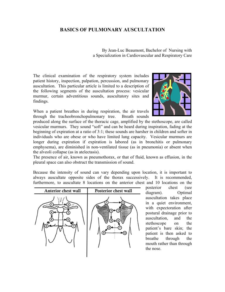
Because the intensity of sound can vary depending upon location, it is important to always auscultate opposite sides of the thorax successively. It is recommended, furthermore, to auscultate 8 locations on the anterior chest and 10 locations on the posterior chest (see diagram). Optimal auscultation takes place in a quiet environment, with expectoration after postural drainage prior to auscultation, and the stethoscope on the patient’s bare skin; the patient is then asked to breathe through the mouth rather than through the nose.
Adventitious breath sounds, commonly called rales, are detected along with vesicular murmurs. Rales are classified as either continuous or discontinuous. Continuous or
“bronchial rales” are produced when there is a buildup of mucus in the bronchi; they are most commonly heard during expiration and change after a cough. Different types of continuous rales include those heard in sibilant episodes in asthma crises, sonorous rales, and mucous rales (rhonchi) which are presented by patients with bronchitis or bronchiolitis. Discontinuous (or parenchymatous) rales differ from continuous rales in that they sound “softer” and are usually heard on inspiration. Their sound is much less obvious, is produced in a localized area, and is not affected by cough. Discontinuous rales are most often found in the pathologies of left ventricular insufficiency, pneumonia and acute pulmonary edema.
Verbal or written notes for all ausculatory findings must accompany all effective pulmonary auscultation, and should be of a descriptive rather than of a diagnostic nature.
They must specify whether the vesicular murmur is normal as well as whether there has been a change during the respiratory phases. In addition, these notes must specify whether there are new, abnormal breath sounds.
The following model describes, in order, the necessary parameters of an adequate chart note: the anomaly presented; a description of respiratory phase auscultation; the upper, middle or lower sections of the anterior and posterior thoracic regions; as well as the right or left hemithorax in question. Furthermore, the term “bottom of lung” (posteroinferior region) is unequivocally used in and specific to pulmonary auscultation terminology.
For example:
-Vesicular murmur – longer in expiratory phase, with mucous rales at base of lungs.
-Normal vesicular murmur and diffuse expiratory sibilants in middle and lower sections of posterior thorax.
-Diminished vesicular murmur and inspiratory crackle in middle section of right anterior hemithorax.
Pain, dyspnea, coughing and palpitations are the most common reasons for office visits to specialists in cardiology and respirology. As a member of a multidisciplinary team, the nurse is able to detect anomalies, provide clinical data to the physician, prevent complications and, as a result, adequately care for patients suffering from cardiorespiratory problems.
Reference
Beaumont, Jean-Luc. L’examen clinique respiratoire avec cassette audio des bruits respiratoires normaux et anormaux. 2 nd
ed., October 1999. Revised in August 2003.
Published by Gestion J.L. Beaumont
316 – 3111 avenue des Hôtels
Ste-Foy (QC) G1W 4W7
Email : gest.jlb@sympatico
