Biological Molecules
advertisement
Q1.
Lactose is a disaccharide sugar which can be broken down by the enzyme lactase into two
monosaccharides, glucose and galactose.
lactase
lactose+ water
(a)
glucose + galactose
The formula for galactose is C6H12O6. What is the formula for lactose?
......................................................................................................................
(2)
(b)
A solution containing the enzyme lactase was added to a lactose solution. The solution
was incubated at 40 °C for one hour. Sample A was removed from the tube before
incubation. Sample B was removed after one hour.
(i)
Describe a chemical test you could carry out on sample A to show that lactose is a
reducing sugar.
.............................................................................................................
.............................................................................................................
.............................................................................................................
.............................................................................................................
(2)
(ii)
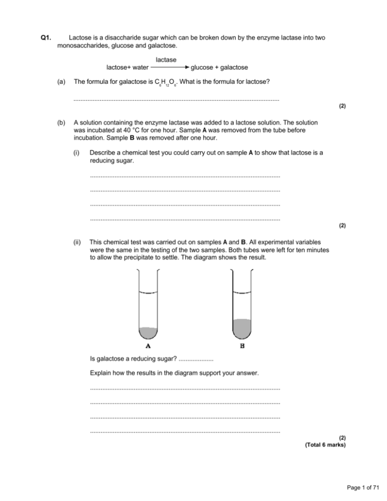
This chemical test was carried out on samples A and B. All experimental variables
were the same in the testing of the two samples. Both tubes were left for ten minutes
to allow the precipitate to settle. The diagram shows the result.
Is galactose a reducing sugar? ....................
Explain how the results in the diagram support your answer.
.............................................................................................................
.............................................................................................................
.............................................................................................................
.............................................................................................................
(2)
(Total 6 marks)
Page 1 of 71
Q2.
A student carried out an investigation into the mass of product formed in an enzymecontrolled reaction at three different temperatures. Only the temperature was different for each
experiment. The results are shown in the graph.
(a)
Use your knowledge of enzymes to explain
(i)
why the initial rate of reaction was highest at 55 °C;
.............................................................................................................
.............................................................................................................
.............................................................................................................
.............................................................................................................
(2)
(ii)
the shape of the curve for 55 °C after 20 minutes.
.............................................................................................................
.............................................................................................................
.............................................................................................................
.............................................................................................................
.............................................................................................................
.............................................................................................................
(3)
Page 2 of 71
(b)
Explain why the curves for 27 °C and 37 °C level out at the same value.
......................................................................................................................
......................................................................................................................
......................................................................................................................
......................................................................................................................
(2)
(Total 7 marks)
Q3.
(a)
Figure 1 shows the structure of a molecule of glycerol and a molecule of fatty acid.
Figure 1
Draw a diagram to show the structure of a triglyceride molecule.
(2)
(b)
Explain why triglycerides are not considered to be polymers.
......................................................................................................................
......................................................................................................................
(1)
Page 3 of 71
(c)
Figure 2 shows two types of fat storage cell. Mammals living in cold conditions have more
brown fat cells than mammals living in tropical conditions.
Figure 2
Using evidence from Figure 2 to support your answer, suggest how the function of brown
fat cells differs from that of white fat cells.
......................................................................................................................
......................................................................................................................
......................................................................................................................
......................................................................................................................
......................................................................................................................
......................................................................................................................
(3)
(Total 6 marks)
Q4.
(a)
Explain how the shape of an enzyme molecule is related to its function.
......................................................................................................................
......................................................................................................................
......................................................................................................................
......................................................................................................................
......................................................................................................................
......................................................................................................................
(3)
Page 4 of 71
(b)
Bacteria produce enzymes which cause food to decay. Explain how vinegar, which is
acidic, can prevent the action of bacterial enzymes in some preserved foods.
......................................................................................................................
......................................................................................................................
......................................................................................................................
......................................................................................................................
......................................................................................................................
......................................................................................................................
(3)
(Total 6 marks)
Q5.
The diagrams show four types of linkage, A to D, which occur in biological molecules.
(a)
Name the chemical process involved in the formation of linkage B.
......................................................................................................................
(1)
(b)
Give the letter of the linkage which
(i)
occurs in a triglyceride molecule;
.............................................................................................................
(1)
(ii)
might be broken down by the enzyme amylase;
.............................................................................................................
(1)
(iii)
may occur in the tertiary, but not the primary structure of protein.
.............................................................................................................
(1)
Page 5 of 71
(c)
Describe how a saturated fatty acid differs in molecular structure from an unsaturated fatty
acid.
......................................................................................................................
......................................................................................................................
......................................................................................................................
......................................................................................................................
(2)
(Total 6 marks)
Q6.
In an investigation, the rate at which phenol was broken down by the enzyme phenol oxidase
was measured in solutions with different concentrations of phenol. The experiment was then
repeated with a non-competitive inhibitor added to the phenol solutions. The graph shows the
results.
(a)
Explain why an increase in concentration of phenol solution from 2.0 to 2.5 mmol dm–3 has
no effect on the rate of the reaction without inhibitor.
......................................................................................................................
......................................................................................................................
......................................................................................................................
......................................................................................................................
(2)
Page 6 of 71
(b)
Explain the effect of the non-competitive inhibitor.
......................................................................................................................
......................................................................................................................
......................................................................................................................
......................................................................................................................
(2)
(c)
Calculate the percentage decrease in the maximum rate of the reaction when the inhibitor
was added. Show your working.
Percentage decrease ...........................................
(2)
(d)
Draw a curve on the graph to show the results expected if a competitive inhibitor instead of
a non-competitive inhibitor had been used.
(1)
(Total 7 marks)
Page 7 of 71
Q7.
Sucrose is a disaccharide. It is formed from two monosaccharides P and Q. The diagram
shows the structure of molecules of sucrose and monosaccharide P.
(a)
(i)
Name monosaccharide Q.
.............................................................................................................
(1)
(ii)
Draw the structure of a molecule of monosaccharide Q in the space above.
(1)
(b)
The enzyme sucrase catalyses the breakdown of sucrose into monosaccharides. What
type of reaction is this breakdown?
......................................................................................................................
(1)
(c)
The diagram shows apparatus used in breaking down sucrose. The enzyme sucrase is
fixed to inert beads. Sucrose solution is then passed through the column.
Page 8 of 71
Describe a biochemical test to find out if the solution collected from the apparatus contains
(i)
the products;
.............................................................................................................
.............................................................................................................
.............................................................................................................
.............................................................................................................
(2)
(ii)
the enzyme.
.............................................................................................................
.............................................................................................................
.............................................................................................................
.............................................................................................................
(2)
(Total 7 marks)
Page 9 of 71
Q8.
In an investigation into carbohydrase activity, the contents from part of the gut of a small
animal were collected. The contents were added to starch solution at pH 7 and kept in a water
bath at 25°C. At one-minute intervals, samples were removed and added to different test tubes
containing dilute iodine solution. The colour intensity of each sample was determined. The graph
shows the results.
(a)
Explain the change in colour intensity.
......................................................................................................................
......................................................................................................................
......................................................................................................................
......................................................................................................................
(2)
(b)
Draw clearly labelled curves on the graph to show the expected result if the experiment
was repeated
(i)
at 35 °C;
(ii)
at pH 2.
(2)
Page 10 of 71
(c)
Explain how
(i)
raising the temperature to 35 °C affects carbohydrase activity;
.............................................................................................................
.............................................................................................................
.............................................................................................................
.............................................................................................................
.............................................................................................................
.............................................................................................................
.............................................................................................................
.............................................................................................................
(ii)
decreasing the pH affects carbohydrase activity.
.............................................................................................................
.............................................................................................................
.............................................................................................................
.............................................................................................................
.............................................................................................................
.............................................................................................................
.............................................................................................................
.............................................................................................................
.............................................................................................................
.............................................................................................................
(7)
(Total 11 marks)
Page 11 of 71
Q9.
Two samples of the roots of pea plants were placed in solutions containing potassium ions.
An inhibitor to prevent respiration was added to one solution. The concentrations of potassium
ions in the two solutions were measured at regular intervals. The graph shows the results.
(a)
Explain the decrease in the concentrations of potassium ions in the two solutions between
0 and 30 minutes.
(i)
With inhibitor
.............................................................................................................
.............................................................................................................
.............................................................................................................
.............................................................................................................
(2)
(ii)
Without inhibitor
.............................................................................................................
.............................................................................................................
(1)
(b)
Explain why there is no further decrease in the concentration of potassium ions in the
solution with the inhibitor after 60 minutes.
......................................................................................................................
......................................................................................................................
......................................................................................................................
......................................................................................................................
(2)
Page 12 of 71
(c)
The substance malonate is an inhibitor of respiration. It has a structure very similar to the
substrate of an enzyme that catalyses one of the reactions of respiration. Explain how
malonate inhibits respiration.
......................................................................................................................
......................................................................................................................
......................................................................................................................
......................................................................................................................
(2)
(Total 7 marks)
Q10.
The diagram shows different structures present in the wall of part of the ileum.
(i)
Describe the function of part X.
......................................................................................................................
......................................................................................................................
(1)
Page 13 of 71
(ii)
Suggest an advantage of having muscle cells in the villi.
......................................................................................................................
......................................................................................................................
......................................................................................................................
......................................................................................................................
(2)
(Total 3 marks)
Q11.
The diagram shows the structure of the amino acid serine.
(a)
(i)
Draw a box on the diagram around the R group of serine and label the box with the
letter R.
(1)
(ii)
Draw a circle around each of the parts of the serine molecule which would be
removed when two other amino acid molecules join directly to it.
(1)
(b)
(i)
Which two substances are formed when two amino acid molecules join together?
1 ..........................................................................................................
2 ..........................................................................................................
(1)
(ii)
Name the type of bond formed between the joined pair of amino acid molecules.
.............................................................................................................
(1)
Page 14 of 71
(c)
Explain how a change in the primary structure of a globular protein may result in a different
three-dimensional structure.
......................................................................................................................
......................................................................................................................
......................................................................................................................
......................................................................................................................
......................................................................................................................
......................................................................................................................
(3)
(Total 7 marks)
Q12.
The diagram represents an enzyme molecule and three other molecules that could
combine with it.
(a)
Which molecule is the substrate for the enzyme? Give a reason for your answer.
......................................................................................................................
......................................................................................................................
......................................................................................................................
(1)
(b)
Use the diagram to explain how a non-competitive inhibitor would decrease the rate of
the reaction catalysed by this enzyme.
......................................................................................................................
......................................................................................................................
......................................................................................................................
......................................................................................................................
......................................................................................................................
......................................................................................................................
(3)
Page 15 of 71
(c)
Lysozyme is an enzyme. A molecule of lysozyme is made up of 129 amino acid
molecules joined together. In the formation of its active site, the two amino acids that are at
positions 35 and 52 in the amino acid sequence need to be close together.
(i)
Name the bonds that join amino acids in the primary structure.
.............................................................................................................
(1)
(ii)
Suggest how the amino acids at positions 35 and 52 are held close together to form
the active site.
.............................................................................................................
.............................................................................................................
.............................................................................................................
.............................................................................................................
(2)
(Total 7 marks)
Q13.
(a) Amylase is an enzyme which hydrolyses starch to maltose. Some amylase and
starch were mixed and the mixture incubated at 37 °C until the reaction was complete.
(i)
Sketch a curve on the axes below to show the progress of this reaction.
(1)
(ii)
Explain why the rate of the reaction decreases as the reaction progresses.
.............................................................................................................
.............................................................................................................
.............................................................................................................
(2)
Page 16 of 71
The effect of temperature on the rate of reaction of an enzyme was investigated. A test tube
containing the enzyme and a test tube containing the substrate were incubated separately at
each of the temperatures being investigated. After 5 minutes, they were mixed and the rate of
reaction was determined. The experiment was repeated but, this time, the enzyme and the
substrate were left for 60 minutes before they were mixed. The results of the investigation are
shown in the graph.
(b)
The enzyme solution used in this investigation was made by dissolving a known mass of
enzyme in a buffer solution. Explain why a buffer solution was used.
......................................................................................................................
......................................................................................................................
(1)
(c)
(i)
Use the graph to describe how incubation time affects the rate of the reaction.
.............................................................................................................
.............................................................................................................
.............................................................................................................
.............................................................................................................
(2)
Page 17 of 71
(ii)
The maximum rate of reaction with an incubation time of 60 minutes is less than the
maximum rate of reaction with an incubation time of 5 minutes. Explain why.
.............................................................................................................
.............................................................................................................
.............................................................................................................
.............................................................................................................
.............................................................................................................
.............................................................................................................
(3)
(d)
Explain how inhibitors affect the rate of enzyme-controlled reactions.
......................................................................................................................
......................................................................................................................
......................................................................................................................
......................................................................................................................
......................................................................................................................
......................................................................................................................
......................................................................................................................
......................................................................................................................
......................................................................................................................
......................................................................................................................
......................................................................................................................
(6)
(Total 15 marks)
Page 18 of 71
Q14.
Read the following passage.
5
Human milk contains all the nutrients a young baby needs in exactly the right
proportions. It is formed in the mammary glands by small groups of milk-producing cells.
These cells absorb substances from the blood and use them to synthesise the lipids,
carbohydrates and proteins found in milk. Milk-producing cells are roughly cube-shaped
and have a height to breadth ratio of approximately 1.2 : 1.
The main carbohydrate in milk is lactose. Lactose is a disaccharide formed by the
condensation of two monosaccharides, glucose and galactose. (A molecule of galactose
has the same formula as a molecule of glucose – the atoms are just arranged in a different
way.)
10 Lactose is synthesised in the Golgi apparatus and transported in vesicles through the
cytoplasm. Because lactose is unable to escape from these vesicles, they increase in
diameter as they move towards the plasma membrane. The vesicle membranes fuse with
the plasma membrane and the vesicles empty their contents out of the cell.
Use the information from the passage and your own knowledge to answer the following
questions.
(a)
(i)
The breadth of a milk-producing cell is 26 µm. Calculate the height of this cell.
Height = .......................... µm
(1)
(ii)
Describe and explain how you would expect the height to breadth ratio of an epithelial
cell from a lung alveolus to differ from the height to breadth ratio of a milk-producing
cell.
.............................................................................................................
.............................................................................................................
.............................................................................................................
.............................................................................................................
(2)
(b)
How many oxygen atoms are there in a molecule of
(i)
galactose;
.............................................................................................................
(1)
(ii)
lactose?
.............................................................................................................
(1)
Page 19 of 71
(c)
The lactose-containing vesicles increase in diameter as they move towards the plasma
membrane of the milk-producing cell (lines 11-12). Use your knowledge of water potential
to explain why.
......................................................................................................................
......................................................................................................................
......................................................................................................................
......................................................................................................................
(2)
(d)
Suggest one advantage of milk-producing cells containing large numbers of mitochondria.
......................................................................................................................
......................................................................................................................
......................................................................................................................
......................................................................................................................
(2)
(e)
Some substances pass through the plasma membrane of a milk-producing cell by
diffusion. Describe the structure of a plasma membrane and explain how different
substances are able to pass through the membrane by diffusion.
......................................................................................................................
......................................................................................................................
......................................................................................................................
......................................................................................................................
......................................................................................................................
......................................................................................................................
......................................................................................................................
......................................................................................................................
......................................................................................................................
......................................................................................................................
......................................................................................................................
(6)
(Total 15 marks)
Page 20 of 71
Q15.
Read the following passage.
5
Job’s Tears is a cereal plant which grows in the tropics. An unusual protein has been found in
its grains. This protein is unusual because it has two functions. It acts as both an enzyme
inhibitor and as an enzyme. As an inhibitor, the protein reduces the activity of starch-digesting
enzymes. The protein acts as an enzyme by breaking down chitin, a polysaccharide found in
the walls of many fungi, to its monomers. Because of the resulting more negative water
potential in the cytoplasm of the fungus, this effectively leads to “death by osmosis” of any
fungus attacking the grain.
Our knowledge of the relationship between protein structure and function has led to the
development of the new technology of protein engineering. This involves changing the amino
10 acid sequence of a protein and altering its tertiary structure. Altering the tertiary structure
changes the protein’s properties. So far, we have been unable to produce a protein with more
than one function such as that found in Job’s Tears. We have had success, though, in making
some enzymes more stable and less prone to heat denaturation. We have done this by
substituting amino acids and allowing the formation of additional chemical bonds.
Use information from the passage and your own knowledge to answer the following questions.
(a)
(i)
The protein found in Job’s Tears breaks down chitin (line 4). What type of chemical
reaction is involved in breaking down chitin?
.............................................................................................................
(1)
(ii)
Breakdown of chitin leads to “death by osmosis” of fungi attacking the grain
(lines 6 - 7). Explain how.
.............................................................................................................
.............................................................................................................
.............................................................................................................
.............................................................................................................
(2)
(iii)
This protein does not break down the cell walls of the Job’s Tears plant.
Explain why.
.............................................................................................................
.............................................................................................................
(1)
(b)
Explain what is meant by the tertiary structure of a protein (line 10).
.............................................................................................................
.............................................................................................................
(1)
Page 21 of 71
(c)
(i)
Explain how heating an enzyme leads to it being denatured.
.............................................................................................................
.............................................................................................................
.............................................................................................................
.............................................................................................................
(2)
(ii)
How can protein engineering make enzymes more stable and less prone to heat
denaturation (line 13)?
.............................................................................................................
.............................................................................................................
.............................................................................................................
.............................................................................................................
(2)
(d)
Describe how the sequence of amino acids in part of the protein from Job’s Tears could
enable this protein to act as an enzyme inhibitor.
......................................................................................................................
......................................................................................................................
......................................................................................................................
......................................................................................................................
......................................................................................................................
......................................................................................................................
......................................................................................................................
......................................................................................................................
......................................................................................................................
......................................................................................................................
......................................................................................................................
......................................................................................................................
(6)
(Total 15 marks)
Page 22 of 71
Q16.
(a) Describe how you would use a biochemical test to show that a solution contained
protein.
......................................................................................................................
......................................................................................................................
......................................................................................................................
......................................................................................................................
(2)
The diagram shows the structure of two amino acid molecules, tyrosine and phenylalanine.
(b)
Copy from the diagram the R group in the phenylalanine molecule.
(1)
(c)
(i)
In the space below, draw the chemical bond formed when these two amino acids are
joined by condensation. You need only draw the parts of the molecules shown in the
box.
(2)
(ii)
Name this bond.
.............................................................................................................
(1)
Page 23 of 71
(d)
Tyrosine can be made in the body by hydroxylating phenylalanine. Use the diagram to
explain the meaning of hydroxylating.
......................................................................................................................
......................................................................................................................
(1)
(Total 7 marks)
Q17.
(a)
Starch and protein are biologically important polymers.
(i)
Explain what is meant by a polymer.
.............................................................................................................
.............................................................................................................
(1)
(ii)
Give one example of a biologically important polymer other than starch or protein.
.............................................................................................................
(1)
(b)
In an investigation, the enzyme amylase was mixed in a test tube with a buffer solution and
a suspension of starch. The amylase broke down the starch to maltose. When all the
starch had been broken down, a sample was removed from the test tube and tested with
biuret reagent.
(i)
Explain why a buffer solution was added to the amylase-starch mixture.
.............................................................................................................
.............................................................................................................
.............................................................................................................
(2)
(ii)
What colour would you expect the sample to go when tested with biuret reagent?
.............................................................................................................
(1)
(iii)
Give an explanation for your answer to part (ii)
.............................................................................................................
.............................................................................................................
.............................................................................................................
(2)
(Total 7 marks)
Page 24 of 71
Q18.
Catalase is an enzyme. It catalyses the breakdown of hydrogen peroxide in the reaction:
2H2O2
→
hydrogen
peroxide
2H2O
water
+
O2
oxygen
In an investigation, samples of different substances were added to hydrogen peroxide in a series
of test tubes. The rate of reaction was measured by recording the rate at which bubbles of
oxygen were produced. A scale going from 0 for no bubbles to 5 for the maximum rate of
bubbling was used to measure this. The results are shown in the table.
Tube
(a)
Substance added
Rate at which bubbles of
oxygen were produced
A
Piece of liver
4
B
Ground liver and sand
5
C
Sand
0
D
Piece of cooled, boiled liver
0
Explain the difference between the rate at which bubbles were produced in.
(i)
tubes A and B;
..............................................................................................................
.............................................................................................................
.............................................................................................................
.............................................................................................................
(2)
(ii)
tubes A and D.
.............................................................................................................
.............................................................................................................
.............................................................................................................
.............................................................................................................
.............................................................................................................
.............................................................................................................
(3)
(b)
Explain the purpose of tube C.
......................................................................................................................
......................................................................................................................
(1)
Page 25 of 71
(c)
The graph shows the energy changes which take place during the reaction in which
hydrogen peroxide is converted to water and oxygen.
Use the graph to explain why
(i)
hydrogen peroxide breaks down at a lower temperature when catalase is present
than when it is not present;
.............................................................................................................
.............................................................................................................
(1)
(ii)
test tubes A and B became warmer when the reaction was taking place.
.............................................................................................................
.............................................................................................................
.............................................................................................................
.............................................................................................................
(2)
(Total 9 marks)
Page 26 of 71
Q19.
(a)
The diagrams represent an enzyme, its substrate and two other molecules, A and B.
The addition of a non-competitive inhibitor will prevent the formation of an enzymesubstrate complex. Draw a labelled diagram based on relevant molecules selected from
the diagram above to explain how this occurs.
(2)
(b)
A decrease in temperature decreases the kinetic energy of molecules in a solution. Explain
how a decrease in temperature decreases the rate of an enzyme-controlled reaction.
......................................................................................................................
......................................................................................................................
......................................................................................................................
......................................................................................................................
(2)
(c)
Urea breaks hydrogen bonds. Explain how the addition of urea would affect the rate of an
enzyme-controlled reaction.
......................................................................................................................
......................................................................................................................
......................................................................................................................
......................................................................................................................
......................................................................................................................
......................................................................................................................
(3)
(Total 7 marks)
Page 27 of 71
Q20.
Read the following passage.
During the course of a day, we come into contact with many poisonous substances.
These include industrial and household chemicals. The skin acts as a barrier and prevents
many of these substances entering and harming the body.
5
The skin is one of the largest organs in the body. It is composed of several layers of
tissue. The outer layer consists of dead cells packed with keratins. Keratins are a group of
proteins that differ from each other in their primary structure. Each keratin molecule
consists of several polypeptide chains, each individual chain wound into a spiral or helix.
The polypeptide chains include many sulphur-containing amino acids and these help to
give the keratin molecules their characteristic strength.
Use information from the passage and your own knowledge to answer the questions.
(a)
What is the evidence from the passage that keratin molecules have a quaternary
structure?
......................................................................................................................
......................................................................................................................
(1)
(b)
Explain how sulphur-containing amino acids help to give keratin molecules their
characteristic strength (lines 8–9).
......................................................................................................................
......................................................................................................................
......................................................................................................................
......................................................................................................................
(2)
(c)
Explain why differences in primary structure result in keratins with different properties
(line 6).
......................................................................................................................
......................................................................................................................
......................................................................................................................
......................................................................................................................
(2)
Page 28 of 71
(d)
The skin prevents poisonous substances entering and harming the body (line 3). Explain
why these substances are unable to pass through the outer layer of skin cells by active
transport.
......................................................................................................................
......................................................................................................................
......................................................................................................................
......................................................................................................................
......................................................................................................................
......................................................................................................................
(3)
(e)
Skin cells may be studied with a transmission electron microscope or an optical
microscope. Explain the advantages and limitations of using a transmission electron
microscope to study cells.
......................................................................................................................
......................................................................................................................
......................................................................................................................
......................................................................................................................
......................................................................................................................
......................................................................................................................
......................................................................................................................
......................................................................................................................
......................................................................................................................
......................................................................................................................
......................................................................................................................
......................................................................................................................
......................................................................................................................
......................................................................................................................
(6)
(Total 14 marks)
Page 29 of 71
Q21.
Uric acid is produced in the body. One of the reactions involved in the production of uric
acid is catalysed by xanthine oxidase.
xanthine
oxidase
xanthine
(a)
uric acid
A sample of xanthine oxidase was tested by mixing with biuret reagent. Describe and
explain the result of this test.
......................................................................................................................
......................................................................................................................
......................................................................................................................
......................................................................................................................
(2)
(b)
Explain why xanthine oxidase is able to catalyse this reaction but it is not able to
catalyse other reactions.
......................................................................................................................
......................................................................................................................
......................................................................................................................
......................................................................................................................
(2)
Page 30 of 71
(c)
Gout is a painful condition caused by uric acid crystals in the joints. It is often treated with a
drug that inhibits xanthine oxidase. The diagram shows a molecule of xanthine and a
molecule of this drug.
Xanthine
Drug used
to treat gout
Use the diagram to explain why this drug is effective in the treatment of gout.
......................................................................................................................
......................................................................................................................
......................................................................................................................
......................................................................................................................
......................................................................................................................
......................................................................................................................
(3)
(Total 7 marks)
Q22.
Some enymes digest protein. They hydrolyse the peptide bonds between amino acids. The
extent to which a protein is digested is called the degree of hydrolysis (DH). The DH value may
be calculated from the equation:
(a)
(i)
A protein molecule contains 151 amino acids. What is the total number of peptide
bonds in this molecule?
.............................................................................................................
(1)
(ii)
A molecule of this protein is digested. The DH value of the digested protein is 18.
Calculate the number of peptide bonds that have been hydrolysed.
Answer ......................................
(1)
Page 31 of 71
(b)
What would be the DH value of a protein if it were completely hydrolysed to amino acids?
Explain how you arrived at your answer.
DH value ......................................................................................................
Explanation ..................................................................................................
......................................................................................................................
......................................................................................................................
(2)
Enzymes A and B digest protein. The graph shows the effect of pH on the rates of reaction of
these enzymes.
(c)
Pepsin is a protein-digesting enzyme found in the stomach. It has an optimum pH of 2 and
is fully denatured at pH 6. Sketch a curve on the graph to show the effect of pH on the rate
of reaction of pepsin.
(1)
(d)
Explain why the rate of reaction of enzyme B is low at pH 5.
......................................................................................................................
......................................................................................................................
......................................................................................................................
......................................................................................................................
......................................................................................................................
......................................................................................................................
(3)
Page 32 of 71
(e)
Enzyme A is present in some washing powders used for cleaning clothes. Use the graph
to suggest why enzyme A would be of more use in washing clothes than enzyme B.
......................................................................................................................
......................................................................................................................
......................................................................................................................
(1)
(f)
Use your knowledge of protein structure to explain why enzymes are specific and may be
affected by non-competitive inhibitors.
......................................................................................................................
......................................................................................................................
......................................................................................................................
......................................................................................................................
......................................................................................................................
......................................................................................................................
......................................................................................................................
......................................................................................................................
......................................................................................................................
......................................................................................................................
......................................................................................................................
......................................................................................................................
......................................................................................................................
......................................................................................................................
(6)
(Total 15 marks)
Page 33 of 71
Q23.
Amylase is an enzyme that breaks down starch. A student investigated the effect of pH on
amylase activity by using a starch agar plate. Six circular wells were cut into the agar plate.
Each well contained the same concentration and volume of amylase, and a buffer solution of
different pH. The agar plate was then left for 24 hours. The diagram shows the results
(a)
Describe how the student could have used these results to compare the activity of the
enzyme at different pH values.
......................................................................................................................
......................................................................................................................
......................................................................................................................
......................................................................................................................
(2)
(b)
The student concluded that the optimum pH for amylase activity was 7. This conclusion
may not be valid. Explain why.
......................................................................................................................
......................................................................................................................
(1)
(c)
Using your knowledge of enzyme structure, explain the result obtained at pH 11.
......................................................................................................................
......................................................................................................................
......................................................................................................................
......................................................................................................................
(2)
Page 34 of 71
(d)
Describe a control that would be necessary for this investigation.
......................................................................................................................
......................................................................................................................
......................................................................................................................
......................................................................................................................
(2)
(Total 7 marks)
Q24.
(a)
Sucrose, maltose and lactose are disaccharides.
(i)
Sucrase is an enzyme. It hydrolyses sucrose during digestion. Name the products of
this reaction.
................................................... and ..................................................
(2)
(ii)
Sucrase does not hydrolyse lactose. Use your knowledge of the way in which
enzymes work to explain why.
.............................................................................................................
.............................................................................................................
.............................................................................................................
.............................................................................................................
(2)
(b)
A woman was given a solution of sucrose to drink. Her blood glucose concentration was
measured over the next 90 minutes. The results are shown on the graph.
Page 35 of 71
(i)
Describe how the woman’s blood glucose concentration changed in the period
shown in the graph.
.............................................................................................................
.............................................................................................................
.............................................................................................................
.............................................................................................................
(2)
(ii)
Explain the results shown on the graph.
.............................................................................................................
.............................................................................................................
.............................................................................................................
.............................................................................................................
(2)
(iii)
This woman was lactose intolerant.
On the graph, sketch a curve to show what would happen to her blood glucose
concentration if she had been given a solution of lactose to drink instead of a
sucrose solution.
(1)
(Total 9 marks)
Q25.
A glucose biosensor is an instrument used to measure glucose concentration. It contains
an enzyme called glucose oxidase.
(a)
A glucose biosensor detects only glucose. Use your knowledge of the way in which
enzymes work to explain why.
......................................................................................................................
......................................................................................................................
......................................................................................................................
......................................................................................................................
......................................................................................................................
......................................................................................................................
(3)
Page 36 of 71
(b)
It is better to use a biosensor than the Benedict’s test to measure the concentration of
glucose in a sample of blood. Suggest two reasons why.
1 ...................................................................................................................
......................................................................................................................
2 ...................................................................................................................
......................................................................................................................
(2)
(c)
(i)
Diabetes mellitus is a disease that can lead to an increase in blood glucose
concentration. Some diabetics need insulin injections. Insulin is a protein so it cannot
be taken orally. Suggest why insulin cannot be taken orally.
.............................................................................................................
.............................................................................................................
.............................................................................................................
(1)
(ii)
A drug company produced a new type of insulin. Scientists from the company carried
out a trial in which they gave this new type of insulin to rats. They reported that the
results of this trial on rats were positive. A newspaper stated that diabetics would
benefit from this new drug. Suggest two reasons why this statement should be
viewed with caution.
1 ..........................................................................................................
.............................................................................................................
2 ..........................................................................................................
.............................................................................................................
(2)
(Total 8 marks)
Page 37 of 71
Q26.
The graph shows the effect of substrate concentration on the rate of an enzyme-controlled
reaction.
(a)
(i)
Describe what the graph shows about the effect of substrate concentration on the
rate of this enzyme-controlled reaction.
.............................................................................................................
.............................................................................................................
.............................................................................................................
.............................................................................................................
(2)
(ii)
What limits the rate of this reaction between points A and B? Give the evidence from
the graph for this.
.............................................................................................................
.............................................................................................................
.............................................................................................................
.............................................................................................................
(2)
(iii)
Suggest a reason for the shape of the curve between points C and D.
.............................................................................................................
.............................................................................................................
(1)
(b)
Sketch a curve on the graph to show the rate of this reaction in the presence of a
competitive inhibitor.
(1)
Page 38 of 71
(c)
Methotrexate is a drug used in the treatment of cancer. It is a competitive inhibitor and
affects the enzyme folate reductase.
(i)
Explain how the drug lowers the rate of reaction controlled by folate reductase.
.............................................................................................................
.............................................................................................................
.............................................................................................................
.............................................................................................................
.............................................................................................................
(2)
(ii)
Methotrexate only affects the rate of the reaction controlled by folate reductase.
Explain why this drug does not affect other enzymes.
.............................................................................................................
.............................................................................................................
(1)
(Total 9 marks)
Q27.
Gangliosides are lipids found in the cell surface membranes of nerve cells.
Hexosaminidase is an enzyme present in blood that breaks down gangliosides. If gangliosides
are not broken down, they damage nerve cells.
(a)
Hexosaminidase only breaks down gangliosides. It does not break down other lipids.
Explain why this enzyme only breaks down gangliosides.
......................................................................................................................
......................................................................................................................
......................................................................................................................
......................................................................................................................
......................................................................................................................
......................................................................................................................
(3)
(b)
Hexosaminidase is found in the blood of healthy people. People with Tay Sachs disease
do not have this enzyme in their blood.
Doctors confirm Tay Sachs disease by using a blood test. The technician carrying out the
test adds a solution containing a high concentration of gangliosides to a sample of blood
from the person being tested. The technician then measures the concentration of
gangliosides in the person’s blood at regular intervals.
Page 39 of 71
(i)
Complete the graph below by sketching a curve to show the results you would
expect for a person with Tay Sachs disease. Label this curve T.
(1)
(ii)
Sketch a curve on the same graph to show the results you would expect for a healthy
person who does not have Tay Sachs disease. Label this curve H.
(1)
(c)
Scientists are trying to find a way to give the missing enzyme to people with Tay Sachs
disease. Suggest why they cannot give the enzyme as a tablet that is swallowed.
......................................................................................................................
......................................................................................................................
......................................................................................................................
......................................................................................................................
(2)
(Total 7 marks)
Page 40 of 71
Q28.
A protease is an enzyme that digests protein. The graph shows how the activity of a
protease varies with temperature.
(a)
(i)
Describe what the graph shows about the effect of temperature on the rate of
reaction.
.............................................................................................................
.............................................................................................................
.............................................................................................................
(1)
(ii)
Explain the shape of the curve between 30 °C and 50 °C.
.............................................................................................................
.............................................................................................................
.............................................................................................................
.............................................................................................................
.............................................................................................................
.............................................................................................................
(Extra space) ......................................................................................
.............................................................................................................
(3)
Page 41 of 71
(b)
Students investigated the effect of pH on the activity of the protease.
•
The students used agar plates containing protein. The protein made the agar cloudy.
•
They made four wells of equal size in the agar of each plate.
•
They added a drop of protease solution to each of the wells. The protease solution in
each well was at a different pH.
•
The students incubated the agar plates for 4 hours at a constant temperature.
The diagram shows the agar plates after they were incubated and the pH of the protease
solution in each well.
(i)
How should the students make sure that the pH of the protease solution did not
change?
.............................................................................................................
(1)
(ii)
Use the graph to suggest a suitable temperature for incubating the agar plates.
Explain your answer.
.............................................................................................................
.............................................................................................................
.............................................................................................................
(1)
(iii)
Use the diagram to describe the effect of pH on the activity of this protease.
.............................................................................................................
.............................................................................................................
.............................................................................................................
(1)
(Total 7 marks)
Page 42 of 71
Q29.
The equation shows the breakdown of lactose by the enzyme lactase.
Lactose + water
(a)
(i)
galactose + monosaccharide X
Name the type of reaction catalysed by the enzyme lactase.
.............................................................................................................
(1)
(ii)
Name monosaccharide X.
.............................................................................................................
(1)
(b)
(i)
Describe how you would use a biochemical test to show that a reducing sugar is
present.
.............................................................................................................
.............................................................................................................
.............................................................................................................
.............................................................................................................
(2)
.............................................................................................................
(ii)
Lactose, galactose and monosaccharide X are all reducing sugars.
After the lactose has been broken down there is a higher concentration of reducing
sugar. Explain why.
.............................................................................................................
(1)
(c)
A high concentration of galactose slows down the breakdown of lactose by lactase.
Use your knowledge of competitive inhibition to suggest why.
......................................................................................................................
......................................................................................................................
......................................................................................................................
......................................................................................................................
......................................................................................................................
(2)
Page 43 of 71
(d)
People who are lactose intolerant are not able to produce the enzyme lactase.
Explain why these people get diarrhoea when they drink milk containing lactose.
......................................................................................................................
......................................................................................................................
......................................................................................................................
......................................................................................................................
......................................................................................................................
(2)
(Total 9 marks)
Q30.
Read the following passage.
Aspirin is a very useful drug. One of its uses is to reduce fever and
inflammation. Aspirin does this by preventing cells from producing
substances called prostaglandins. Prostaglandins are produced by
an enzyme-controlled pathway. Aspirin works by inhibiting one of the
enzymes in this pathway. Aspirin attaches permanently to a
chemical group on one of the monomers that make up the active site
of this enzyme.
The enzyme that is involved in the pathway leading to the production
of prostaglandins is also involved in the pathway leading to the
production of thromboxane. This is a substance that promotes blood
clotting. A small daily dose of aspirin may reduce the risk of
myocardial infarction (heart attack).
5
10
Use information from the passage and your own knowledge to answer the following
questions.
(a)
Name the monomers that make up the active site of the enzyme (lines 6 – 7).
........................................................................................................................
(1)
Page 44 of 71
(b)
The diagram shows the pathways by which prostaglandins and thromboxane
are formed.
(i)
Aspirin only affects one of the enzymes in this pathway. Use information in lines
5 – 7 to explain why aspirin does not affect the other enzymes.
...............................................................................................................
...............................................................................................................
...............................................................................................................
...............................................................................................................
...............................................................................................................
(2)
(ii)
Which enzyme, X, Y or Z, is inhibited by aspirin? Explain the evidence from the
passage that supports your answer.
Enzyme ................................................................................................
Explanation ...........................................................................................
...............................................................................................................
...............................................................................................................
(2)
(c)
Aspirin is an enzyme inhibitor. Explain how aspirin prevents substrate molecules being
converted to product molecules.
........................................................................................................................
........................................................................................................................
........................................................................................................................
........................................................................................................................
........................................................................................................................
(2)
Page 45 of 71
(d)
Aspirin may reduce the risk of myocardial infarction (lines 8 – 12). Explain how.
...............................................................................................................
...............................................................................................................
...............................................................................................................
...............................................................................................................
...............................................................................................................
...............................................................................................................
(Extra space) ........................................................................................
...............................................................................................................
...............................................................................................................
(3)
(Total 10 marks)
Q31.
(a) Some seeds contain lipids. Describe how you could use the emulsion test to show
that a seed contains lipids.
........................................................................................................................
........................................................................................................................
........................................................................................................................
........................................................................................................................
........................................................................................................................
........................................................................................................................
........................................................................................................................
(Extra space) .................................................................................................
........................................................................................................................
........................................................................................................................
(3)
Page 46 of 71
(b)
A triglyceride is one type of lipid. The diagram shows the structure of a triglyceride
molecule.
(i)
A triglyceride molecule is formed by condensation. From how many molecules
is this triglyceride formed?
(1)
(ii)
The structure of a phospholipid molecule is different from that of a triglyceride.
Describe how a phospholipid is different.
...............................................................................................................
...............................................................................................................
...............................................................................................................
...............................................................................................................
...............................................................................................................
(2)
(iii)
Use the diagram to explain what is meant by an unsaturated fatty acid.
...............................................................................................................
...............................................................................................................
...............................................................................................................
...............................................................................................................
...............................................................................................................
(2)
(Total 8 marks)
Page 47 of 71
Q32.
(a) A student investigated the effect of pH on the activity of the enzyme amylase.
She set up the apparatus shown in the diagram.
The tubes were made from Visking tubing. Visking tubing is partially permeable.
She added an equal volume of amylase solution and starch to each tube.
•
She added a buffer solution at pH2 to tube A.
•
She added an equal volume of buffer solution at pH8 to tube B.
After 30 minutes, she measured the height of the solutions in both tubes.
She then tested the solutions in tubes A and B for the presence of reducing sugars.
Describe how the student would show that reducing sugars were present in a solution.
........................................................................................................................
........................................................................................................................
........................................................................................................................
........................................................................................................................
........................................................................................................................
........................................................................................................................
(Extra space) .................................................................................................
........................................................................................................................
........................................................................................................................
(3)
Page 48 of 71
(b)
After 30 minutes, the solution in tube B was higher than the solution in tube A.
(i)
Explain why the solution in tube B was higher.
...............................................................................................................
...............................................................................................................
...............................................................................................................
...............................................................................................................
...............................................................................................................
...............................................................................................................
(Extra space) ........................................................................................
...............................................................................................................
...............................................................................................................
(3)
(ii)
The student concluded from her investigation that the optimum pH of amylase
was pH8. Is this conclusion valid? Explain your answer
...............................................................................................................
...............................................................................................................
...............................................................................................................
(1)
(Total 7 marks)
Page 49 of 71
M1.
(a)
C12 ; H22O11 ;
2
(b)
(i)
heat with Benedict’s;
yellow/brown/orange/red;
2
(ii)
(yes)
(may appear on second line)
more precipitate in sample B;
both sugars are reducing sugars/ give a positive test;
2
[6]
M2.
(a)
(i)
substances/molecules have more (kinetic) energy/moving faster;
(reject vibrate)
increased collisions / enzyme substrate complexes formed;
2
(ii)
causes denaturation/tertiary structure/shape change;
H+/ionic bonds break;
(shape) of active site changed;
substrate no longer binds/not complementary to (active site);
3 max
(b)
all substrate changed into product / reaction is complete;
same amount of product formed;
same initial substrate concentration;
2 max
[7]
M3.
(a) 3 fatty acids attached;
ester bond correct;
(H on glycerol component, O attached to carbon, R at other end)
2
Page 50 of 71
(b)
not made of monomers/many repeating units;
1
(c)
(many) mitochondria present in brown fat cells;
mitochondria release heat/energy; (ignore ATP)
white fat cells for fat storage / reduced fat storage in brown fat cells;
3
[6]
M4.
(a) specific 3D tertiary structure/shape;
substrate complementary shape;
(reject same shape)
substrate (can bind) to active site/ can fit into each active site;
3
(b)
(bacterial) active site/enzymes/proteins denatured /
tertiary 3D structure disrupted/changed;
(ionic) bonds broken;
(reject peptide bonds)
(ignore other bonds)
no enzyme substrate complex formed / substrate no longer fits;
3
[6]
M5.
(a)
(i)
condensation;
1
(b)
(i)
D;
1
(ii)
C;
1
(iii)
A;
1
(c)
absence of a double bond;
in the (hydrocarbon) chain;
unable to accept more hydrogen / saturated with hydrogen;
2 max
[6]
Page 51 of 71
M6.
(a) maximum rate at which enzyme can combine with substrate /
form enzyme-substrate complexes / substrate no longer limiting /
enzyme is a limiting factor;
(active site of) enzyme saturated with substrate
(disqualify active sites/enzymes ‘used up’);
2
(b)
inhibitor attaches to enzyme away from the active site;
changes shape of active site;
prevents formation of enzyme-substrate complex;
2 max
(c)
x 100;
= 26.32%; (accept 26% or 26.3%)
(correct answer = 2 marks)
(principle –
× 100 = 1 mark)
2
(d)
curve below top curve (without inhibitor) joining to top curve /
continues to increase to end of x-axis
(must not exceed or level out below ‘without inhibitor curve’ and
must start from origin);
1
[7]
M7.
(a)
(i)
fructose;
1
(ii)
correctly drawn (OH group at bottom left);
1
(b)
hydrolysis;
1
(c)
(i)
heat with Benedict’s solution (disqualify if HCl added);
orange/brown/brick red/green/yellow colour or precipitate;
2
(ii)
biuret test / NaOH + CuSO4;
purple / violet / lilac / mauve;
2
[7]
Page 52 of 71
M8.
(a) colour results from starch-iodine reaction;
decrease due to breakdown of starch by carbohydrase/enzyme;
2
(b)
(i)
curve drawn below curve on graph and starting at same point;
1
(ii)
curve drawn above curve on graph and starting at same point but
finishing above;
(allow curve or horizontal line)
(allow alternative curve for pH if explanation in (ii) is consistent)
1
(c)
(i)
1
2
3
(ii)
4
5
6
7
8
9
10
increase in temperature increases kinetic energy;
increases collisions (between enzyme/active site and substrate) /
increases formation of enzyme/substrate complexes;
increases rate of breakdown of starch /rate of
reaction/carbohydrase activity;
(decrease in pH) increases H+ ions/protons;
attach/attracted to amino acids;
hydrogen/ionic bonds disrupted/broken;
denatures enzyme / changes tertiary structure;
changes shape/charge of active site;
active site/enzyme unable to combine/fit with starch/
enzyme-substrate complex no longer able to form;
decreases rate of breakdown of starch/rate of reaction
/carbohydrase activity;
(allow alternative explanation for pH if consistent with line
drawn in (ii))
7 max
[11]
M9.
(a)
(i)
absorbed by diffusion;
no energy/ATP available / active transport requires energy/ATP;
2 max
(disqualify energy made)
(allow energy reference in either (i) or (ii))
(ii)
absorbed by active transport;
1
(b)
(absorption by) diffusion no longer occurs / diffusion/movement
of ions equal in both directions;
because no concentration/diffusion gradient / reached equilibrium;
2
Page 53 of 71
(c)
malonate fits into/blocks active site of enzyme / complementary to active site;
(prevents fitting neutral)
competes with substrate / is a competitive inhibitor / prevents substrate
forming enzyme-substrate complex;
2
[7]
M10.
(i)
absorbs/transports triglycerides/fats/lipids/chylomicrons;
1
(ii)
enables villi to move;
increased contact with food;
2
[3]
M11.
(a)
(i)
box drawn around R group (i.e. CH2OH group)
(allow circle if labelled R);
1
(ii)
circle drawn around either of the Hs on NH2 group and circle drawn
around the OH;
1
(b)
(i)
(di)peptide and water;
1
(ii)
peptide;
1
(c)
sequence of amino acids changes;
tertiary structure changes/folds in a different way;
bonds form in different places;
(Reject peptide bonds)
3
[7]
M12.
(a)
A and structure(of A) is complementary to that of the active site;
1
(b)
idea that non-competitive inhibitor(C) binds at a site not the active
site; binding causes a change in the shape of the active site;
substrate is no longer able to bind to the active site;
3
Page 54 of 71
(c)
(i)
peptide;
1
(ii)
idea that amino acid chain folds/tertiary structure;
named bond holding tertiary structure e.g. ionic disulphide hydrogen;
{reject peptide)
2
[7]
M13.
(a)
(i)
Curve rising and levelling out;
1
(ii)
Substrate becomes limiting/falls/gets less;
Fewer collisions/complexes formed;
2
(b)
To keep pH the same / optimum pH / so change
in pH does not affect reaction;
1
(c)
(i)
For temperature up to 40 – 50 °C has no effect;
Over temperature (of 40 – 50 °C) reduces rate of reaction;
Note. Award one mark for general statement about the
longer the incubation time, the slower the rate of reaction.
2
(ii)
Bonds (holding tertiary structure) broken;
More enzyme denatured / tertiary structure destroyed;
Active sites lose shape/no longer fit;
Fewer enzyme-substrate complexes formed;
Note. Award marks if clearly in the context of more denaturation.
Allow credit here for converse relating to exposure for 5 minutes.
max 3
(d)
1 Statement about two types, competitive and non-competitive;
Note. Award points 2 –5 only in context of competitive and noncompetitive inhibition
Competitive
2 Similarity of shape of inhibitor and substrate;
3 Inhibitor can enter/bind with active site (of enzyme);
Non-competitive
4 Affect/bind to enzyme other than at active site;
5 Distorts shape of active site;
Inhibitors
6 Prevent entry of/binding of substrate to active site;
7 Therefore fewer/no enzyme-substrate complexes formed;
max 6
[15]
Page 55 of 71
M14.
(a)
(i)
31/31.2;
1
(ii)
Ratio would be less/smaller;
Cell is thin / has large surface area / (adapted) for diffusion;
Accept converse. Must relate to concept of ratio.
2
(b)
(i)
6;
1
(ii)
11;
1
(c)
Water potential inside vesicle more negative/lower;
Water moves into vesicle by osmosis/diffusion;
2
(d)
Mitochondria supply energy/ATP;
For active transport / absorption against concentration
gradient / synthesis / anabolism / exocytosis / pinocytosis;
Do not credit references to making,
creating or producing energy.
2
(e)
1 Phospholipids forming bilayer/two layers;
2 Details of arrangement with “heads” on the outside;
3 Two types of protein specified;
e.g. passing right through or confined to one layer /
extrinsic or intrinsic /
channel proteins and carrier proteins /
two functional types
4 Reference to other molecule e.g. cholesterol or glycoprotein;
5 Substances move down concentration gradient/from high to low
concentration;
Reject references to across or along a gradient
6 Water/ions through channel proteins/pores;
7 Small/lipid soluble molecules/examples pass between phospholipids/
through phospholipid layer;
8 Carrier proteins involved with facilitated diffusion;
Ignore references to active transport.
Credit information in diagrams.
max 6
[15]
Page 56 of 71
M15.
(a)
(i)
Hydrolysis;
1
(ii)
Water enters fungus (by osmosis);
Increases pressure inside fungus;
Cell wall no longer strong enough/present so cannot
withstand this;
max 2
(iii)
Cell wall (of plant) not made of chitin/made of cellulose;
Enzyme is specific to chitin / will not break down cellulose;
1
(b)
Way in which the whole protein/polypeptide is folded / shape adopted by
whole protein molecule / further folding of 2° structure;
Do not credit unqualified reference to three-dimensional shape.
Reject third level /third sort.
1
(c)
(i)
More (kinetic) energy;
Bonds/specified bonds (holding tertiary structure) break;
2
(ii)
Change amino acids;
Allowing formation of more hydrogen bonds/disulphide bridges;
2
(d)
1
2
3
4
5
6
7
Sequence of amino acids gives shape;
This is tertiary structure;
Has similar shape to substrate;
Fits / competes for active site;
Fits at site other than active site;
Distorting active site;
Therefore substrate will not fit (active site);
max 6
[15]
M16.
(a)
(i)
Biuret / alkali + copper sulphate;
Lilac/purple/mauve/violet;
Do not give credit for blue or pink. Ignore references to heating.
2
(b)
R group of phenylalanine copied accurately;
1
(c)
(i)
Bond shown linking carbon and nitrogen;
OH and H removed, =O and –H remaining;
2
(ii)
Peptide bond;
1
Page 57 of 71
(d)
Addition of hydroxyl/OH group;
Candidate must distinguish clearly between hydroxylation and
hydrolysis
1
[7]
M17.
(a)
(i)
(Molecule) made up of many identical/similar molecules/monomers/
subunits;
Not necessary to refer to similarity with monomers.
1
(ii)
Cellulose / glycogen / nucleic acid / DNA / RNA;
1
(b)
(i)
To keep pH constant;
A change in pH will slow the rate of the reaction / denature
the amylase / optimum for reaction;
2
(ii)
Purple/lilac/mauve/violet;
Do not allow blue or pink.
1
(iii)
Protein present;
The enzyme/amylase is a protein;
Not used up in the reaction / still present at the end of
the reaction;
max 2
[7]
M18.
(a)
(i)
(Grinding) breaks open cells / increases surface area (of liver);
Releases catalase/enzyme/more catalase /
allows more hydrogen peroxide into liver;
2
(ii)
Heating causes bonds (maintaining tertiary structure) to break;
Denatures / changes tertiary structure;
Active site changed;
Substrate no longer fits / ES complex not formed;
max 3
Page 58 of 71
(b)
(Control) to show that sand did not affect reaction (with ground liver);
1
(c)
(i)
Lower activation energy / less energy required to bring
about reaction;
1
(ii)
Energy in products/water and oxygen less than energy in
substrate/reactants/hydrogen peroxide;
(Difference) given out as heat / exothermic;
2
[9]
M19.
(a) diagram showing molecule A fitting in inhibition site; distortion
of active site;
2
(b)
molecules moving less/slower; reduces chance of collision
(between enzyme and substrate)/of enzyme-substrate
complexes being formed; (reject converse)
2
(c)
these bonds hold/maintain tertiary/globular structure (of enzyme);
enzyme denatured/tertiary structures destroyed; (shape of) active site
distorted/changes;
substrate no longer fits/enzyme-substrate complex not formed;
3 max
[7]
M20.
(a)
Several/more than one polypeptide chain in molecule;
Evidence must only relate to 4º structure
1
(b)
Chemical bonds formed between sulphur-containing groups/
R-groups/form disulphide bonds; Stronger bonds; Bind chain(s)
to each other;
max 2
(c)
Different number of amino acids; Different sequence of amino
acids; Bonds in different places; Gives different shape;
max 2
(d)
Outer layer of skin cells are dead; Do not respire/Do not contain
mitochondria; Do not produce ATP/release energy; Cells do not
have required proteins/carriers;
max 3
Page 59 of 71
(e)
1
TEM uses (beam of) electrons;
2
These have short wavelength;
3
Allow high resolution/greater resolution/Allow more detail to
be seen/greater useful magnification;
4
Electrons scattered (by molecules in air);
5
Vacuum established;
6
Cannot examine living cells;
7
Lots of preparation/procedures used in preparing specimens
/ fixing/staining/sectioning;
8
May alter appearance/result in artefacts;
max 6
[14]
M21.
(a)
Lilac/purple/mauve/violet;
Xanthine oxidase is a protein;
Reject pink or blue as the resulting colour with biuret.
2
(b)
Substrate has specific shape;
Allows binding/fitting/forms ES complex with active site;
Or
Active site has specific shape;
Allows binding/fitting/forms ES complex with substrate;
Accept structure ≡ shape
2
(c)
Xanthine similar shape to drug;
Drug fits active site/competes for active site/is a competitive inhibitor;
Less/no uric acid formed;
3
[7]
Page 60 of 71
M22.
(a)
(i)
150;
1
(ii)
27;
1
(b)
100;
number of peptide bond hydrolysed = total number present / all peptide
bonds have been hydrolysed;
accept calculation showing same number top and bottom.
2
(c)
curve rising to peak at pH 2 and falling to zero by pH 6;
1
(d)
(change in pH) leads to breaking of bonds holding tertiary structure
/ changes charge on amino acids;
enzyme/protein/active site loses shape/denatured;
substrate will not bind with/fit active site;
fewer/no ES complexes formed;
3 max
(e)
more resistant to changes in pH and washing conditions variable/
works in alkaline pH and washing powders alkaline;
mark awarded for indicating aspect of effect of pH and advantage of this
in terms of washing powder and conditions in wash.
1
(f)
maximum of three marks for specificity, points 1 - 4.
Can only be given credit in context of specificity
1
each enzyme/protein has specific primary structure / amino
acid sequence;
2
folds in a particular way/ has particular tertiary structure;
3
active site with unique structure;
4
shape of active site complementary to/ will only fit that of substrate;
maximum of three marks for inhibition, points 5 – 8
5
inhibitor fits at site on the enzyme other than active site;
6
determined by shape;
7
distorts active site;
8
so substrate will no longer fit / form enzyme-substrate complex;
6 max
[15]
Page 61 of 71
M23.
(a) Measure diameter / radius / area of clear zone;
Detail of method e.g. determine mean diameter of each clear zone /
use of graph paper to determine area;
2
(b)
No measurements at intermediate pH values i.e. 5-7 / 7-9;
1
(c)
Enzyme denatured / tertiary structure altered;
Ionic / hydrogen bonds broken;
Substrate cannot bind to active site;
Q To gain first marking point, answer should use terminology
specified in scheme
2 max
(d)
Use of denatured / boiled enzyme;
At all pH values;
2
[7]
M24.
(a)
(i)
Glucose;
Fructose;
Any order.
2
(ii)
Lactose has a different shape/structure;
Does not fit/bind to active site of enzyme/sucrase;
Only allow a second mark if reference is made to the active site.
Max 1 mark if active site is described as being on the substrate.
OR
Active site of enzyme/sucrase has a specific shape/structure;
Does not fit/bind to lactose;
Do not accept same shape.
2
Page 62 of 71
(b)
(i)
Rose and fell;
Peak at 45 (minutes) / concentration of 6.6 (mmol dm–3);
2
(ii)
Glucose (produced by digestion) is absorbed / enters blood;
Decrease as used up/stored;
2
(iii)
Curve roughly parallel to the x-axis or falling, starting
from approximately the same point;
1
[9]
M25.
(a)
Enzyme/active site has a (specific) tertiary structure;
Only glucose has correct shape / is complementary / will bind/fit;
To active site;
(Forming) enzyme-substrate complex;
Q Allow second mark if candidate refers to correct shape or
complementary in terms of the enzyme. Do not allow ‘same’ shape
Q Do not allow third mark if active site is described as being on
substrate.
3 max
(b)
(Only detects glucose whereas) Benedict’s detects (all) reducing
sugars/named examples;
Provides a reading / is quantitative / Benedict’s only provides a colour /
doesn’t measure concentration / is qualitative/semiquantitative;
Is more sensitive / detects low concentration;
Red colour/colour of blood masks result;
Can monitor blood glucose concentration continuously;
Q Do not credit quicker/more accurate unless qualified.
Q Allow Benedict’s detects monosaccharides for first mark point.
2 max
Page 63 of 71
(c)
(i)
Broken down by enzymes / digested / denatured (by pH) too
large to be absorbed;
1
(ii)
Study not carried out on humans / only carried out on rats;
Long-term/side effects not known;
Scientists have vested interest;
Study should be repeated / further studies / sample size not known;
2 max
[8]
M26.
(a)
(i)
Increases then plateaus/constant/steady/rate does not change;
Neutral: ‘peaks’/‘reaches a maximum’/‘stops increasing’/‘no effect’
instead of ‘plateaus’
Reject: rate decreases/reaction stops
Correct reference. to 27/28 units;
e.g. increases up to/plateaus at 27/28
2
(ii)
Substrate concentration/amount of substrate;
As substrate concentration increases, rate increases/positive
correlation (between rate and substrate concentration);
2
(iii)
All active sites occupied/saturated/enzyme limiting (rate of
reaction)/maximum number of E-S complexes;
Reject: enzymes used up
Reject: substrate limits rate of reaction
Neutral: substrate no longer limits the reaction
Neutral: reference to temperature
1
(b)
Curve is lower and plateaus at a higher substrate concentration
(it must also start at zero);
Accept: curve lower and joins existing curve at final point (with no
plateau)
Reject: if curve plateaus before original
Reject: if curve plateaus lower than original
1
(c)
(i)
Methotrexate/drug is a similar shape/structure to substrate;
Q Reject: same structure/shape
Binds to/fits/is complementary to active site;
Q Reject: reacts with active site
Less substrate binds/less enzyme-substrate complexes formed;
Accept: substrate cannot bind/enzyme-substrate complex not
formed
2 max
Page 64 of 71
(ii)
Methotrexate/drug is only similar shape to specific substrate/
only fits this active site;
Assume that ‘it’ refers to the drug
OR
Methotrexate/drug is a different shape to other substrates/will
not fit other active sites;
1
[9]
M27.
(a) Active site;
(Complementary/specific) structure/shape;
(Only) fits/binds to gangliosides;
Forms enzyme-substrate complexes;
OR
Active site;
(Complementary/specific) structure/shape;
(Does not) fit/bind with other lipids;
Does not form enzyme-substrate complexes;
Note: ‘active site has a specific shape’ = 2 marks;
Reject: same shape
Second mark for either route can refer to the enzyme or the
substrate
Accept: converse of second mark point and (different)
structure/shape if referring to other lipids
3 max
(b)
(i)
No change/substrate remains high/horizontal line;
Curve should be labelled
If curve H correctly labelled then assume other is curve T
Reject: obvious rise or fall/rise then plateau
1
(ii)
Curve decreases rapidly at first then more slowly;
Curve should be labelled
If curve T correctly labelled then assume other is curve H
Reject: falling at a slower rate initially
1
Page 65 of 71
(c)
(Enzymes are) proteins;
Digested/broken down/destroyed (by enzymes/acid);
OR
(Enzymes are) too large;
To cross cell membranes/be absorbed/enter the bloodstream;
Accept: denatured (by acid)
Neutral: digested by saliva
Reject: digested by amylase
Neutral: will not reach the bloodstream
2
[7]
M28.
(a)
(i)
Increase to 30 °C/31 °C and then decreases / optimum or max
rate at 30 °C/31 °C;
Accept: peak at 30 °C/31 °C
1
(ii)
1. Enzyme denatured / hydrogen bonds/bonds holding tertiary
structure broken / tertiary structure changed;
2. Change in shape of active site (of enzymes);
3. Substrate / protein no longer fits / binds (into active site) / few or no ES
complexes;
4. More enzyme (molecules) denatured as temperature increased;
1. Reject: Peptide bonds broken
Denatures active site = 2 marks for mp 1 and 2
2. Q Only allow second point if active site is used correctly
Accept: active site no longer complementary
3. Accept: Substrate cannot bind to enzyme
3 max
(b)
(i)
Use buffer / test pH (at end/ at intervals);
Accept a method of measuring pH.
Reject litmus.
1
(ii)
(30 °C/31 °C) Maximum rate / optimum temperature;
Accept other valid answers e.g. temp below
30 °C as enzyme not denatured.
1
Page 66 of 71
(iii)
Works best at pH 6 / at higher pH activity decreases;
Accept converse
Insufficient: pH 6 had largest clear area
1
[7]
M29.
(a)
(i)
Hydrolysis;
Accept phonetic spelling.
Ignore reaction.
1
(ii)
(Alpha) glucose;
Accept α glucose.
Reject β glucose / beta glucose
1
(b)
(i)
Add Benedict’s (reagent) and heat / warm;
Red/orange/yellow/green (colour);
Reject Add HCl
Accept brown, reject other colours
2
(ii)
2 products / 2 sugars produced;
Look for idea of two
Accept named monosaccharides produced.
“More” insufficient for mark
Neutral if incorrect products named
Neutral “lactose is a polysaccharide”
Neutral “lactose is not a reducing sugar”
Neutral: Reference to surface area.
1
(c)
1. Galactose is a similar shape / structure to lactose/both complementary;
2. (Inhibitor / Galactose) fits into / enters / binds with active site (of enzyme);
3. Prevents/less substrate fitting into / binding with (active site) / fewer or no E-S
complexes;
1. Q Reject: Same shape / structure
2. Accept blocks active site
Look for principles:
1 Shape
2 Binding to active site
3 Consequence
2 max
Page 67 of 71
(d)
Low / decreased water potential (in gut);
Water enters gut / lumen / leaves cells by osmosis;
Neutral ref to concentrations
Accept ψ for water potential
2
[9]
M30.
(a)
Amino acid / amino acids ;
If anything else is given as well do not award mark.
1
(b)
(i)
1.
Affects one monomer / amino acid;
i.e. What is affected
2.
Not found in all active sites;
i.e. Where it is found.
2. Must relate to active site. Enzyme is insufficient.
2
(ii)
1.
X;
2.
Enzyme in both pathways;
2 Award independently
2
(c)
1.
Occupies / blocks / binds to active site;
i.e. What it does in terms of the active site.
2.
Substrate will not fit / does not bind / no longer complementary to /
enzyme-substrate complex not formed;
1. Ignore references to change in shape and shape of aspirin
molecule.
Ignore reference to competitive inhibitor i.e. Consequence
required
2
(d)
1.
Prevents / reduces formation of thromboxane;
1. Must prevent/reduce production.
2.
Blood clots do not form / less likely to form;
2. Accept converse from this point onwards
3.
(Do not block) coronary arteries / vessels;
4.
Heart muscle / wall gets oxygen;
4. Reference to heart must be qualified.
3 max
[10]
Page 68 of 71
M31.
(a)
1.
Crush / grind;
2.
With ethanol / alcohol;
3.
Then add water/then add to water;
2 Water must be added after ethanol for third mark.
4.
Forms emulsion / goes white / cloudy;
4 Do not accept carry out emulsion test.
3
(b)
(i)
4 / four;
1
(ii)
1.
Phosphate / PO4;
“It” refers to phospholipid.
2.
Instead of one of the fatty acids / and two fatty acids;
1 Accept minor errors in formula. Do not accept phosphorus /
phosphorus group.
2
(iii)
1.
Double bonds (present);
Answer refers to unsaturated unless otherwise clearly indicated.
2.
Some / two carbons with only one hydrogen / (double bonds)
between carbon atoms / not saturated with hydrogen;
1 and 2. May be shown in appropriate diagram.
3.
In (fatty acid) C / 3;
2 max
[8]
M32.
(a)
1.
2.
Heat;
Add Benedict’s;
Hydrolyse with acid negates mp1
2. Accept warm, but not an unqualified reference to water bath
3.
Red / orange / yellow / green (shows reducing sugar present);
3. Accept brown
3
(b)
(i)
1.
Starch hydrolysed / broken down / glucose / maltose produced;
1. Neutral: Sugar produced
2.
Lower water potential;
3.
Water enters by osmosis;
3
Page 69 of 71
(ii)
Only 2 pHs studied / more pHs need to be tested;
Accept: different amylase may have a different optimum pH
1
[7]
Page 70 of 71
Page 71 of 71

