A 65-Year-Old Woman With a Left Occipital Lobe Tumor
advertisement
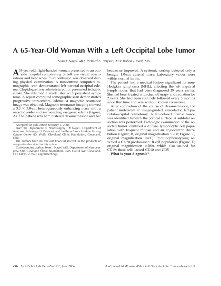
A 65-Year-Old Woman With a Left Occipital Lobe Tumor Sean J. Nagel, MD; Richard A. Prayson, MD; Robert J. Weil, MD A 65-year-old, right-handed woman presented to an outside hospital complaining of left eye visual obscurations and headaches; mild confusion was observed during physical examination. A noncontrast computed tomographic scan demonstrated left parietal-occipital edema. Clopidogrel was administered for presumed ischemic stroke. She returned 1 week later with persistent symptoms. A repeat computed tomographic scan demonstrated progressive intracerebral edema; a magnetic resonance image was obtained. Magnetic resonance imaging showed a 3.0 3 3.0-cm heterogeneously enhancing mass with a necrotic center and surrounding vasogenic edema (Figure, A). The patient was administered dexamethasone and her Accepted for publication February 2, 2006. From the Department of Neurosurgery (Dr Nagel), Department of Anatomic Pathology (Dr Prayson), and the Brain Tumor Institute, Taussig Cancer Center (Dr Weil), Cleveland Clinic Foundation, Cleveland, Ohio. The authors have no relevant financial interest in the products or companies described in this article. Corresponding author: Sean J. Nagel, MD, Department of Neurosurgery, S80, Cleveland Clinic Foundation, 9500 Euclid Ave, Cleveland, OH 44195 (e-mail: nagels@ccf.org). e96 Arch Pathol Lab Med—Vol 130, June 2006 headaches improved. A systemic workup detected only a benign, 1.0-cm adrenal mass. Laboratory values were within normal limits. The patient had a medical history significant for nonHodgkin lymphoma (NHL), affecting the left inguinal lymph nodes, that had been diagnosed 29 years earlier. She had been treated with chemotherapy and radiation for 2 years. She had been routinely followed every 6 months since that time and was without known recurrence. After completion of the course of dexamethasone, the patient underwent an image-guided, stereotactic, left parietal-occipital craniotomy. A tan-colored, friable tumor was identified beneath the cortical surface. A subtotal resection was performed. Pathologic examination of the resected tumor identified a diffuse, lymphocytic cell population with frequent mitosis and an angiocentric distribution (Figure, B, original magnification 3200; Figure, C, original magnification 3400). Immunophenotyping revealed a CD20-predominant B-cell population (Figure, D, original magnification 3200), which also stained for CD19; these cells lacked CD10 and CD5. What is your diagnosis? A 65-Year-Old Woman With a Left Occipital Lobe Tumor—Nagel et al Arch Pathol Lab Med—Vol 130, June 2006 A 65-Year-Old Woman With a Left Occipital Lobe Tumor—Nagel et al e97 Pathologic Diagnosis: Diffuse Large B-Cell, Primary Central Nervous System Lymphoma Abstract A 65-year-old woman with a medical history significant for remote non-Hodgkin lymphoma presented with confusion and visual complaints. Magnetic resonance imaging showed a parietal-occipital mass with surrounding vasogenic edema. The patient underwent a stereotactic, parietal-occipital craniotomy and biopsy of the lesion. A diagnosis of primary central nervous system lymphoma was made on the basis of histopathologic features and immunophenotyping. A recent increase in the incidence of primary central nervous system lymphoma has been observed. These lymphomas are typically angiocentric with infiltration of intervening parenchyma by atypical lymphoic cells. Patients with previously diagnosed non-Hodgkin lymphoma are at increased risk for a second malignancy. Distinguishing primary central nervous system lymphomas from brain metastasis or other tumors is essential to therapy selection. Advances in the treatment of primary central nervous system lymphoma, specifically with high-dose methotrexate, has significantly improved long-term, disease-free survival. Although histologically similar to other non-Hodgkin types of lymphomas, primary central nervous system lymphoma (PCNSL) is a clinically distinct disease entity, both in behavior and treatment. Primary central nervous system lymphoma is currently reported to account for up to 5% of primary brain neoplasms.1 It is more common in patients who are immunosupressed; for example, its incidence in patients with acquired immunodeficiency syndrome is as high as 6%.2 However, immunocompromised patients, including those with the human immunodeficiency virus as well as immunosuppressed transplant recipients, only partially account for a recent increased incidence of PCNSL.1–3 In immunocompetent individuals, PCNSL has a peak incidence in the fifth to seventh decades of life.2 Patients with PCNSL often present with cognitive disturbance; however, a new focal deficit and/or seizure may be the first symptom of disease. The disease has suggestive, but not pathognomonic, neuroimaging features. In a recent retrospective review, 35% of patients with PCNSL had multifocal disease at presentation.4 Strong contrast enhancement and edema are characteristic of most lesions. Central necrosis, as seen on the magnetic resonance image of this patient, is more commonly a feature in patients with human immunodeficiency virus, but is infrequently observed in immunocompetent patients. In most instances, stereotactic biopsy is sufficient to establish a diagnosis. If possible, steroid administration in clinically stable patients should be avoided prior to biopsy because steroids may induce tumor cell apoptosis and alter the pathologic picture.2,5 Occasionally, a cerebrospinal fluid sample may be used in place of tissue biopsy. Distinguishing PCNSL from brain metastasis or other tumors is essential to the therapy that is selected. Given the presence of necrosis, a feature seen in only 6% of PCNSLs not associated with human immunodeficiency virus, but seen commonly in high-grade glial neoplasms such as glioblastoma, a craniotomy was performed in this patient.4 Once a diagnosis of lymphoma was established by frozen section, the tumor was only debulked. e98 Arch Pathol Lab Med—Vol 130, June 2006 Microscopically, PCNSLs are typically angiocentric, with infiltration of intervening parenchyma by atypical lymphoic cells. Follicle formations are not present. Lymphomatous foci may be present at distant sites within the brain that are not apparent on radiographic studies. There are usually frequent mitoses and an admixture of apoptotic cells or necrotic foci. Primary central nervous system lymphomas usually express B-cell markers, including CD19, CD20, and CD79a. Only 2% of PCNSLs are of Tcell origin, and these often occur in the cerebellum.2 Although most primary lymphomas of the central nervous system (CNS) are of the diffuse large cell type, the pathologic subtype has not been shown to confer a survival advantage to date.6 Patients with previously diagnosed NHL are at increased risk for a second malignancy. This risk remains elevated even 2 decades after diagnosis.7 Lymphoreticular neoplasms, such as leukemia and lymphoma, are the most common second cancer to occur in patients who have undergone radiation and/or chemotherapy for prior malignancy, including NHL.8 The occurrence of a secondary PCNSL is less frequently observed in these patients. Diagnosing PCNSL in patients with a history of NHL is especially challenging because the cancers may be remarkably similar. Only 5% of patients with an indolent variant of NHL will demonstrate CNS involvement. This number approaches 30% in more aggressive systemic lymphomas.9 Recurrences in the CNS are observed most often during the first 14 months after initial diagnosis.9 Disease recurrence in the CNS of patients with a history of extracerebral NHL cannot always reliably be excluded. However, previous investigators have used the typical radiographic features, response to steroids, absence of systemic disease, and disease-free interval from the initial malignancy to diagnose and initiate treatment.10 Additionally, leptomeningeal spread, rather than intraparenchymal lesions, more often portends metastatic involvement of extacerebral disease. Regrettably in this patient, the original tumor specimen from 29 years earlier had been discarded by the outside hospital, so it was not possible to assess tumor clonality or compare immunophenotyping markers. Patients with a diagnosis of PCNSL who are not treated or are treated only with steroids survive an average of 3 to 6 months.2,5 Advances in the treatment of PCNSL during the past several decades have significantly improved long-term, disease-free survival to 12 to 24 months or longer. Current treatment options include radiotherapy, corticosteroids, chemotherapy, and stem-cell rescue and immunotherapy, either alone or in combination. Age, Karnofsky performance status, and extent of disease are significant prognostic markers; survival is poorer for patients aged 60 years and older, those with a poor Karnofsky performance status, and in those patients with more extensive or multifocal CNS disease.1,6 Corticosteroids have been shown to induce cellular lysis in mouse lymphoma cells. This experimental observation may explain tumor regression on radiographic images following steroid administration.5 Corticosteroids alone, however, have a negligible effect on patient survival. Whole-brain radiation therapy in conjunction with corticosteroids improves the median survival to 22 months; a dose of approximately 45 Gy is recommended.6 Wholebrain radiation therapy in patients older than age 60 should be limited because of the neurotoxicitiy. Because patients with PCNSL typically have microscopic disease A 65-Year-Old Woman With a Left Occipital Lobe Tumor—Nagel et al beyond the limits of neuroimaging-defined tumor, focal irradiation is not recommended. Chemotherapy, and specifically methotrexate when used in high doses (.1 g/m2), is currently the mainstay in the treatment of PCNSL. Intrathecal methotrexate (injected into the lumbar subarachnoid space or via an indwelling Ommaya reservoir) has been used in patients with cytologic evidence of lymphoma cells in the cerebrospinal fluid. Blood-brain barrier disruption is another method used to increase the concentration of methotrexate in the CNS. The chemotherapy regimen selected should be completed prior to initiating whole-brain radiation therapy. Radiation disrupts the blood-brain barrier and potentiates the neurotoxic effects of chemotherapy on normal brain.5 Blood-brain barrier disruption may obviate the need for whole-brain radiation therapy; studies are ongoing to test this hypothesis.11 References 1. Hoang-Xuan K, Camilleri-Broet S, Soussain C. Recent advance in primary CNS lymphoma. Curr Opin Oncol. 2004;16:601–606. Arch Pathol Lab Med—Vol 130, June 2006 2. Schlegel U, Schmidt-Wolf IGH, Deckert M. Primary CNS lymphoma: clinical presentation, pathological classification, molecular pathogenesis and treatment. J Neurol Sci. 2000;181:1–12. 3. Batara JF, Grossman SA. Primary central nervous system lymphomas. Curr Opin Neurol. 2003;16:671–675. 4. Küker W, Nägele T, Korfel A, et al. Primary central nervous system lymphoma (PCNSL): MRI features at presentation in 100 patients. J Neurooncol. 2005; 72:169–177. 5. DeAngelis LM, Hormigo A. Treatment of primary central nervous system lymphoma. Semin Oncol. 2004;31:684–692. 6. DeAngelis LM, Yahalom J. Lymphomas, section 4: Primary central nervous system lymphoma. In: DeVita VT, Hellman S, Rosenberg SA, eds. Cancer: Principles and Practice of Oncology. 7th ed. Philadelphia, Pa: Lippincott Williams & Wilkins; 2005:2012–2020. 7. Travis LB, Curtis RE, Glimelius B. Second cancers among long-term survivors of non Hodgkin’s lymphoma. J Natl Cancer Inst. 1993;85:1932–1937. 8. Reni M, Ferreri AJM, Zoldan MC, Villa E. Primary brain lymphomas in patients with a prior or concomitant malignancy. J Neurooncol. 1997;32:135–142. 9. Lossos A, Ashhab Y, Sverdin E, et al. Late-delayed cerebral involvement in systemic non-Hodgkin lymphoma: a second primary tumor or a tardy recurrence? Cancer. 2004;101:1843–1849. 10. DeAngelis LM. Primary central nervous system lymphoma as a secondary malignancy. Cancer. 1991;67:1431–1435. 11. Blood-brain barrier disruption trial, Cleveland Clinic IRB no. 3669B. Available at: http://www.clevelandclinic.org/neuroscience/trials/brain/3669.htm. Accessed June 8, 2005. A 65-Year-Old Woman With a Left Occipital Lobe Tumor—Nagel et al e99
