CHAPTER 6: INTEGUMENTARY SYSTEM OBJECTIVES: 0. Name
advertisement
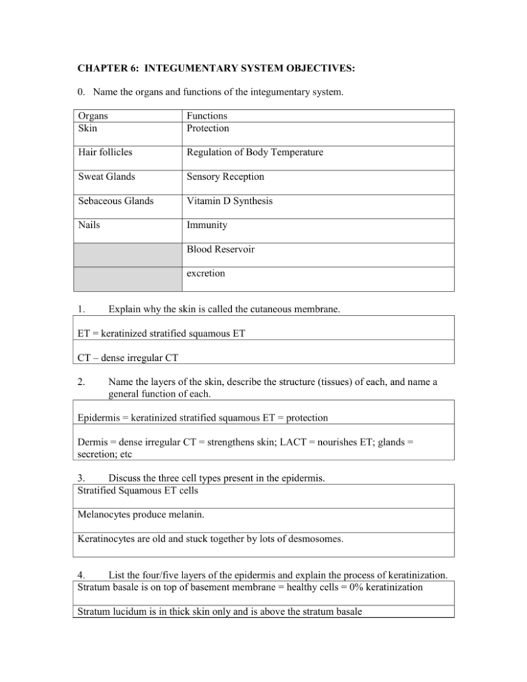
CHAPTER 6: INTEGUMENTARY SYSTEM OBJECTIVES: 0. Name the organs and functions of the integumentary system. Organs Skin Functions Protection Hair follicles Regulation of Body Temperature Sweat Glands Sensory Reception Sebaceous Glands Vitamin D Synthesis Nails Immunity Blood Reservoir excretion 1. Explain why the skin is called the cutaneous membrane. ET = keratinized stratified squamous ET CT – dense irregular CT 2. Name the layers of the skin, describe the structure (tissues) of each, and name a general function of each. Epidermis = keratinized stratified squamous ET = protection Dermis = dense irregular CT = strengthens skin; LACT = nourishes ET; glands = secretion; etc 3. Discuss the three cell types present in the epidermis. Stratified Squamous ET cells Melanocytes produce melanin. Keratinocytes are old and stuck together by lots of desmosomes. 4. List the four/five layers of the epidermis and explain the process of keratinization. Stratum basale is on top of basement membrane = healthy cells = 0% keratinization Stratum lucidum is in thick skin only and is above the stratum basale Stratum spinosum are cells that have pushed toward to surface and have started to accumulate keratin Stratum granulosum are a few layers of squamous cells that contain granules of keratin Stratum corneum is the outermost thick layer of dead cell full of keratin = 100% keratinization 5. Explain the protective role of keratin, and in turn, the epidermis. Keratin protects from mechanical injury, water-loss, effects from harsh chemicals, and against harmful pathogens that can cause disease. 6. Name the pigment responsible for skin and hair color, and explain how people of different races (i.e. and skin color) differ in regards to it, and the cell that produces it. Melanin - melanocyte 7. List some factors that promote the production of melanin (besides DNA). Sunlight, UV rays, X-rays, some drugs 8. Distinguish between the papillary layer and reticular layer of the dermis, and locate the appropriate sensory receptor in each of these layers. Papillary is top 20% of dermis beneath Bottom 80% of dermis with DICT and basement membrane with LACT and it more forms finger-like projections. Meissner’s Corpuscle Pacinian Corpuscle Compare and contrast tactile (Meissner’s) and lamellated (Pacinian) corpuscles in terms of their structure, function, and location. Tactile Meissner’s Corpuscle lamellated (Pacinian) corpuscles 9. Specialized end of a dendrite that resembles a Q-tip Fine touch receptor Specialized end of a dendrite that resembles onion Pressure receptor Located in dermal papillae Located deep in dermis and into subcutaneous layer 10. Describe the structure and function of the subcutaneous layer. The subcutaneous layer lies beneath the dermis (hypodermis) and is composed of adipose tissue = energy store; protection; and cushioning 11. Explain what is meant by the term epidermal derivative, and list four examples. An epidermal derivative originated from the epidermis = epithelium. The four examples are hair follicles, merocrine sweat glands, apocrine sweat glands, sebaceous glands. 12. Describe the general structure of a hair follicle and identify two other structures that are usually associated with them. Root is down deep in dermis, the follicle runs up toward the surface, the shaft is the exposed potion of hair. They are composed of keratinized stratified squamous ET with melanocytes. 13. Distinguish between merocrine (eccrine) and apocrine sweat glands in terms of structure, secretion content and odor, activation, and major body locations. Merocrine (Eccrine) Sweat Glands Apocrine Sweat Glands Respond to an increase in body temperature Respond to stress Secretion is watery with water, salts, and Secretion is thicker than merocrine sweat wastes (urea and uric acid) due to cellular debris plus water, salts, and wastes (urea and uric acid) No odor Secretion does have odor Run from coil to surface Empty into a hair follicle Function throughout life Function from puberty and then through life Are widely distributed but are abundant on Axillary and Inguinal region the forehead, neck m and back 14. Name two modified apocrine glands of the skin. Ceruminous glands in external ear = protective wax Mammary glands of breasts = nourishing milk 15. Describe the structure, function, secretion, and location of sebaceous glands. Sebaceous glands are holocrine glands that produce sebum (oil). They surround hair follicles and deposit the sebum into it. The sebum keeps skin and hair soft, pliable, and waterproof. 16. Discuss the many functions of skin. Protection (see above) Regulation of body temperature with sweating and activation/deactivation of superficial blood vessels Sensory reception with tactile Meissner’s and lamellated Pacinian Corpuscles Vitamin D synthesis with sunlight (Vit D is needed for the absorption of dietary calcium) Immunity with Langerhan cells (macrophages) and some T-cells Blood reservoir with 10% of blood vessels located in skin Excretion, along with kidneys. Wastes are urea from amino acid metabolism and uric acid from nucleotide metabolism. 17. Describe some major homeostatic imbalances of the skin. See chapter. Burns Wounds Acne Skin Cancers 18. Sketch a typical layer of skin and label each layer and all structures. Then in complete sentences, discuss the function of each layer and structure. 19. Discuss heat production and loss as it relates to the integumentary system. When body temperature rises, the hypothalamus causes sweating which cools in two ways, and dilation of superficial blood vessels, so that the warm blood on the skin’s surface can lose its heat to the environment. When body temperature fall, the hypothalamus deactivates sweat glands, and causes constriction of superficial blood vessels, so the warm blood stays deep. 20. Define the terms hyperthermia and hypothermia and discuss the cause(s) of each condition. N/A 21. N/A Discuss the healing of cuts/wounds that occur to the skin. 22. Distinguish between 1st degree, 2nd degree, and 3rd degree burns, and discuss the process by which each burn heals. 1st – penetrates epidermis; no bleeding; least harmful 2nd = penetrated dermis and its tissues; bleeding; harmful 3rd = penetrated subcutaneous tissue and muscle below; most harmful


