respiratory alkalosis
advertisement

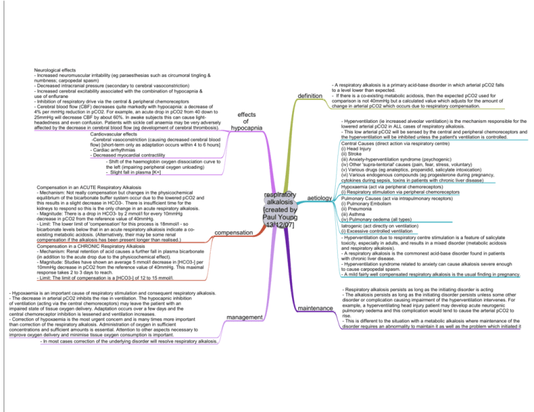
Neurological effects - Increased neuromuscular irritability (eg paraesthesias such as circumoral tingling & numbness; carpopedal spasm) - Decreased intracranial pressure (secondary to cerebral vasoconstriction) - Increased cerebral excitability associated with the combination of hypocapnia & use of enflurane - Inhibition of respiratory drive via the central & peripheral chemoreceptors - Cerebral blood flow (CBF) decreases quite markedly with hypocapnia: a decrease of 4% per mmHg reduction in pCO2. For example, an acute drop in pCO2 from 40 down to 25mmHg will decrease CBF by about 60%. In awake subjects this can cause lightheadedness and even confusion. Patients with sickle cell anaemia may be very adversely affected by the decrease in cerebral blood flow (eg development of cerebral thrombosis). Cardiovascular effects -Cerebral vasoconstriction (causing decreased cerebral blood flow) [short-term only as adaptation occurs within 4 to 6 hours] - Cardiac arrhythmias - Decreased myocardial contractility - Shift of the haemoglobin oxygen dissociation curve to the left (impairing peripheral oxygen unloading) - Slight fall in plasma [K+] Compensation in an ACUTE Respiratory Alkalosis - Mechanism: Not really compensation but changes in the physicochemical equilibrium of the bicarbonate buffer system occur due to the lowered pCO2 and this results in a slight decrease in HCO3-. There is insufficient time for the kidneys to respond so this is the only change in an acute respiratory alkalosis. - Magnitude: There is a drop in HCO3- by 2 mmol/l for every 10mmHg decrease in pCO2 from the reference value of 40mmHg. - Limit: The lower limit of 'compensation' for this process is 18mmol/l - so bicarbonate levels below that in an acute respiratory alkalosis indicate a coexisting metabolic acidosis. (Alternatively, their may be some renal compensation if the alkalosis has been present longer than realised.) Compensation in a CHRONIC Respiratory Alkalosis - Mechanism: Renal retention of acid causes a further fall in plasma bicarbonate (in addition to the acute drop due to the physicochemical effect). - Magnitude: Studies have shown an average 5 mmol/l decrease in [HCO3-] per 10mmHg decrease in pCO2 from the reference value of 40mmHg. This maximal response takes 2 to 3 days to reach. - Limit: The limit of compensation is a [HCO3-] of 12 to 15 mmol/l. definition - A respiratory alkalosis is a primary acid-base disorder in which arterial pCO2 falls to a level lower than expected. - If there is a co-existing metabolic acidosis, then the expected pCO2 used for comparison is not 40mmHg but a calculated value which adjusts for the amount of change in arterial pCO2 which occurs due to respiratory compensation. effects of hypocapnia respiratory alkalosis [created by Paul Young 13/12/07] aetiology compensation - Hypoxaemia is an important cause of respiratory stimulation and consequent respiratory alkalosis. - The decrease in arterial pCO2 inhibits the rise in ventilation. The hypocapnic inhibition of ventilation (acting via the central chemoreceptors) may leave the patient with an impaired state of tissue oxygen delivery. Adaptation occurs over a few days and the central chemoreceptor inhibition is lessened and ventilation increases. - Correction of hypoxaemia is the most urgent concern and is many times more important than correction of the respiratory alkalosis. Administration of oxygen in sufficient concentrations and sufficient amounts is essential. Attention to other aspects necessary to improve oxygen delivery and minimise tissue oxygen consumption is important. - In most cases correction of the underlying disorder will resolve respiratory alkalosis. maintenance management - Hyperventilation (ie increased alveolar ventilation) is the mechanism responsible for the lowered arterial pCO2 in ALL cases of respiratory alkalosis. - This low arterial pCO2 will be sensed by the central and peripheral chemoreceptors and the hyperventilation will be inhibited unless the patient's ventilation is controlled. Central Causes (direct action via respiratory centre) (i) Head Injury (ii) Stroke (iii) Anxiety-hyperventilation syndrome (psychogenic) (iv) Other 'supra-tentorial' causes (pain, fear, stress, voluntary) (v) Various drugs (eg analeptics, propanidid, salicylate intoxication) (vi) Various endogenous compounds (eg progesterone during pregnancy, cytokines during sepsis, toxins in patients with chronic liver disease) Hypoxaemia (act via peripheral chemoreceptors) (i) Respiratory stimulation via peripheral chemoreceptors Pulmonary Causes (act via intrapulmonary receptors) (i) Pulmonary Embolism (ii) Pneumonia (iii) Asthma (iv) Pulmonary oedema (all types) Iatrogenic (act directly on ventilation) (i) Excessive controlled ventilation - Hyperventilation due to respiratory centre stimulation is a feature of salicylate toxicity, especially in adults, and results in a mixed disorder (metabolic acidosis and respiratory alkalosis). - A respiratory alkalosis is the commonest acid-base disorder found in patients with chronic liver disease. - Hyperventilation syndrome related to anxiety can cause alkalosis severe enough to cause carpopedal spasm. - A mild fairly well compensated respiratory alkalosis is the usual finding in pregnancy. - Respiratory alkalosis persists as long as the initiating disorder is acting - The alkalosis persists as long as the initiating disorder persists unless some other disorder or complication causing impairment of the hyperventilation intervenes. For example, a hyperventilating head injury patient may develop acute neurogenic pulmonary oedema and this complication would tend to cause the arterial pCO2 to rise. - This is different to the situation with a metabolic alkalosis where maintenance of the disorder requires an abnormality to maintain it as well as the problem which initiated it


