Loss of melanocytes in epidermis affected by vitiligo
advertisement
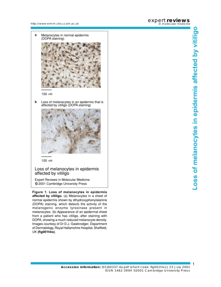
a expert reviews in molecular medicine Melanocytes in normal epidermis (DOPA staining) 100 µm b Loss of melanocytes in an epidermis that is affected by vitiligo (DOPA staining) 100 µm Loss of melanocytes in epidermis affected by vitiligo Expert Reviews in Molecular Medicine c 2001 Cambridge University Press Loss of melanocytes in epidermis affected by vitiligo http://www-ermm.cbcu.cam.ac.uk Figure 1. Loss of melanocytes in epidermis affected by vitiligo. (a) Melanocytes in a sheet of normal epidermis shown by dihydroxyphenylalanine (DOPA) staining, which detects the activity of the melanogenic enzyme tyrosinase present in melanocytes. (b) Appearance of an epidermal sheet from a patient who has vitiligo, after staining with DOPA, showing a much reduced melanocyte density. Images courtesy of Dr D.J. Gawkrodger, Department of Dermatology, Royal Hallamshire Hospital, Sheffield, UK (fig001hks). Accession information: (01)00337-4a.pdf (short code: fig001hks); 23 July 2001 ISSN 1462-3994 ©2001 Cambridge University Press 1

