A low-lying placenta (placenta praevia) after 20 weeks
advertisement
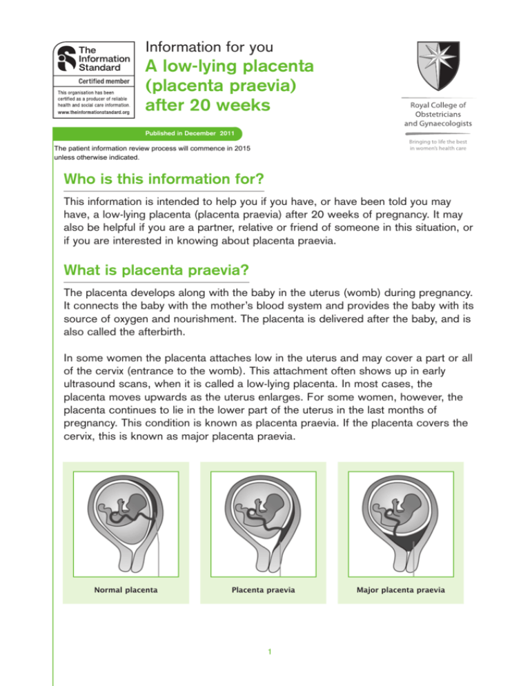
Information for you A low-lying placenta (placenta praevia) after 20 weeks Published in December 2011 The patient information review process will commence in 2015 unless otherwise indicated. Who is this information for? This information is intended to help you if you have, or have been told you may have, a low-lying placenta (placenta praevia) after 20 weeks of pregnancy. It may also be helpful if you are a partner, relative or friend of someone in this situation, or if you are interested in knowing about placenta praevia. What is placenta praevia? The placenta develops along with the baby in the uterus (womb) during pregnancy. It connects the baby with the mother’s blood system and provides the baby with its source of oxygen and nourishment. The placenta is delivered after the baby, and is also called the afterbirth. In some women the placenta attaches low in the uterus and may cover a part or all of the cervix (entrance to the womb). This attachment often shows up in early ultrasound scans, when it is called a low-lying placenta. In most cases, the placenta moves upwards as the uterus enlarges. For some women, however, the placenta continues to lie in the lower part of the uterus in the last months of pregnancy. This condition is known as placenta praevia. If the placenta covers the cervix, this is known as major placenta praevia. Normal placenta Placenta praevia 1 Major placenta praevia What are the risks to me and my baby? Because the placenta is in the lower part of the womb, there is a risk that you may bleed in the second half of pregnancy. Bleeding from placenta praevia can be heavy, and so put the life of the mother and baby at risk. However deaths from placenta praevia are rare. You are more likely to need a caesarean section because the placenta is in the way of your baby being born. How is placenta praevia diagnosed? A low-lying placenta may be suspected during the routine 20-week ultrasound scan. Most women who have a low-lying placenta at the routine 20-week scan will not go on to have a low-lying placenta later in the pregnancy – only 1 in 10 (10%) go on to have a placenta praevia. Overall this is a very small percentage of pregnant women. However if you have had a caesarean section before, the placenta is less likely to move upwards, and half (50%) of women who have had a caesarean section and have a low-lying placenta at the routine 20 week scan will go on to have placenta praevia. The best way to confirm whether or not you have placenta praevia is with a transvaginal ultrasound scan (where the probe is placed inside the vagina). This is safe for you and your baby. Placenta praevia may be suspected if you have bleeding in the second half of pregnancy. The bleeding is usually painless and may occur after sexual intercourse. Occasionally, placenta praevia may be suspected later in pregnancy if the baby is found to be lying in an unusual position, for example bottom first (breech) or lying across the womb (transverse). What extra antenatal care can I expect if I have a low-lying placenta? If your placenta remains low-lying in the second half of pregnancy, you will have at least one more scan to check whether the position of the placenta has moved with the development and stretching of the uterus. 2 If your placenta does not cover the cervix and you have no bleeding during your pregnancy, your repeat ultrasound scan should be at 36 weeks. However, a repeat ultrasound is recommended at 32 weeks if: ● ● your placenta covers the cervix at the 20-week scan you have had a caesarean section before and your placenta is low-lying at the front part of the uterus. Additional care will be given based on your individual circumstances. If you have major placenta praevia (the placenta covers the cervix) you may be offered admission to hospital after 34 weeks of pregnancy. Even if you have had no symptoms before, there is a small risk that you could bleed suddenly and severely, which may mean that you need an urgent caesarean section. You should always contact the hospital if you have a low-lying placenta and you have any bleeding, contractions or pain. If your placenta praevia is confirmed, you and your partner should have the opportunity to discuss the options for delivery with your doctor. Depending on your circumstances, you may be advised to have a planned caesarean section. In a few instances, a blood transfusion is essential to save your life and the life of your baby. If you feel that you could never accept a blood transfusion, then you should explain this to your obstetrician and midwife as early as possible. You can then discuss any objections or particular questions that you may have. You may need to have your baby in a hospital which has additional expertise available, should the need arise. What will happen at the birth? Your healthcare team will recommend the best way for you to give birth based on your own individual circumstances. If you have a major placenta praevia, your baby will need to be delivered by caesarean section. If the edge of your placenta is less than 2 centimetres from the entrance to the cervix on your scan at 32 to 36 weeks, you are still likely to need a caesarean section. A consultant obstetrician and anaesthetist should be in attendance at the time of your delivery. This is particularly important if you have previously had a caesarean section. Unless there is severe bleeding or another indication, delivery by caesarean section should be performed after 38 weeks. You will usually have a course of antenatal corticosteroids to help your baby (see RCOG Patient Information: Corticosteroids in pregnancy to reduce complications from being born premature). 3 Your anaesthetist will discuss the options for anaesthesia if you need a caesarean section. You may need to have a general anaesthetic, especially in an emergency situation. If you have a placenta praevia, you are more likely to need a blood transfusion. Blood supplies should be available, as necessary, for your individual circumstances. In extreme cases, if the bleeding continues and cannot be controlled, a hysterectomy (removal of the womb) may be the only means of controlling the bleeding. If there is bleeding before your due date, you may have to be delivered earlier than planned. What is placenta accreta? Rarely, placenta praevia may be complicated by a problem known as placenta accreta. This is when the placenta grows into the muscle of the uterus, making separation at the time of birth difficult. Placenta accreta is more commonly found in women with placenta praevia who have previously had a caesarean section. Placenta accreta may be suspected in the antenatal period when undergoing an ultrasound scan, but while additional tests such as magnetic resonance imaging (MRI) scans may help with the diagnosis, your doctor will only be able to tell for sure if you have this condition at the time of your caesarean section. Placenta accreta causes bleeding when an attempt is made to remove your placenta. The bleeding may be severe and you may require a hysterectomy (removal of the womb) to stop the bleeding. It may be possible to leave the placenta in place after birth, to allow it to absorb over a few weeks and months. Unfortunately this latter type of treatment is not always successful and some women will still need a hysterectomy. If placenta accreta is suspected before your baby is born, your doctor will discuss your options and the extra care that you will need at delivery. Delivery may be planned earlier – for example between 36 and 37 weeks, depending on individual circumstances. You may need to have your baby in a hospital which has additional facilities such as interventional radiology available. Your consultant will discuss this with you. Is there anything else I should know? ● You may be advised to avoid having sexual intercourse during pregnancy, particularly if you have been bleeding. 4 ● ● You may be offered an examination with a speculum (a plastic or metal instrument used to separate the walls of the vagina) to see how much and where your bleeding is coming from. This is an entirely safe examination. If you have a low-lying placenta you should eat a healthy diet rich in iron to reduce the risk of anaemia. 5 Sources and acknowledgements This information has been developed by the RCOG Patient Information Committee. It is based on the RCOG guideline Placenta Praevia, Placenta Praevia Accreta and Vasa Praevia: Diagnosis and Management (January 2010). The guideline contains a full list of the sources of evidence we have used. You can find it online at: http://www.rcog.org.uk/womens-health/clinical-guidance/placenta-praevia-andplacenta-praevia-accreta-diagnosis-and-manageme. The RCOG produces guidelines as an educational aid to good clinical practice. They present recognised methods and techniques of clinical practice, based on published evidence, for consideration by obstetricians and gynaecologists and other relevant health professionals. The ultimate judgement regarding a particular clinical procedure or treatment plan must be made by the doctor or other attendant in the light of clinical data presented by the patient and the diagnostic and treatment options available. This means that RCOG Guidelines are unlike protocols or guidelines issued by employers, as they are not intended to be prescriptive directions defining a single course of management. Departure from the local prescriptive protocols or guidelines should be fully documented in the patient’s case notes at the time the relevant decision is taken. This information has been reviewed before publication by women attending clinics in London. A glossary of all medical terms is available on the RCOG website at: http://www.rcog.org.uk/womens-health/patient-information/medical-terms-explained. A final note The Royal College of Obstetricians and Gynaecologists produces patient information for the public. This information is based on guidelines which present recognised methods and techniques of clinical practice, based on published evidence. The ultimate judgement regarding a particular clinical procedure or treatment plan must be made by the doctor or other attendant in the light of the clinical data presented and the diagnostic and treatment options available. © Royal College of Obstetricians and Gynaecologists 2011 6
