European Heart Journal - Cardiovascular Imaging Advance Access published May 22, 2013
IMAGE FOCUS
doi:10.1093/ehjci/jet100
.............................................................................................................................................................................
STEMI revealing an exceptional variant of single right coronary artery
Geoffrey C. Colin1*, Olivier Gurné2, Emmanuel Coche1, Olivier Van Caenegem2, and Benoit Ghaye1
1
Department of Medical Imaging, Catholic University of Louvain, Cliniques Universitaires St-Luc, Avenue Hippocrate 10, Brussels 1200, Belgium and 2Department of
Cardiovascular Diseases, Catholic University of Louvain, Cliniques universitaires St-Luc, Avenue Hippocrate 10, Brussels 1200, Belgium
* Corresponding author. Tel: +32 27642900, Email: geoffreycolin1@hotmail.com
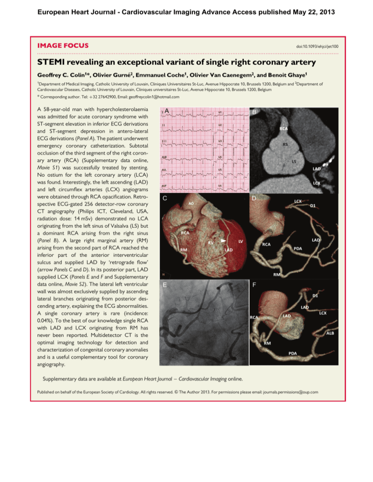
A 58-year-old man with hypercholesterolaemia
was admitted for acute coronary syndrome with
ST-segment elevation in inferior ECG derivations
and ST-segment depression in antero-lateral
ECG derivations (Panel A). The patient underwent
emergency coronary catheterization. Subtotal
occlusion of the third segment of the right coronary artery (RCA) (Supplementary data online,
Movie S1) was successfully treated by stenting.
No ostium for the left coronary artery (LCA)
was found. Interestingly, the left ascending (LAD)
and left circumflex arteries (LCX) angiograms
were obtained through RCA opacification. Retrospective ECG-gated 256 detector-row coronary
CT angiography (Philips ICT, Cleveland, USA,
radiation dose: 14 mSv) demonstrated no LCA
originating from the left sinus of Valsalva (LS) but
a dominant RCA arising from the right sinus
(Panel B). A large right marginal artery (RM)
arising from the second part of RCA reached the
inferior part of the anterior interventricular
sulcus and supplied LAD by ‘retrograde flow’
(arrow Panels C and D). In its posterior part, LAD
supplied LCX (Panels E and F and Supplementary
data online, Movie S2). The lateral left ventricular
wall was almost exclusively supplied by ascending
lateral branches originating from posterior descending artery, explaining the ECG abnormalities.
A single coronary artery is rare (incidence:
0.04%). To the best of our knowledge single RCA
with LAD and LCX originating from RM has
never been reported. Multidetector CT is the
optimal imaging technology for detection and
characterization of congenital coronary anomalies
and is a useful complementary tool for coronary
angiography.
Supplementary data are available at European Heart Journal – Cardiovascular Imaging online.
Published on behalf of the European Society of Cardiology. All rights reserved. & The Author 2013. For permissions please email: journals.permissions@oup.com


