notes
advertisement
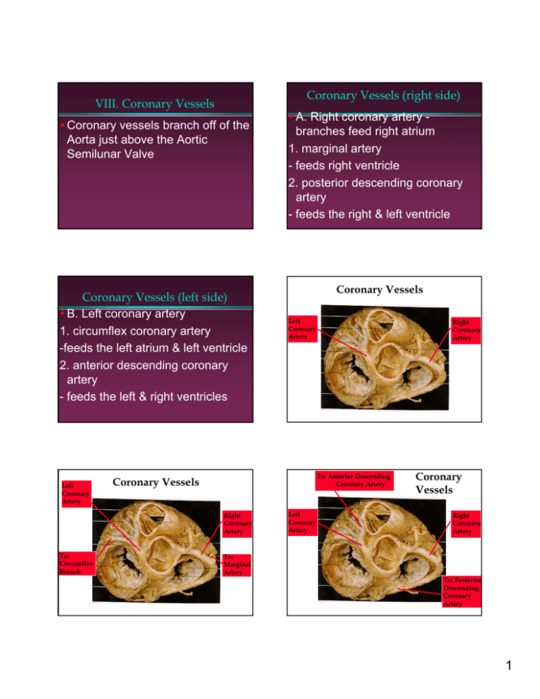
Coronary Vessels (right side) VIII. Coronary Vessels y Coronary vessels branch off of the Aorta just above the Aortic Semilunar Valve Coronary Vessels (left side) y B. Left coronary artery 1. circumflex coronary artery -feeds the left atrium & left ventricle 2. anterior descending coronary artery - feeds the left & right ventricles Left Coronary Artery Coronary Vessels Left Coronary Artery Right Coronary Artery To: Anterior Descending Coronary Artery Coronary Vessels Right Coronary Artery To: Circumflex Branch y A. Right coronary artery branches feed right atrium 1. marginal artery - feeds right ventricle 2. posterior descending coronary artery - feeds the right & left ventricle To: Marginal Artery Left Coronary Artery Coronary Vessels Right Coronary Artery To: Posterior Descending Coronary Artery 1 Conduction System: Components of Conduction System O Specialized cardiac muscle tissue (contraction) O Generates & distributes electrical impulses (conduction) O as a result heart can contract without neural stimulationintrinsic rhythm. Sinoatrial Components of Conduction System Components of Conduction System Atrioventricular (AV) node – O Rt. Atrial wall/inferior interatrial septum O Last portion to receive impulse from SA node O Stimulated & passes on impulse Atrioventricular Components of Conduction System Components of Conduction System a) right branch of AV bundles (Bundle Bundle of His) His b) left branch of AV bundles (Bundle Bundle of His) His O Carry impulse down the interventricular septum Purkinje (SA) node O Right Atrial wall, inferior to opening of superior vena cava O Initiates cardiac cycle – sets pace (pacemaker) O Impulse spreads over both atria resulting in contraction Bundle (Bundle of His) O Carry the impulse from AV node to superior part of interventricular septum fibers – lateral branches that pass through the walls of the ventricles O stimulate myocardial cells in ventricles to contract. O After short delay SA node fires again – cycle repeats. 2 Cardiac Cycle: O Complete series of events occurring within the heart beginning with depolarization of SA node. O Involves specific length of time, pressure & volume changes, electrical activity & heart sounds. I. Electrocardiogram (ECG) (EKG) O Impulses through the conduction system generate electrical currents – O detected at body surface. O ECG is the recording of this electrocardiograph is the instrument. O Each part of cardiac cycle produces a different electrical impulse. ECG First wave: P (small upward) O atrial depolarization, (excitation) passage of impulse from SA node through both atria = result atrial contraction ECG ’PR interval O start of P wave to start of Q wave O from the start of atrial excitation to start of ventricular excitation, passage of impulse from the SA node to AV node to Purkinje fibers in ventricles ECG QRS complex – small downward deflection, large triangular upward deflection, ends w/ small downward deflection. O Depolarization of ventricles, (excitation) § ventricular contraction. ECG ’ST segment – end of S to start of T wave O end of ventricular depolarization (spread of impulse through vent.) to start of repolarization of ventricles 3 ECG T wave O ventricular repolarization O atrial repolarization occurs in the midst of the QRS complex & is masked by that event O This cycle of events repeats. II. Heart & Blood Pressure O Systole: contraction phase of cardiac cycle – atria/ ventricles O Diastole: relaxation phase of cardiac cycle – atria/ ventricles O Pressure changes: related to 1) chamber size & 2.) volume of blood it contains O Greater vol = higher pressure O Blood flow: Higher to lower pressure Blood Pressure O B.P. = pressure exerted by blood on wall of any blood vessel O Clinically: pressure in arteries exerted by left ventricle (systole) and pressure remaining when ventricle is in diastole O Systolic = (Force of left vent. Contraction) Diastolic = (Pressure in blood vessel walls during left vent. relaxation) III. Heart Sounds O Typically 2: closing of AV valves – longer (ventricle contraction) “Lubb” closing of semilunar valves – shorter, sharper “Dupp” O Provide information about valves: O Lubb Dupp Pause Lubb Dupp Pause 1 2 3&4 1 2 3&4 IV. Pulse O Pulse: expansion & recoil of artery due to: rhythmic injections of blood from Left ventricle into aorta -> arteries expansion and contraction of arterial walls Pulse O Pulse: farther from heart = fainter O Related to ventricular contraction/ does not coincide. 4


