URINARY SYSTEM – Chapter 25
advertisement
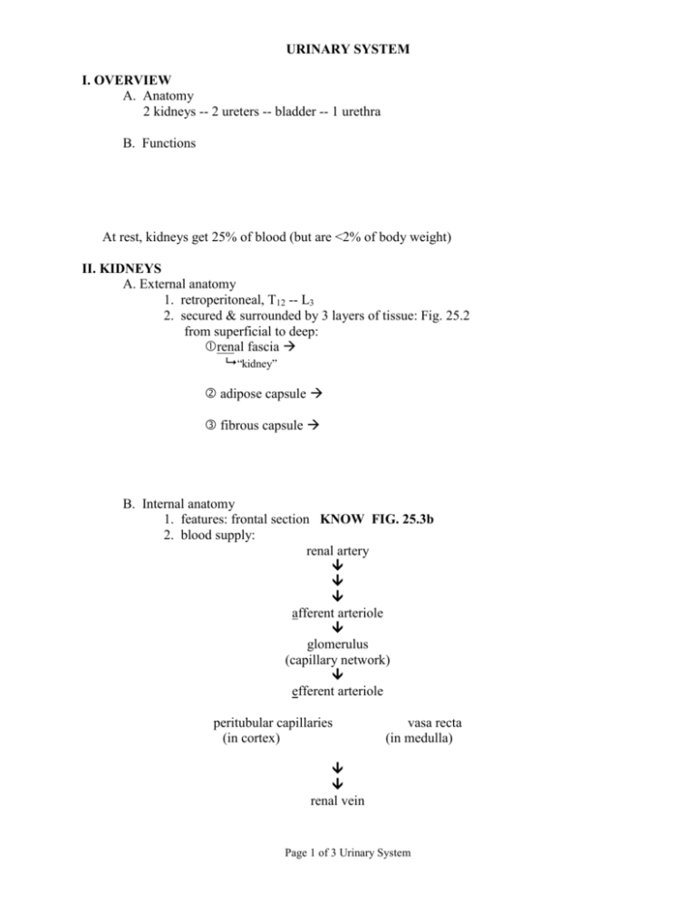
URINARY SYSTEM I. OVERVIEW A. Anatomy 2 kidneys -- 2 ureters -- bladder -- 1 urethra B. Functions At rest, kidneys get 25% of blood (but are <2% of body weight) II. KIDNEYS A. External anatomy 1. retroperitoneal, T12 -- L3 2. secured & surrounded by 3 layers of tissue: Fig. 25.2 from superficial to deep: renal fascia “kidney” adipose capsule fibrous capsule B. Internal anatomy 1. features: frontal section KNOW FIG. 25.3b 2. blood supply: renal artery afferent arteriole glomerulus (capillary network) efferent arteriole peritubular capillaries (in cortex) vasa recta (in medulla) renal vein Page 1 of 3 Urinary System C. Nephron – KNOW Fig. 25.6, 25.5 1. functional unit of kidney: 2. components: renal corpuscle = glomerulus + glomerular (Bowman’s) capsule filtration occurs at the renal corpuscle renal tubule = proximal convoluted tubule + nephron loop (of Henle) + distal convoluted tubule filtrate is modified by reabsorption/secretion as it passes through renal tubule Note the renal tubule includes the collecting duct, but that portion is not part of the nephron 3. two types of nephrons cortical nephrons (85%) – juxtamedullary nephrons (15%) – Page 2 of 3 Urinary System 4. Path of filtrate: nephron collecting duct papillary duct minor calyx major calyx renal pelvis -- filtrate is considered “urine” once it enters collecting duct III. TRANSPORT AND STORAGE ORGANS A. Ureters (2) 1. kidney bladder 2. 3. 4. B. Bladder -- KNOW Fig. 25.11 1. hollow, muscular bag, retroperitoneal 2. 3. Histology (from internal external) mucosa – muscularis – adventitia – C. Urethra (1) 1. bladder exterior 2. external urethral sphincter 3. external urethral orifice Page 3 of 3 Urinary System
