Bone and Cartilage
advertisement

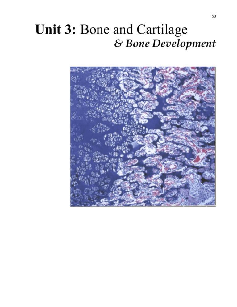
53 Unit 3: Bone and Cartilage & Bone Development 54 55 Introduction and objectives Cartilage is a specialized connective tissue composed of cells, fibers and ground substance. The unique properties of cartilage play an important role in the structure and function of movable joints such as the glenohumeral joint. The growth in length of long bones is totally dependent upon the unique properties of cartilage. With the exception of certain membranous bones, the entire skeletal system develops first as a cartilaginous model, which is later replaced by bone. One of the most incapacitating and painful diseases man is subjected to, osteoarthritis, involves a degenerative change in the ground substance of articular cartilage such that collagen fibrils are unmasked and freely exposed on the joint surfaces. The importance of rigid calcified tissue (bone) as a support for the body and limbs should be obvious. Not so obvious is its role as a reservoir of calcium ions. Calcium ions are exchanged between the blood and bone compartments of the body under the influence of parathyroid hormone and calcitonin on various bone cells such as osteoblasts, osteocytes and osteoclasts. Cartilage & Bone Tissue with WebMic There are five specimens each for the study of cartilage and bone tissue. Hyaline cartilage, elastic cartilage and fibrous cartilage are demonstrated from the overall organization down to the individual cells, the chondrocytes, which form and maintain cartilage as a viable tissue. Adult compact and cancellous lamellar bone are demonstrated in two specimens. Finally, the two types of ossification are demonstrated, endochondral and intramembranous ossification. After completing this unit, you should be able to identify and distinguish between the following: 1. 2. 3. 4. 5. 6. 7. 8. 9. 4. 5. 6. 7. the three types of cartilage: hyaline, elastic, and fibro-cartilage chondroblasts chondrocytes perichondrium bone lamella canaliculi Haversian system (osteon) Haversian canal osteoblasts osteocytes osteoclasts periosteum Also, after completing this unit in conjunction with reading a chapter on connective tissue in a histology textbook and/or hearing a lecture on connective tissue, you should understand: 1. 2. 3. 4. 5. how cartilage grows the differences between cartilage and bone the remodeling of bone the process of intramembranous bone formation the process of endochondral bone formation 56 Cartilage Before we begin a study of the histology of cartilage it will be helpful to know some features that cartilage and bone have in common. The cells of cartilage and bone are found in spaces called lacunae (small pits) and thus are separated from the surrounding extracellular material. The extracellular matrix (matrix is composed of fibers and ground substance) of both bone and cartilage is composed of collagen fibers and glycosaminoglycans. Cartilage collagen is type II and bone collagen is type I. Cartilage ground substance (glycosaminoglycans) is not calcified as compared to bone which is calcified. Bone can grow in the adult by one method and that is appositional growth which is the deposition of bone matrix by osteoblasts in the periosteum onto the old bone matrix. Cartilage can grow by two methods, appositional and interstitial which is the process whereby cartilage cells within the cartilage matrix can divide resulting in growth of cartilage within itself. Hyaline Cartilage To begin our study of cartilage go to the general histology section and, under cartilage, choose: Hyaline Cartilage (Rib)-Hematoxylin-Eosin Scan the 5x image noting that the overall staining reaction is blue. Can you observe the many small spaces? These are the lacunae and, in each one, is a chondrocyte. Find the label perichondrium at the edge of the cartilage. The perichondrium as its name suggests surrounds cartilage. It is composed of a dense connective tissue. The innermost layer of that surrounding connective tissue has stem cells that can divide and provide a reservoir of cells that can secrete cartilage matrix. This is called the chondrogenic layer. The line emerging from the label ends right at the point where new cartilage cells are being formed. On the cartilage side the stained matrix is blue because of the highly sulfated glycosaminoglycans being produced in contrast to the pink stained outer perichondrium (called the fibrous perichondrium) which has a matrix composed predominately of collagen type I with little ground substance. Within the cartilage in addition to the glycosaminoglycans the matrix contains type II collagen in the form of fine fibers often referred to as fibrils. Hyaline cartilage as is present in this specimen of a rib appears rather smooth because of the small dimension of its collagen fibers (ground substance masks the fibers). Hence the term hyaline which means glass-like. Now scan into the center of the image and find the label interterritorial matrix. There are two named regions of the matrix. The other named region is territorial. Territorial matrix is the matrix that is closely associated with each lacunae containing a chondrocyte. Interterritorial matrix is the remaining matrix. Select the 20x image. Find the interterritorial label and read the text related to it. In this specimen the contrast in staining between the territorial and interterritorial matrix is not very obvious. There is more ground substance in territorial matrix hence it will stain more blue than interterritorial which contains a higher percentage of collagen which is responsible for it staining more pink. This difference will be better demonstrated in the specimen we use to study elastic cartilage. The other feature important to learn in this specimen is the morphological feature related to interstitial growth of cartilage: the isogenous group. Find the label isogenous group and read the related text. Observe that there are two chondrocytes that look like they came from a dividing cell. Isogenous groups are groups of daughter cells resulting from mitosis. This is the way the cartilage grows interstitially. This method of cartilage growth can occur in the adult to a limited degree, but is one of the main growing mechanisms in the developing embryo and fetus. 57 Elastic Cartilage In the general histology section under cartilage choose: Elastic Cartilage (Epiglottis) – Elastin (Verhoeff’s)-Nuclear Red-Picroindigo In this specimen nuclei stain light red, cytoplasm stains red, collagen fibers stain yellow, and elastic fibers stain brown-black. Unlike the rib, the cartilage of the epiglottis is elastic. When food is swallowed the epiglottis is forced downward covering the glottis resulting in food being directed into the esophagus. When food passes the epiglottis it rebounds to its resting form because of its elastic nature. The predominant fiber in the interterritorial matrix of cartilage is composed of elastin (the elastic fiber). Scan the 5x image noting the cartilage, its perichondrium and the elastic cartilage. Again, as in hyaline cartilage, the spaces are the lacunae each of which contains a chondrocyte. The line in the image that indicates the perichondrium ends at precisely the point at which there is a transition from fibrous to chondrogenic perichondrium. Stem cells located in the chondrogenic perichondrium give rise to chondroblasts. Chondroblasts then secrete matrix. As soon as the chondroblasts surround themselves with matrix they are then called chondrocytes. The outer layer, the fibrous perichondrium is yellow indicating a dominance of collagen fibers. This layer functions like a capsule which defines the outer limit of the cartilage. From the chondrogenic region inward the staining of the matrix becomes darker. The dark staining reaction is because of the presence of elastic fibers. As the new chondrocytes mature, they secrete more elastic fibers until finally the matrix is very rich in elastic fibers. Select the 20x image. Scan it noting the labels perichondrium, chondrocyte, isogenous group, and interterritorial matrix. As you scan from the perichondrium inward observe the increase in density of elastic fibers. Read the text related to the interterritorial matrix. This is the matrix containing the highest density of elastic fibers. Similarly, in hyaline cartilage, this is the region that contains the highest density of type II collagen fibers. Although not labeled in this specimen, observe the matrix immediately surrounding the labeled chondrocyte and the chondrocyte below the ‘interterritorial matrix label’. This matrix, called the territorial matrix, stains lighter because it has more ground substance than elastic fibers. Now choose the specimen of elastic cartilage stained with hematoxylin and eosin: Elastic Cartilage (Epiglottis) – Hematoxylin-Eosin In the 5x image note the perichondrium and the main mass of elastic cartilage. If you look closely in the region of the perichondrium you can see how the cells get larger as they are located deeper in the cartilage. This is because they are maturing from chondroblasts to chondrocytes where finally the chondrocyte is surrounded by a clear region (the lacuna). Scan the 20x image and study the perichondrium label region to see this more clearly demonstrated. Also in this image, note and read the text related to the labels interterritorial matrix and territorial matrix. Note that the territorial matrix stains bluish whereas the interterritorial matrix stains pink due to the higher density of elastic fibers. Note that the matrix of this elastic cartilage is less homogeneous (glassy like) than the hyaline cartilage due to the larger and more refractile elastic fibers. However, when you compare the appearance of the matrix of this specimen with the previous one you studied stained with a special elastic fiber stain, the elastic fibers do not appear obvious. To complete your study of this specimen select the 40x image and note the two regions of the matrix and the labeled chondrocyte. Read the text label related to the chondrocyte. 58 Fibrocartilage Fibrocartilage is the tissue that makes up the structure of menisci in joints, tendons as they approach their insertions into bone, and the capsule surrounding the intervertebral discs. Begin your study with the specimen stained with a special stain. In the general histology section under cartilage choose: Fibrocartilage (Meniscus) – Hematoxylin-Eosin-Metanil Yellow Nuclei stain blue-black, collagen stains yellow, and ground substance stains light blue. Scan the 5x image. Note the labeled collagenous fibers (made up of collagen type I) that stain yellow. Look between the collagen fibers, especially in the upper half of this image, and see if you can make out the darkly stained strands. These are a series of isogenous groups of chondrocytes. This is the main characteristic diagnostic feature of fibrocartilage. Recall that you did not observe such a physical arrangement of chondrocytes in either hyaline or elastic cartilage. The chondrocytes and collagen fibers are organized in a fashion parallel to the line of force. Fibrocartilage in tendons, for example, has its collagen continue in the same orientation as the tendon, parallel to the line of force. To observe this more closely, select the 20x image and find the labeled chondrocyte and collagenous fiber located in the middle just to the left of the center of the image. Now you can observe several strands of chondrocytes (isogenous groups) running parallel between collagen fibers. The longitudinal strands of chondrocytes making up the isogenous groups got that way because the parent cells divided in one direction. Note the bluish stain immediately surrounding the chondrocytes. This is the territorial matrix that has very little to no collagen. Read the text related to the labels. Select and scan the 100x image where you can observe the chondrocytes and matrix in more detail. This now concludes your study of cartilage. You have learned that there are three types. You have learned that the difference between the three types mainly lies in the difference in the type of fibers present and how those fibers are oriented. In hyaline cartilage the fibers are collagen type II and they are oriented in all directions. In elastic cartilage, the fibers are elastic and they too are oriented in all directions. In fibrocartilage, the collagen fibers are made up of type I collagen, the fibers are oriented in one direction, parallel to the line of force generation. You have learned that cartilage can grow in two ways, appositional and interstitial. Finally, you have learned that the perichondrium serves both as a capsule enclosing the cartilage and as a reservoir of stem cells. New cartilage cells, chondroblasts, are formed by mitosis which then differentiate to secrete the matrix of cartilage. Now let’s study the histology of bone and see how it differs from cartilage. Bone From its earliest beginnings bone is more rigid than cartilage. Its matrix becomes calcified and therefore it cannot expand from within, as is the case of interstitial growth in cartilage. Bone can only grow by deposition of cells and matrix upon its surface, appositional growth. Bone is similar to cartilage in that its forming cells, the osteoblasts, are quickly surrounded by matrix (secreted by osteoblasts) and then the name of the cells changes to osteocytes which are contained in lacunae. There are several different types of bone. Bone which is composed of interconnecting strands (called spicules or trabeculae) is known as cancellous (also called 59 trabeculated) bone. This kind of bone has a spongy appearance and is present in the center of bone, the medullary region, which is where the bone marrow is also located. Another kind of bone is compact bone. In this type of bone, the matrix is not arranged in trabeculae but is solid being penetrated throughout by blood vessels running in structures called Haversian canals. This is the kind of bone that is present in the outer part of long bones (the cortex region of a bone). Let’s begin our study of adult bone with one that is stained with a special stain to demonstrate all the features. Go to the general histology section, and under bone choose: Lamellar Bone (Femur) – Thionin-Picric Acid This specimen has been prepared by taking a rather thick section of undecalcified bone and grinding it until it is very thin, about 50 microns (compared to a histology section which is on the order of 5-10 microns thick). Nuclei and cytoplasm stain red. Collagen stains yellow. Small canals in the bone, canaliculi, appear dark due to refraction of the light as it passes from matrix into the empty space of the small canals. Lamellar bone is so named because the collagen fibers are arranged in layers between which there are rows of osteocytes in lacunae. Scan the 5x image of this specimen with the labels turned on. As you scan the image note that there are dark round stained areas and circular areas that have no stain. These are the Haversian canals that normally contain blood vessels. The empty circular areas are Haversian canals from which the blood vessels have been lost in the preparation of this tissue. Find the labels Haversian canal and osteon. The osteon is a name given to the structure made up of concentrically arranged lamellae and osteocytes arranged around a center, the Haversian canal. Another term equal to osteon is Haversian system. Even at this magnification, if you look carefully, you should be able to observe the concentric appearance of the layers around the Haversian canals. The lamellae, which surround the Haversian canals, are called concentric lamellae. Now scan to the upper edge of the image and find the label ‘outer circumferential lamellae’. This is the outer most part of the bone cortex where the lamella is running in parallel with the surface of the bone rather than around Haversian canals. This is typical of all major bones in the body. Now we shall see this in more detail. Scan the first 20x image and find the labeled osteon. Note that it is a discrete unit of bone matrix oriented around a Haversian canal. Examine the osteon to the right of this one where you can find one of the lamella labeled. Now you can see the layered arrangement including the dark small areas which are the lacunae containing osteocytes. Can you see the fine dark lines that are mostly oriented across the lamellae from the Haversian canal outward? These are the small channels in matrix that contain the processes of osteocytes and where fluid can move from the Haversian canal outward into the outermost perimeter of the osteon. These small channels are called canaliculi. Before you leave this image, find the two regions labeled ‘cementing substance’. Cementing substance is deposited at the very beginning of the formation of an osteon. Note that the cementing substance forms a boundary between an osteon and interstitial lamellae. Interstitial lamellae are either primitive lamellae formed before osteons, or represent in the adult, portions of osteons left in the process of bone remodeling (bone remodeling should be studied by consulting your lecture notes and textbook). An osteon is formed by osteoblasts secreting matrix that is rich in ground substance initially to form the cement line and then secreting matrix rich in collagen entrapping osteoblasts which are then called osteocytes. More osteoblasts are generated by mitosis of osteoprogenitor cells between the blood vessels in the forming Haversian canal then secreting another layer of matrix in which they are trapped. Next study the second 20x image. Observe the difference between the periosteum (similar to cartilage in that it has a fibrous portion surrounding an osteogenic portion in which osteoprogenitor cells are located), the outer 60 circumferential lamellae, and the osteon in which an osteocyte and Haversian canal are labeled. Now select the 100x image where you can study the components of an osteon in detail. Note the osteocyte processes in the canaliculi and read the text related to the label, locate the Haversian canal, observe the two labeled lamellae, and note the osteocytes in lacunae. To conclude your study of compact bone in the adult under bone choose: Lamellar Bone (Femur) – Hematoxylin-Eosin This specimen was prepared by removing the calcium using a process called decalcification. After removing a sample of bone and placing it in a fixative, the tissue is decalcified by immersion in weak acid over a period of days. Finally the sample is embedded in paraffin and sectioned routinely at 5 – 10 microns in thickness. One difference is striking between this sample and the ground section. You will not be able to make out the osteons and lamella as clearly in this specimen. You can observe lacunae but you will not be able to observe the canaliculi because the refractive index of the canaliculi containing the osteocyte cell process is very similar to the refractive index of the surrounding matrix. In the ground section the air in the canaliculi was responsible for the change in refractive index from the matrix rendering them readily visible. Scan all three images noting the structures and recalling what you learned in the previous specimen. Now we will turn out attention to learning about how bone develops. Bone Development Bone develops in the embryo and fetus in two ways. One involves a cartilaginous precursor and is called endochondral bone formation. The other has no cartilage precursor with the bone forming directly in mesenchyme and is called intramembranous bone formation. Both means of bone formation continue into the adult, but endochondral bone formation is more extensive and prominent in the adult throughout the growth in length of long bones until the end of puberty, even into the early 20’s of postnatal life. Intramembranous bone formation involves growth of flat bones like the bones making up the calvarium. For example, the temporal, frontal and parietal bones form first by intramembranous formation in the embryo and fetus and continue to growth in size and thickness in the adult. First we will study the histology of intramembranous bone formation. Intramembranous Bone Formation Begin by choosing a specimen in the general histology section under bone. Choose: Intramembranous Ossification (Skull) – Hematoxylin-Eosin-Methylene Blue Nuclei are stained blue, cytoplasm is stained red, collagen is stained blue and calcified matrix is stained red. Beginning with this special stain will permit us to readily observe the features of intramembranous bone formation. Scan the 5x image noting the label bone spicules and the surrounding connective tissue. Note that most of the bone matrix stains red indicating that it is mineralized. Examine carefully the lower surface of either labeled bone spicule. Can you make out the row of circular objects (small) just below the red stained spicule? These are osteoblasts lined up on the surface of the bone. If you take note of the left bone spicule that is labeled and look directly below it you can see a much smaller spicule and to the left of it is a linear region where you can also see 61 osteoblasts. This is how intramembranous bone formation begins: differentiation of mesenchymal cells into osteoblasts secretion of bone matrix by osteoblasts, osteoblasts then surrounded by the matrix and now called osteocytes, etc. You will see this in greater detail by examining the 20x image. In this image you can easily see osteocytes entrapped in the lacunae in bone matrix. Observe the osteoid, osteoblasts, osteoclasts reading all of the text related to the labels. Note how the osteoid stains blue due to the presence of more collagen than calcified ground substance. Note that the bone spicule is labeled as woven bone. Woven bone is not arranged in strict layers (lamellae). As it matures it becomes lamellated and will eventually be lamellar bone. Woven bone can be remodeled into circumferential bone as in the cortex of the femur (outer circumferential lamella) or into compact bone as in the main mass of the cortex of long bones. Note also the osteoclasts which are important in the remodeling and shaping of adult bone. Now select the first 100x image and scan it to observe the osteoclast, the lacuna in which it resides, Howships’s lacuna, bone matrix, and an osteocyte in a lacuna. Note the multinucleated osteocyte. These are large cells that always reside on the surface of bone. The lacuna in which an osteoclast resides is merely a depression on the surface of bone formed by the bone resorbing action of the osteoclast itself. The osteoclast on the right is separated from its lacuna by a large space which is an artifactual displacement. Read the text related to the labeled osteoclast and Howship’s lacuna to learn more. Note the osteocyte within the lacuna in the bone matrix. In the second 100x image you will be able to observe in more detail osteoblasts, osteoclasts, a lacuna with two canaliculi projecting from it, and the bone matrix. Find the two osteoid labels and read one of them. Then look for the osteocyte between the two labeled osteoid areas. This osteocyte has just become surrounded with matrix it has secreted. Now find the labeled osteocyte just above the right region of osteoid that is labeled. You can observe two canaliculi projecting from the lacuna in which this osteocyte is entrapped. Now read all of the text related to the labels. Conclude your study of intramembranous bone formation by choosing: Intramembranous Ossification (Skull) – Hematoxylin-Eosin In this hematoxylin – eosin stained specimen the features of intramembranous bone formation are represented and labeled but you will find that they are not as easily contrasted as in the special stained specimen you just studied. However, using what you learned in studying the previous specimen you should be able to recognize the features of intramembranous bone formation. Endochondral Bone Formation You have one specimen to study the histology of endochondral bone formation. In the general histology section under bone choose: Chondral Ossification (Finger) – Hematoxylin – Eosin Nuclei are stained blue. Cytoplasm and other acidophilic regions are stained red. Basophilic regions are stained blue. By inspecting and observing the rectangular boxes in the 5x image you can see that the higher magnified images will be showing you the details of regions going from left to right. This is a specimen of a developing finger, as it would appear in a fetus. Scan this image from left to right across the middle and note that the staining is intense blue on the left 62 extreme with numerous spaces. This is hyaline cartilage. In the middle of this image you will note that the solid blue staining ceases and is replaced by strands and islands of blue stain surrounded by light pink staining tissue. The strands and islands are composed of calcified cartilage surrounded by bone marrow. Finally if you scan to the extreme right you will observe strands of blue staining surrounded by a pink layer of tissue outside of which is the highly cellular (observe the nuclei) bone marrow. The pink layer of tissue on the calcified cartilage is the newly formed matrix (osteoid) formed by the osteoblasts. Now return to the left side and slowly scan from left to right again. At the extreme left part of the image you are looking at the last bit of the zone of reserve non-proliferating cartilage that resembles ordinary hyaline cartilage. As you slowly scan to the right notice how the cartilage cells are now lined up in parallel, particularly in the region where you find the label ‘proliferating chondrocytes’. Notice next how the cartilage cells are enlarged. This is where the cartilage cells are beginning to die and the matrix of the cartilage is becoming calcified. Next you see the strands of calcified cartilage upon which osteoblasts are depositing bone. Can you now understand that, in endochondral bone formation, it begins with cartilage, which is calcified and partially resorbed? The remaining strands of calcified cartilage serve as a template upon which bone is deposited to form the bond spicules (trabeculae) that are seen in the adult. In this finger the growth in length is achieved by the proliferation of chondrocytes in a linear fashion in the zone of proliferation. In the higher magnified images you will inspect the zone of proliferation, zone of hypertrophied and calcified cartilage, and the zone of bone deposition. The first 20x image is the zone of proliferation. Note how the isogenous groups of chondrocytes are arranged in linear arrays. The second 20x image illustrates in more detail the zone of hypertrophic cartilage. The next 20x image illustrates the zone of calcified cartilage which is being shaped by the action of chondroclasts (same as osteoclasts but resorb cartilage) to prepare the templates for forming the bone spicules. The last 20x image illustrates the zone of bone deposition where you can observe and study osteoblasts which have recently deposited bone matrix (osteoid because it is pink indicating collagen and ground substance that has not yet become calcified), an osteoclast (chondroclast) participating in the modeling of the calcified cartilage into appropriate sized and shaped templates for spicule formation, osteocytes, and bone marrow. This now concludes your study of bone formation. You have learned that there are two ways in which bone forms. The formation of bone in mesenchyme from mesenchymal cell derived osteoblasts is intramembranous bone formation. This occurs without any involvement or presence of cartilage. The formation of bone that does involve cartilage is endochondral bone formation. This is the way that most long bones form. The cartilage involvement provides two important steps in bone formation by this method. First the proliferating cartilage is the means whereby bone grows in length. As long as cartilage is proliferating, the bone is growing in length. When the proliferation stops due to the decrease of growth hormone at the end of puberty, bone cannot grow in length anymore. The other way in which cartilage is important is that it provides first the model of the bone and then the templates for the formation of bone trabeculae. 63 Sample Practical Questions In the image above the structure labeled 1 is which of the following? A. osteoblast B. osteocyte C. osteoclast D. osteoid E. canaliculus In the image above the structure labeled 2 is which of the following? A. osteoid B. interstitial lamellae C. osteon D. Haversian canal E. bond spicule 64 Connective Tissue Section Labeled Structures The tables below are arranged by sections listing the specimens in the bone tissue collection with the labeled structures by magnification. In reviewing you will find it easy to find a structure by consulting these tables. Intramembranous Ossification (Skull)-Staining: Hematoxylin-Eosin-Methylene Blue 20X Connective Tissue Bone Spicule 80X Osteocyte Osteoblast Osteoclast Woven bone Osteoid Blood vessel Connective tissue 400X-A Nucleus Osteocyte Bone matrix Osteoclast Howship’s lacuna 400X-B Osteoid Osteoblast Bone matrix Osteocyte Osteoid Lacuna Intramembranious Ossification (Skull)-Staining: Hematoxylin-Eosin 20X Bone spicule Connective tissue 80X Osteoclast OsteocyteOsteoblast Woven bone Osteoid Blood vessel Connective tissue 160X Osteoblast Osteocyte Osteoid Bone matrix Osteoclast Blood vessel Chondral Ossification (Finger)-Staining: Hematoxylin-Eosin 20X 80X-A 80X-B 80X-C 80X-D Proliferating chondrocytes Interterritorial matrix Hypertrophic chondrocyte Calcified interterritorial matrix Bone matrix Hypertrophic chondrocytes Chondrocyte Interterritorial matrix Bone marrow Osteocyte Bone spicule Hypertrophic chondrocyte Osteoblasts Osteoclast Interterritorial matrix Bone marrow Lamellar Bone (Femur)-Staining: Thionin-Picric Acid 20X Osteon Haversian canal Periosteum Outer circumferential lamellae 80X-A Haversian canal Interstitial lamellae Osteon Lamella Osteocyte Cementing substance 80X-B Outer circumferential lamellae Osteocyte Haversian canal Periosteum 400X Haversian canal Osteocyte Osteocyte process Lamella 65 Lamellar Bone (Femur)-Staining: Hematoxylin-Eosin 20X Osteon Haversian canal 80X Osteon Haversian canal Osteocyte in lacuna Lamella 160X Haversian canal Blood vessel Lacuna Lamella Hyaline Cartilage (Rib)-Staining: Hematoxylin-Eosin 20X Chondrocytes Interterritorial matrix Perichondrium 80X Isogenous group Chondrocyte Interterritorial matrix Elastic Cartilage (Epiglottis)-Staining: Elastin (Verhoeff's)-Nuclear Red-Picroindigo 20X Epiglottis - elastic cartilage Epithelium Perichondrium - connective Tissue Glandular tissue 80X Perichondrium Isogenous group Interterritorial matrix Chondrocyte 160X Interterritorial matrix Chondrocyte Elastic fibers Isogenous group Elastic Cartilage (Epiglottis)-Staining: Hematoxylin-Eosin 20X Perichondrium Elastic cartilage Glandular tissue 80X Perichondrium Interterritorial matrix Chondrocyte Isogenous group 160X Interterritorial matrix Chondrocyte Territorial matrix Fibrocartilage (Meniscus)-Staining: Hematoxylin-Metanil Yellow 20X Chondrocyte Collagenous fiber 80X Collagenous fiber Chondrocyte 400X Isogenous group Chondrocyte Nucleus Collagenous fiber Fibrocartilage (Meniscus)-Staining: Hematoxylin-Eosin 20X Collagen 80X Collagenous fiber Chondrocytes 160X Collagenous fiber Chondrocytes 66


