Chicken Lab Conclusion (pdf file)
advertisement
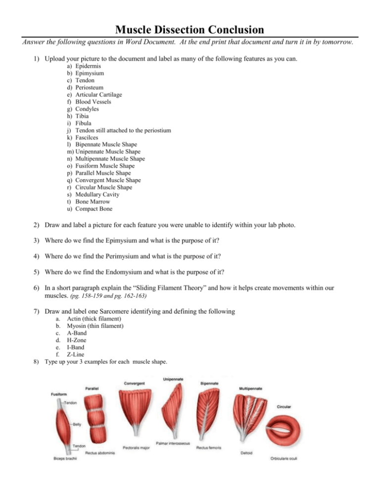
Muscle Dissection Conclusion Answer the following questions in Word Document. At the end print that document and turn it in by tomorrow. 1) Upload your picture to the document and label as many of the following features as you can. a) Epidermis b) Epimysium c) Tendon d) Periosteum e) Articular Cartilage f) Blood Vessels g) Condyles h) Tibia i) Fibula j) Tendon still attached to the periostium k) Fascilces l) Bipennate Muscle Shape m) Unipennate Muscle Shape n) Multipennate Muscle Shape o) Fusiform Muscle Shape p) Parallel Muscle Shape q) Convergent Muscle Shape r) Circular Muscle Shape s) Medullary Cavity t) Bone Marrow u) Compact Bone 2) Draw and label a picture for each feature you were unable to identify within your lab photo. 3) Where do we find the Epimysium and what is the purpose of it? 4) Where do we find the Perimysium and what is the purpose of it? 5) Where do we find the Endomysium and what is the purpose of it? 6) In a short paragraph explain the “Sliding Filament Theory” and how it helps create movements within our muscles. (pg. 158-159 and pg. 162-163) 7) Draw and label one Sarcomere identifying and defining the following a. Actin (thick filament) b. Myosin (thin filament) c. A-Band d. H-Zone e. I-Band f. Z-Line 8) Type up your 3 examples for each muscle shape.


