Rhodopsin, a G-Protein Coupled Receptor
advertisement
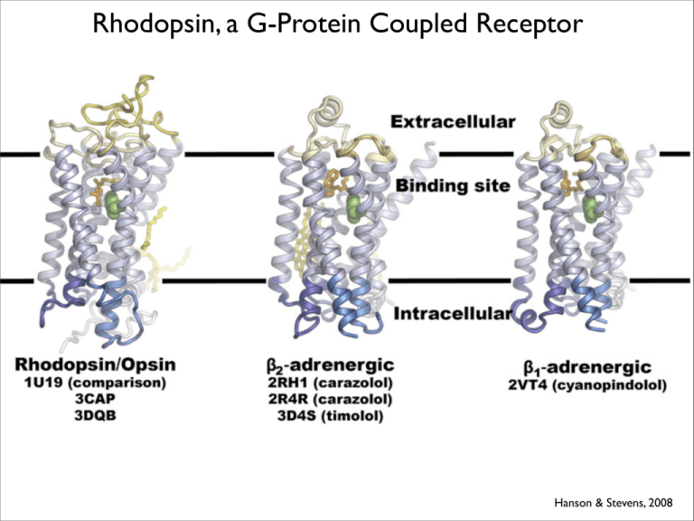
Rhodopsin, a G-Protein Coupled Receptor Hanson & Stevens, 2008 Signal Transduction & G-Protein Coupled Receptors Sigma-Aldrich Physiological Roles 1) Vision (Rhodopsin in Rods); (Photopsins in Cones) 2) Olfaction (Several hundred receptors) 3) Behavior & mood regulation (receptors for neurotransmitters including dopamine, serotonin, GABA, opiates,glutamate) 4) Immune system & inflammation (e.g. histamine receptors) 5) Autonomic nervous transmission, blood pressure, heart rate, digestion (e.g. beta adrenergic receptors) Morphine diacetylmorphine (heroin) adrenaline Cloning & Early Structural Model of Rhodopsin (the Light Absorbing Molecule of Vertebrate Rod Cells) Ovchinnikov 1982, Hargrave 1982 K296 Hargrave 2001 In 1986 it was discovered that the structure and key amino acids of rhodopsin were shared by the receptor that binds adrenaline......this was astonishing. Hamster βAR1 Bovine rhodopsin Mechanism of Photo-Transduction by Rhodopsin 1) hv discrimination & absorbance 2) Receptor activation 3) G-protein activation Rhodopsin Coupled to its Heterotrimer G-Protein, Transducin Physiology of Photo-Transduction Rhodopsin & Vision Portuguese Neurological Society Dark Light Glutamate Amplification in the Photo-Transduction Pathway 1 photon 1 rhodopsin activated 500 transducin molecules activated 1 cGMP phosphodiesterase activated per transducin 1 cGMP phosphodiesterase activated per transducin 1000 cGMP/s cleaved per phosphodiesterase Fesenko, 1985 Rhodopsin: Structure & Activity Palczewski, 2000 Inactive Receptor (Rhodopsin Model) 1) Inactive receptor 2) Ligand bound (activated) receptor 3)Binding of heterotrimeric G protein (transducin) to activated receptor Dark hv (fs) (us) Retinal MI (Metarhodopsin I) Retinol + opsin The scale of the 11 cis to all trans retinal structural change is significant as illustrated here (but the exact position is unknown) All retinal 11trans cis retinal H+ TMVII Dark hv (fs) Retinal MI (Metarhodopsin I) (us) Deprotonation (ms) H+ MII (Metarhodopsin II) Activated R* Hydrolysis (minutes) Retinol + opsin Orienting cis-11-Retinal in the Inactive Receptor Retinal TMVII Palczewski, 2000 Retinal Dark K 296 (TMVII) 1)Schiff base at K296 TMVII Rhodopsin & Photopsins are Unsual Because Ligand is Tethered & Normally Maintains Inactive Receptor. Another Tethered Ligand: Thrombin Receptor Nature Publishing TMIII TMVII Palczewski, 2000 1)Schiff base at K296 2)Counterion at E113 TMIII TMVI TMVII Palczewski, 2000 1)Tryptophan at W265 TMIII TMVI TMVII Palczewski, 2000 1)Phenylalanine at F261 II)Alanine at A269 TMIII TMVI TMVII Palczewski, 2000 Residues of TMIII Light Absorbance by Rods and the Three Types of Cones Gives Additional Clues to Which Amino Acids Strongly Interact with 11-cis-retinal Blue Cone Green Red Rod Cone Cone 437nm 498nm 533nm 564nm Dowling, 1987 via U. Florida, Physics 400 450 500 550 Wavelength (nm) 600 650 700 1) Localized effects 1I) Delocalized effects ‘HOOP’ Mathies Friends, 1999 Methanol Control Protonated 11 cis retinal (PSB) dissolved in solvent with polar residues Mathies Friends, 1999 Blue Cone 437nm KEY POINTS: 1) No strong location-specific differences relative to methanol 2) No strong delocalized effect relative to chromophore in methanol. Prediction: Blue pigment retinal interacts with polar amino acids in key positions. Mathies Friends, 1999 Green Cone 533nm KEY POINTS: 1) HOOP and ‘fingerprint modes are virtually identical to blue cone. 2) Dramatic shift in C=C stretch. 3) Vibration at the Schiff base changes dramatically relative to blue chromophore. Mathies Friends, 1999 Molecular explanation for blue to green shift: amino acids proximal to Schiff base go from polar to non-polar S90 to A90 S295 to A295 Red Cone 564nm KEY POINTS: 1) Fingerprints nearly identical to blue, green pigments. 2) HOOP mode is identical to blue, green pigments. 3)C=NH unchanged relative to green. 4) Ethylenic mode shifted further down in wavelength. Mathies Friends, 1999 Molecular explanation for green to red shift: amino acids distal from the chromophore go from apolar to polar F261Y and A269T (Apolar to polar residues) Mutations associated with congenital stationary night blindness TMII G90 T94 D I A292 TMVII E Activated Receptor 1) Inactive receptor 2a) Photoisomerization, or ligand binding to receptor 3)Binding of heterotrimeric G protein (transducin) to activated receptor Activated Receptor 1) Inactive receptor 2b) Conformational change transmitted to cytosolic residues 3)Binding of heterotrimeric G protein (transducin) to activated receptor

![Shark Electrosense: physiology and circuit model []](http://s2.studylib.net/store/data/005306781_1-34d5e86294a52e9275a69716495e2e51-300x300.png)
