Chlorophyta macroalgae
advertisement
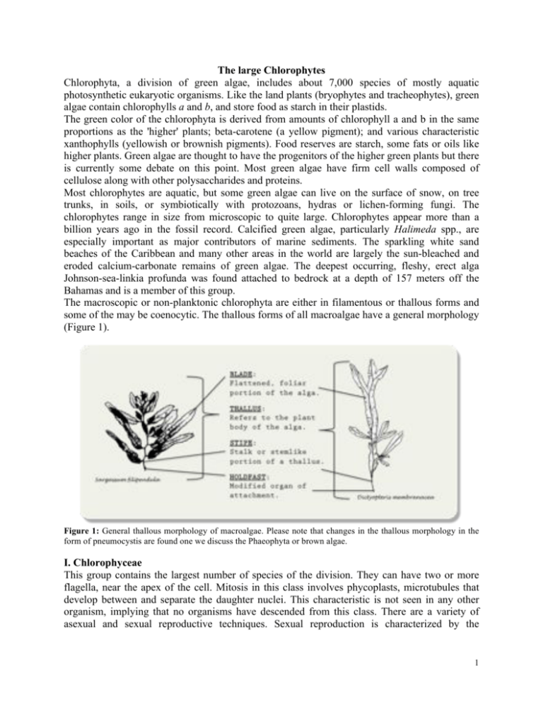
The large Chlorophytes Chlorophyta, a division of green algae, includes about 7,000 species of mostly aquatic photosynthetic eukaryotic organisms. Like the land plants (bryophytes and tracheophytes), green algae contain chlorophylls a and b, and store food as starch in their plastids. The green color of the chlorophyta is derived from amounts of chlorophyll a and b in the same proportions as the 'higher' plants; beta-carotene (a yellow pigment); and various characteristic xanthophylls (yellowish or brownish pigments). Food reserves are starch, some fats or oils like higher plants. Green algae are thought to have the progenitors of the higher green plants but there is currently some debate on this point. Most green algae have firm cell walls composed of cellulose along with other polysaccharides and proteins. Most chlorophytes are aquatic, but some green algae can live on the surface of snow, on tree trunks, in soils, or symbiotically with protozoans, hydras or lichen-forming fungi. The chlorophytes range in size from microscopic to quite large. Chlorophytes appear more than a billion years ago in the fossil record. Calcified green algae, particularly Halimeda spp., are especially important as major contributors of marine sediments. The sparkling white sand beaches of the Caribbean and many other areas in the world are largely the sun-bleached and eroded calcium-carbonate remains of green algae. The deepest occurring, fleshy, erect alga Johnson-sea-linkia profunda was found attached to bedrock at a depth of 157 meters off the Bahamas and is a member of this group. The macroscopic or non-planktonic chlorophyta are either in filamentous or thallous forms and some of the may be coenocytic. The thallous forms of all macroalgae have a general morphology (Figure 1). Figure 1: General thallous morphology of macroalgae. Please note that changes in the thallous morphology in the form of pneumocystis are found one we discuss the Phaeophyta or brown algae. I. Chlorophyceae This group contains the largest number of species of the division. They can have two or more flagella, near the apex of the cell. Mitosis in this class involves phycoplasts, microtubules that develop between and separate the daughter nuclei. This characteristic is not seen in any other organism, implying that no organisms have descended from this class. There are a variety of asexual and sexual reproductive techniques. Sexual reproduction is characterized by the 1 formation of a zygospore (a dormant diploid zygote protected by a thick wall) that later undergoes meiosis. We have already seen the unicellular organisms, member of the phytoplankton, like Chlamydomonas. The most complex of the class, however, are the filamentous members, some of which exhibit features that are seen primarily in plants. Despite this similarity the class is not believed to have been the evolutionary source of plants. 1. Sphaeroplea – Filamentous, cell body very long, cylindrical in shape, with many chloroplasts. Green to red uniseriate filaments with coenocytic cells (often paired nuclei) and annular (ringshaped) chloroplasts separated by clear cytoplasm. Cells are cylindrical, much longer than wide. Generally 10 – 50 µm diameter, but at least one species reported to be up to 170 µm diameter. Crosswalls between adjacent cells vary from thin to much chicker in some species. Asexual division by fragmentation of filaments. Sexual reproduction is oogamous or occasionally anisogamous. Oogonia produce several spherical green oocytes in one or more rows. Antheridia usually are on the same filament (monoecious). All gametes are biflagellate (female as well as male). Zygotes eventually turn orange-red before discharge, and are capable of prolonged dehydration. A. Examine a prepared slide of Sphaeroplea. What is the shape and arrangement of chloroplasts? Are the filaments branched or unbranched and are they enclosed in a gelatinous matrix or not? Which autotrophs already covered show filaments enclosed in a gelatinous matrix? What is the difference between those autotrophs and the Sphaeroplea? Figure 2: Sphaeroplea (left) and Zygnema (right). Sphaeroplea one of the characteristic chloroplast shapes and arrangements found in the organisms. In the picture of Zygnema you can determine the chloroplast shape (cells on the left) and see the oogonia, each with many oocytes. 2. Zygnema - Filamentous thalloid alga comprising about 100 species. Zygnema grows as a freefloating mass of filaments, although young Zygnema may be found anchored to streambeds with a holdfast. The filaments form a yellow-green to bright green colored tangled mat, and are composed of elongate barrel-shaped cells, each with chloroplasts arrayed one after the other along the axis of the cell. A. Examine a prepared slide of Zygnema. Pay special attention to the chloroplasts. What is different regarding the chloroplasts of Zygnema and other chlorophytes? Contemplate the shape and number of chloroplasts. Can you discern a gelatinous sheath? If so, describe it. How do the filaments of Sphaeroplea and Zygnema differ? 2 3. Chaetophora - Plants globose, tuberculose or arbuscular, mucilaginous or somewhat cartilaginous. Filamentous algae attached to a substrate at base, differentiation of axis-branch structure variable; cell body cylindrical, spherical or elongated ellipsoidal, with one or several chloroplasts; pyrenoids one to several in number or absent; terminal cells tapered or rounded at its end. Cells each with parietal chloroplast and one to several pyrenoids. Motile stages produced from peripheral cells, zoospores quadriflagellate and gametes biflagellate and isogamous. Figure 3: Chaetophora entire organism (left), structure of the organism (middle) and individual cells showing characteristic chloroplast shape (right). II. Ulvophyceae Most representatives of the Ulvophyceans are marine, though few occur in freshwater and terrestrial habitats. Body types may vary from filaments, membranous sheets to coenocytes but the majority of the Ulvophyceae are macroscopic. The Ulvophyceans are characterized by the following ancestral characteristics: Closed mitosis, persistent spindles and furrowing at cytokinesis, flagellar bases that are counterclockwise oriented and potentially flagellated gametes. 1. Cladophora – With thalli of uniseriate branched filaments with apical and/or intercalary growth. Branches are sparse to profuse with branches inserted laterally below the apex of a cell or apically on cell. If attached, branching rhizoids arise from basal cell and other cells in basal region, or simple discoid holdfast produced. Chloroplasts parietal, either densely packed discoid and/or united in a reticulum. Pyrenoids are present in multiple chloroplasts and of bilenticular shape, flanked by two bowl-shaped starch bodies. Cells are multinucleate with nuclei dividing more or less synchronously with the nuclear membrane remaining intact. The cell wall is mainly composed of crystalline cellulose I, forming numerous lamellae of microfibrils in a crossed fibrillar pattern. Asexual reproduction by biflagellate or quadriflagellate zoospores is the only method of reproduction in some species while other species only reproduce by thallus fragmentation. Cladophora is cosmopolitan in temperate and tropical regions, occurring in freshwater, brackish and marine conditions. The various species are found intertidally in waveexposed to very sheltered habitats, brackish pools, lagoons and mudflats, and in more or less eutrophic freshwater streams and lakes with pH > 7. 3 A. Examine the prepared and stained slides of Cladophora. Draw a vegetative cell at 10 and 40x magnification and observe and label the general outline as well as the organelles the chloroplast and pyrenoid. Draw a vegetative cell at 10 and 40x magnification and observe and label the general outline as well as the organelles the chloroplast and pyrenoid. What are differences to Rhizoclonium and Ulothrix? Figure 4: Cladophora lifecycle (left) and microscopic structure (right). B Make a wet mount of Cladophora. After you have observed the live, motile cells place a drop of methylene blue near the edge of one side of the coverslip and a paper towel near the opposite edge to draw the solution under the coverslip. Though the methylene blue will kill the cells it will aid you to see the nucleus (nuclei). To another wet mount add a drop of Lugol’s to make the nucleus more discernible. Which works better? Marine species of Cladophora exhibit the type of lifestyle known as alteration of generations. Because the alternating phases, or generations, are morphologically similar in Cladophora, its alternation of generations is considered to be isomorphic. In the lifecycle, which generation is the gametophyte, which the sporophyte? What are the chromosome numbers (n or 2n) of those lifecycle stages? Why? 4 2. Ulothrix - Filaments flaccid, basically unbranched and uniseriate green algae. Cells are always closely adherent. The cell wall in juveniles thin and smooth while more thickened and sometimes roughened later on. Apical cell are rounded and sometimes slightly narrowed. Attachment occurs by simple or rhizoidal basal cells, sometimes by rhizoidal outgrowths projecting from both intercalary and apical cells. Cells are cylindrical and uninucleate with a single chloroplast that is parietal, napkin ring-shaped, partially or fully encircling cell circumference, usually lobed and not enclosed in young cells, usually more irregular and sometimes fully closed in mature cells. There is a single pyrenoid in more mature cells, surrounded by a thin or more conspicuous starch envelope. Asexual reproduction occurs by quadriflagellate zoospores, arising in each cell. Zoospores are positively phototactic, oval, enclosing a cup-shaped parietal chloroplast with distinct stigma. Sexual reproduction occurs by isogamous biflagellate gametes, arising in all differentiated cells. Filaments produced gametes that often coiled and yellowish-green. Life histories are basically heteromorphic and diplobiontic with multicellular filamentous gametophyte and generally a unicellular sporophyte. Ulothrix is cosmopolitan with wide ecological distribution especially in temperate and colder regions. It usually forms strands several cm in length with extensive populations forming tufts or mats of varying size, often in green belt with distinct seasonality. They are present in aerated localities such as shores of eutrophic lakes and are less abundant in stagnant waters such as ditches and pools, and almost absent in boggy habitats. Ulothrix also flourishes in brackish areas with extreme (daily) variation in environmental factors, e.g. near mouth of intertidal freshwater streams where plants are covered with seawater at high tide, in estuaries or tidal rivers, in supralittoral pools of rocky shores, and supralittoral pools exposed to freshwater drip. In marine habitats forming extensive sheets in upper fringe of littoral zone; in mid and low littoral zone of rocky shores often present as important component of pioneer vegetation. Ulothrix grows on hard substrata but is also abundant in salt marshes on soft bottoms. A. Examine a stained, permanent mount with your 10x objective lens. You should be able to find vegetative and both zoospore- and gamete-producing filaments in the same preparation. How can you distinguish between vegetative and zoospore- or gamete-producing cells? Draw and label all three filaments. B. Switch to the 40x magnification and study both a vegetative and zoospore-producing cell. Draw a single vegetative cell to show the following: nucleus, plastids, pyrenoids, terminal vacuoles and label the contents. How can you distinguish zoospore-producing filaments from gamete-producing filaments? See the review of the Ulothrix life history. 3. Rhizoclonium - Filaments are very slender (60 µm in diameter) and loose lying with basal cells, or attached by holdfast with basal lobes. Plants unbranched or with one- to few-celled rhizoidal laterals. Cells are several to many times longer than broad. There are numerous nuclei per cell and chloroplast are reticulate, parietal, with pyrenoids, often densely packed with starch. 5 Reproduction is done by fragmentation. Rhizoclonium is a cosmopolitan species in fresh, brackish and marine waters, often growing entangled with other algae and forms dense mats in salt marshes. A. Examine the prepared and stained slides of Rhizoclonium. Draw a vegetative cell at 10 and 40X and observe and label the general outline as well as the organelles (nucleus, chloroplast and pyrenoid). What are differences between Rhizoclonium and Ulothrix? 4. Acetabularia – The thallus is an unbranched unicell (1-6 cm long), composed of a compact rhizoid, a tubular stalk ca. 1 mm diam., whorls of thrice-branched sterile laterals and a more or less flattened apical cap (0.5-1.5 cm in diameter). The mature cap is composed of 30-75 free or joined, terminally tapered or rounded elongate rays. The thallus may be lightly to heavily calcified. Thallus size, cap diameter and cap morphology are important species characteristics. Numerous parietal, discoid, grass green plastids without pyrenoids circulate in the peripheral cytoplasm that surrounds a large central vacuole. The chloroplast ultrastructure may be variable, without or with stacked grana and variations are generally species specific. Starch grains are present in the chloroplast and cytoplasm, the latter not membrane bound. Acetabularia distribution is pantropical and subtropical, commonly in brackish to hypersaline shallow waters. Thalli are firmly attached to solid substrates such as stones, coral rubble, shells as well as to wood and industrial detritus such as rubber. Seasonal life history is variable, especially at the cool extremes of the range. The development, nucleo-cytoplasmic interactions, biochemistry, photobiology, genetics and molecular biology of Acetabularia species have been extensively studied. Changes in microtubules and microfilaments associated with key phenomena during growth and differentiation. Their taxonomy within the Ulvophyceans remains uncertain. A. Observe a live representative of Acetabularia. Observe and draw the rhizoid, stalk (with hair whorls) and the cap. Where would you expect to find the nucleus of the unicellular organism? What is a difference between this unicell and the unicells we have previously observed? Acetabularia has a sexual life history that is isogamous with a modified heteromorphic alternation of generation. The zygote (l-b) germinates to make a uninucleate sporophyte. The single cell begins to produce branches at its tip and then the diploid nucleus undergoes meiosis ultimately to produce hundreds of haploid nuclei which migrate to the gametangia at the tip of the cell. The nuclei accrete cytoplasm about themselves and produce cysts within which gametes differentiate and are released. B. Take a section of the rim of the cap and prepare a wet mount. You should be able to see a divided cell obscured by chloroplasts but with some bright, oval structures. Unlike in other unicells these organelles are not pyrenoids but cysts. Examine the lifecycle of Acetabularia present as a drawing to understand the significance of these cysts in the organism’s lifecycle. What do you think might activate the cysts to germinate? Why? (Make sure that you check the answer with your lab instructor) 6 Figure 5: Acetabularia lifecycle. 5. Ulva – The mature thallus consists of a flattened distromatic blade in which two cell layers are developmentally independent but closely adherent. Blades can be broadly expanded, irregularly lobed, cuneate, linear, lanceolate or deeply divided into linear lacinae. Growth of the blade occurs through diffuse cell divisions primarily along the margins. Ulva is usually attached to substrate by rhizoidal cells in a basal holdfast and/or rhizoidal extensions of cells in the lower portion of the blade or occasionally extending along part or the entire longitudinal axis of the blade. Rhizoidal extensions run between the two cell layers of the blade. Vegetative cells contain a single chloroplast and 1 or more pyrenoids and are generally uninucleate. In opposition rhizoidal cells are often multinucleate. Ulva is a cosmopolitan genus with species in all oceans and estuaries of the world. Species of Ulva have traditionally been based on morphological, anatomical and cytological characteristics such as shape, size, presence or absence of dentation, thickness, cell dimensions and number of pyrenoids. Many studies have shown that these characteristics can be highly variable within species, varying with age, reproductive state, wave exposure, tidal factors, temperature, salinity, light and biological factors such as grazing. In recent years developmental patterns in culture, reproductive details and the apparent inability of species to interbreed have been used to evaluate species concepts based on morphological and anatomical characteristics. 7 A. Make a cross section of a piece of Ulva and prepare a wet mount (in addition, prepared section slides of Ulva are available in lab). What is the cell arrangement in Ulva? What is the chloroplast arrangement in Ulva? Draw a general outline of an Ulva cross section? What is the chloroplast location in an Ulva leaf? What is the typical chloroplast shape found in Ulva? B. Examine a rehydrated or live Ulva. What is the overall thallous morphology? How does it differ from the other chlorophyta observed? Ulva has a very high surface to volume ratio due to the fact that it is only a few cell layers thick. What might be the advantage of a high surface to volume ratio? Are you looking at the sporophyte of gametophyte of Ulva? How can you determine this? C. Ulva with germlings. The “germlings” are derived from meiospores. With that knowledge, what lifecycle stage do these germlings represent? Figure 6: Ulva lifecycle. 6. Enteromorpha – When Enteromorpha first begins growing, it forms a single row of cells, this structure is monosiphonous. Soon after the monosiphonous filament is formed, longitudinal division of cells creates a two layered filament. Eventually, after more cell division the two cell layers separate to form a tube, forming the adult morphology. The thallus of Enteromorpha is tubular with the wall of the tube a single cell layer thick. The thallus can be branched or unbranched, and there is a wide variety of forms within the genus. Enteromorpha is attached to the substrate by a disc-like holdfast. The holdfast is formed by the basal cell dividing into three or four holdfast cells which elongate and undergo further division. The cells in Enteromorpha can vary in size and shape from species to species, and sometimes they will form regular linear series in a frond, while other times there is an irregular arrangement of the cells. Each cell contains a single chloroplast, varying in size depending on the size of the cell. Enteromorpha and the previous Ulva are not very distinct species. A. Compare the live Enteromorpha to the live or rehydrated Ulva. What parts of the thallus morphology are discernible. How does that differ from other chlorophytes. 8 How can you distinguish between Ulva and Enteromorpha? B. Examine a prepared slide of Enteromorpha. Describe the chloroplast arrangement and consider the reason for this arrangement. What is the chloroplast shape? 7. Codium – The thallus is spongy, anchored to rocks or shells by a weft of rhizoids, varying in size from 1 cm. to 10m long. Their habit varying widely their form may be digitaliform, globular, petaloid, membraniform, or dichotomously branched. The internal structure is composed of a colorless medulla of densely intertwined siphons and a green palisade-like layer of vesicles called utricles. Organelles, including innumerable nuclei and discoid chloroplasts (but no amyloplasts) are confined to a layer of cytoplasm pressed to the cell wall. Chloroplasts lack pyrenoids. Despite the ubiquity of Codium very little is known about its biology. Codium grows intertidally, and to at least -40m. It occurs in all marine waters except the Arctic and Southern Oceans. The largest numbers of species are found in floras that are transitional between temperate and subtropical, namely Japan, South Africa, Australia and California-Mexico. Each of these floras includes a member of most sections of the genus, indicating an ancient dispersal of the progenitors. Because of lack of calcification, there is no fossil record of the genus. A. Take a thin section of a blade of Codium. Observe the multinucleate filaments and distinguish the medulla from the surrounding cortex. How do they differ? Why is their coloration as it is and not reversed. Codium has a life history that is similar to that of Caulerpa. Both are diplontic with anisogamous sexual reproduction. There is not asexual reproduction, though vegetative reproduction does occur. Figure 7: Codium lifecycle. 9 IV. Charophyceae 1. Mougeotia - Thalli unbranched, forming extensive intertwining uniseriate filaments. Cells cylindrical, 5 to 30 µm in diameter, much longer than wide; cell wall two-layered with inner cellulose, outer mucilage layer; end walls plane; no flagellated stages; a few species with one- or two-celled basal branches or rhizoids that anchor filament to substrate. Cell uninucleate; chloroplasts one per cell (sometimes two), axial, uncoiled, plate-like (=wide ribbon) in shape with axial row of pyrenoids. Nucleus one per cell, midway along length of cell, parietal to single chloroplast or in cytoplasmic bridge between two chloroplasts. Chromosomes (n= 24-92) typically small and dotlike (<1.5 µm). Haploid chromosome number 24 to 92. Asexual reproduction common by aplanospores and parthenospores (preempted gametes that become aplanospores). Aplanospores with essentially same wall color and morphology as sexually produced zygospores. Life cycle haplobiontic, meiosis zygotic. Sexual reproduction by conjugation, scalariform in most species, lateral in about a half dozen species. Almost all species homothallic and isogamous; both gametes usually capable of amoeboid movement; zygospore forms within conjugation tube between two gametangia, in both receptive gametangia or with part of zygospore in gametangia and part in tube; transverse sporangial walls cut off zygospore from gametangia ; cytoplasmic residue remains inside spent gametangia. Filaments may adhere, sometimes genuflecting (zig-zag bending of adhering filaments); both processes apparently unrelated to conjugation and of unknown function. Zygospores variously shaped, from spheroid to ovoid to quadrangular (square-shaped with blunt corners). Zygospores and aplanospores commonly with two layers: outer chitinous and inner transparent. Earliest fossil zygospores from Carboniferous. 2. Coleochaete - The "chaete" in the names of the genera refer to long, sheathed hair cells which cover the algal surface. These are believed to defend the alga against potential herbivores. Flagella are present on both sperm cells and zoospores; two are inserted on each of these cell types, and are arranged asymmetrically over a multilayered structure (MLS). Members of the Coleochaetales have a variety of morphological forms. Species of both genera may grow as branched filaments, while other species may be pseudoparenchymatous. Two species of Coleochaete are parenchymatous and disc-shaped; these also have complex gametangia. Colecochaetales may be found growing epiphytically in aqueous habitats, on the stems and leaves of larger aquatic plants, or attached to rocks or soil. A. Examine a slide of a cross section of Coleochaete. What is the overall cell arrangement and Describe the chloroplast arrangement and structure. Are pyrenoids present? How many pyrenoids can you find in each cell? 10
