MUSCLE FATIGUE
advertisement
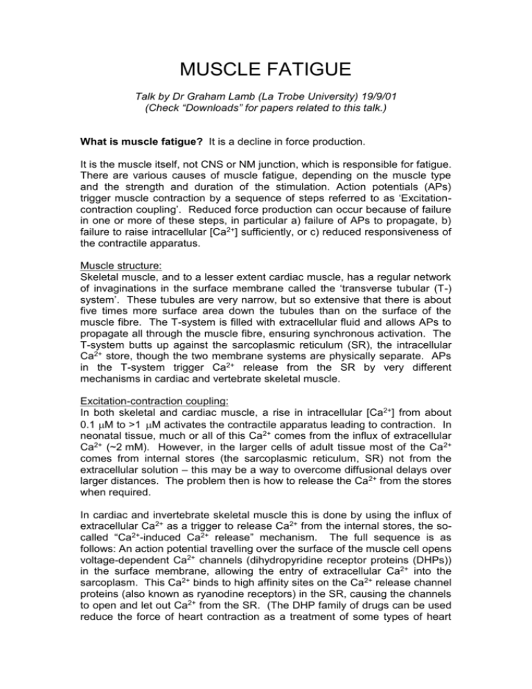
MUSCLE FATIGUE Talk by Dr Graham Lamb (La Trobe University) 19/9/01 (Check “Downloads” for papers related to this talk.) What is muscle fatigue? It is a decline in force production. It is the muscle itself, not CNS or NM junction, which is responsible for fatigue. There are various causes of muscle fatigue, depending on the muscle type and the strength and duration of the stimulation. Action potentials (APs) trigger muscle contraction by a sequence of steps referred to as ‘Excitationcontraction coupling’. Reduced force production can occur because of failure in one or more of these steps, in particular a) failure of APs to propagate, b) failure to raise intracellular [Ca2+] sufficiently, or c) reduced responsiveness of the contractile apparatus. Muscle structure: Skeletal muscle, and to a lesser extent cardiac muscle, has a regular network of invaginations in the surface membrane called the ‘transverse tubular (T-) system’. These tubules are very narrow, but so extensive that there is about five times more surface area down the tubules than on the surface of the muscle fibre. The T-system is filled with extracellular fluid and allows APs to propagate all through the muscle fibre, ensuring synchronous activation. The T-system butts up against the sarcoplasmic reticulum (SR), the intracellular Ca2+ store, though the two membrane systems are physically separate. APs in the T-system trigger Ca2+ release from the SR by very different mechanisms in cardiac and vertebrate skeletal muscle. Excitation-contraction coupling: In both skeletal and cardiac muscle, a rise in intracellular [Ca2+] from about 0.1 M to >1 M activates the contractile apparatus leading to contraction. In neonatal tissue, much or all of this Ca2+ comes from the influx of extracellular Ca2+ (~2 mM). However, in the larger cells of adult tissue most of the Ca 2+ comes from internal stores (the sarcoplasmic reticulum, SR) not from the extracellular solution – this may be a way to overcome diffusional delays over larger distances. The problem then is how to release the Ca2+ from the stores when required. In cardiac and invertebrate skeletal muscle this is done by using the influx of extracellular Ca2+ as a trigger to release Ca2+ from the internal stores, the socalled “Ca2+-induced Ca2+ release” mechanism. The full sequence is as follows: An action potential travelling over the surface of the muscle cell opens voltage-dependent Ca2+ channels (dihydropyridine receptor proteins (DHPs)) in the surface membrane, allowing the entry of extracellular Ca2+ into the sarcoplasm. This Ca2+ binds to high affinity sites on the Ca2+ release channel proteins (also known as ryanodine receptors) in the SR, causing the channels to open and let out Ca2+ from the SR. (The DHP family of drugs can be used reduce the force of heart contraction as a treatment of some types of heart disease – this works because the DHPs partially block the surface Ca 2+ channels (the DHP receptor proteins), thereby reducing Ca2+ influx which in turn reduces Ca2+-induced Ca2+ release from the SR). In vertebrate skeletal muscle, there have been a number of evolutionary changes, probably to enhance the speed and control of Ca 2+ release and hence contraction. Firstly, the influx of extracellular Ca 2+ is not necessary for triggering Ca2+ release from the SR. The DHP receptor/voltage-dependent Ca2+ channels in the T-system (also known as ‘voltage-sensors’, (VS)) physically couple by a protein-protein interaction with the Ca2+ release channels in the SR. Secondly, the sensitivity of the Ca2+ release channels to cytoplasmic Ca2+ is much lower than in cardiac muscle – this is due to a strongly enhanced inhibitory effect of intracellular Mg2+ on the release channels. The overall effect of these two changes is that the DHP receptor/voltage-sensor controls the Ca2+ release channel directly, not by a ‘Ca2+-induced Ca2+ release’ mechanism, with the consequence that Ca 2+ release can not only be activated in milliseconds, but can also be stopped in milliseconds, a necessary requirement for rapid, accurate muscle control. Muscle fibre types: In mammals: slow twitch is Type I and fast twitch is Type II. Slow twitch: - red appearance - many mitochondria, lower force output - fatigue “resistant”, postural or repetitive role. Fast twitch: - white appearance, fewer mitochondria, high force output, faster contractions and relaxation, fatigue faster. - recruit for “explosive” contraction. Types of muscle fatigue High frequency fatigue – onset in seconds. Fast and full recovery in seconds. “Metabolic” fatigue. Long duration fatigue (takes a long time to recover). K+ Na+ Ca2+ VS Transverse tubule in muscle 2 Ca2+ Release Channel Ca2+ store in Sarcoplasmic reticulum High Frequency Fatigue: Each AP leads to Na+/K+ movement. A large number of APs will produce an increase in [K+] in the tubules leading chronic depolarization of the T-system (eg. from –90 mV to ~-60 mV), which prevents the voltage-dependent sodium channels in the T-system from resetting (ie. they stay ‘inactivated’). In the extreme, this would mean APs would not propagate into and along the Tsystem, and hence there would be no activation of the voltage-sensor (VS) coupling to the Ca2+ release channel, and hence no muscle contraction. This is called high frequency fatigue. It is induced rapidly (ie. over seconds) by high frequency stimulation (eg. 100 – 200Hz), but recovery is also very rapid (over seconds) upon cessation of stimulation due to K + both diffusing out of the T-system and being pumped back into the cell by the Na-K-ATPase in the T-system membrane. This type of fatigue is not caused by metabolic changes within the fibre. Metabolic Fatigue: When a muscle has been stimulated sufficiently long and/or vigorously there can be metabolic changes within the fibre. This will occur differently in fast and slow-twitch fibres. The metabolic changes could cause reduced Ca2+ release from the SR and/or reduced responsiveness of the contractile apparatus. Energy production reaction: ATP ADP + Pi Muscles have stores of high-energy phosphates: 8 mM ATP and 50 mM creatine phosphate (CrP). Normally [ATP] is kept close to 8 mM by the resynthesising it from ADP using the CrP. Resynthesis of ATP from ADP CrP + ADP ATP + Cr This is a fast reaction – keeps [ATP] high and [ADP] low. The net reaction: CrP Cr + Pi ATP is used at ~1 to 2 mM/s during maximal contraction (by the contractile apparatus and the SR Ca2+ pump). This means that all the CrP will be exhausted within ~30 to 60s. If this happens [ATP] will fall, and also [Mg 2+] will rise (most cellular Mg2+ is normally bound to ATP), and if unchecked, ultimately the cell would die due to ATP exhaustion and consequent failure to keep cytoplasmic [Ca2+] relatively low. This is why having a ‘fatigue’ mechanism is essential – there is a clear advantage to having a system which will shut itself down before it exhausts the cellular ATP supply. 3 Muscles can function for long periods of activity so long as the energy use is matched to supply. In slow-twitch muscle in general ATP usage is met by aerobic respiration. Aerobic respiration: 36 ATP produced per glucose molecule. ATP can also be produced by: a) Anaerobic respiration: Glucose +2 ADP + 2 Pi 2 ATP + 2 lactic acid b) 2ADP ATP + AMP (AMP is deaminated to IMP and there is reduction in available ATP). This is a very inefficient process. The muscle here is being pushed very hard. Metabolic changes occurring during exercise Fast-twitch muscle undergoing intense exercise (anaerobic) Early term: Cr , Pi , lactate , H+ progressively. Medium term: ATP , Mg2+ , IMP Slow-twitch – low to medium intensity exercise (aerobic) Early to medium term: Cr , Pi progressively, some lactate , H+ Longer term: glycogen Which of the above cause muscle fatigue? Direct effects on the contractile apparatus: The rise in P i will directly reduce the Ca2+ sensitivity of the contractile apparatus to some degree though this is largely offset by the effect of the concomitant reduction in CrP. Overall there is only a small reduction in the maximum force production and Ca 2+ sensitivity of the contractile apparatus – the most dramatic effects are due to reductions in Ca2+ release. Experiments in which lactate is added to isolated muscle fibres has shown that lactate has no effect on Ca2+ release and contractions. The same is also true for raising intracellular [H+] (this has been shown recently to actually reduce high frequency fatigue). Instead, the three primary causes of reduced Ca 2+ release areas follow, the first two probably being most important in slow-twitch fibres and the third in fast-twitch fibres: 4 Increase in [Pi]. Some Pi moves into the Ca2+ store (sarcoplasmic reticulum). This Pi precipitates Ca2+ which then reduces the free Ca2+ available for release from the SR. Glycogen depletion. This is deleterious both because of the limitation of aerobic metabolism and also because of a poorly understood effect in which glycogen depletion directly inhibits the coupling between the VS and the Ca2+ release channels, possibly because of the liberation of associated enzymes. Decrease in [ATP] (and the accompanying increase in [Mg2+] and metabolites of ATP). The Ca2+ release channel has a low affinity binding site for ATP (dissociation constant ~ 1 mM), and only when ATP is on this site can the channel be activated. Thus, if [ATP] drops to <1 mM it is difficult to fully activate the Ca2+ release channel. Any build-up of ADP and AMP will worsen this because they compete for the same site but don’t cause much stimulation. The concomitant increase in free [Mg2+] also inhibits the Ca2+ release channel directly. Long–duration fatigue (also known as ‘low-frequency fatigue’) is caused by Ca2+ induced damage where Ca2+ moves in and out of the SR for too long. This can also lead to structural changes or damage resulting in very prolonged fatigue. Muscle-fatigue – is it all bad? No – it is a vital fail-safe mechanism. If [ATP] decreases and almost runs out then cellular death could occur. Then fatigue is a self-limiting mechanism. That also suggests that there must be sensors for [ATP]. Should creatine be taken as a supplement? Some studies claim creatine supplements can reduce muscle fatigue and others say it has no effect or can be deleterious. Some reports have associated it with undesired weight gain. One would expect it to be of most use for preventing the type of fatigue occurring in fast-twitch fibres with vigorous stimulation. However, it is not clear that ingesting creatine raises muscle creatine levels, nor that this will raise the creatine phosphate levels – raising creatine alone would worsen the energy balance. Muscle meltdown. This happens due to exhaustion and when muscle temperature increases to about 41o. Contractile apparatus becomes dysfunctional, all of the ATP is used up and proteases are activated. Heat-shock proteins are also activated. 5
