I) Why Genetics for Eye Care Practioners
advertisement
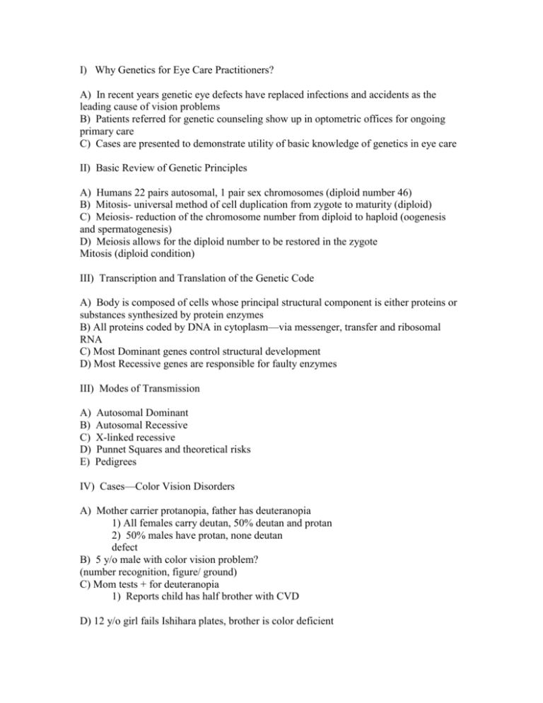
I) Why Genetics for Eye Care Practitioners? A) In recent years genetic eye defects have replaced infections and accidents as the leading cause of vision problems B) Patients referred for genetic counseling show up in optometric offices for ongoing primary care C) Cases are presented to demonstrate utility of basic knowledge of genetics in eye care II) Basic Review of Genetic Principles A) Humans 22 pairs autosomal, 1 pair sex chromosomes (diploid number 46) B) Mitosis- universal method of cell duplication from zygote to maturity (diploid) C) Meiosis- reduction of the chromosome number from diploid to haploid (oogenesis and spermatogenesis) D) Meiosis allows for the diploid number to be restored in the zygote Mitosis (diploid condition) III) Transcription and Translation of the Genetic Code A) Body is composed of cells whose principal structural component is either proteins or substances synthesized by protein enzymes B) All proteins coded by DNA in cytoplasm—via messenger, transfer and ribosomal RNA C) Most Dominant genes control structural development D) Most Recessive genes are responsible for faulty enzymes III) Modes of Transmission A) B) C) D) E) Autosomal Dominant Autosomal Recessive X-linked recessive Punnet Squares and theoretical risks Pedigrees IV) Cases—Color Vision Disorders A) Mother carrier protanopia, father has deuteranopia 1) All females carry deutan, 50% deutan and protan 2) 50% males have protan, none deutan defect B) 5 y/o male with color vision problem? (number recognition, figure/ ground) C) Mom tests + for deuteranopia 1) Reports child has half brother with CVD D) 12 y/o girl fails Ishihara plates, brother is color deficient Color vision deficiencies: A) Are never inherited via an autosomal dominant mode of transmission B) Are of variable prevalence among the different races in females C) Usually involve a protan defect in white males D) Are inherited via an x-linked recessive mode for R/G defects. V) Clinical Case Presentation-- Presenting Visual Fields (central scotoma A) Differential diagnosis—other findings? 1) +APD OD, Fang neg, Media clear, IOP normal 2) Irregular dilatation of the peripapillary capillaries B) Results of referral to Nuero-Opthalmology U of MN? 1) Dx Leber’s Optic Atrophy – onset in the second or third decade with rapid and severe loss of visual acuity (15% females) 2) Pedigrees can resemble X-linked recessive; however mechanism is thought to be cytoplasmic inheritance 3) As is XLR mothers must transmit trait, but no evidence that fathers pass gene to daughters (no carrier status) C) Five genetic optic atrophies “likely” to be seen in primary eye care 1) Leber’s OA 2) OA seen with Diabetes (AR) 3) Behr Syndrome (AR) 4) Simple recessive OA (AR) 5) Dominant OA D) Case of vision loss in a 28yo female (Linda M) 1) Initial Dx Leber’s 2) Pedigree next slide 3) Onset age 13 4) VA decreases to 20/40 by age 19 5) Stable VA of 20/60 by age 38 6) What is your pedigree analysis? 7) Leber’s (cytoplasmic inheritance) 8) Defective cytoplasmic mitochondrial DNA from mother (affects manufacture of ATP) 8) Strict maternal transmission E) Follow up Linda M 1) Alphagan P (brimonidine tartrate oph sol 0.15%) clinical trial 2) Medication increases the amount of brain derived neuro-trophic factor (BDNF) to retinal ganglion cells in rat glaucoma models 3) Proof of benefit in humans remains to be demonstrated 4) 20/300 OD, 20/400 OS, felt Tx delayed onset OD—attended DVR Wausau CCTV, etc (works 12-16 hours per week in medical records) Leber’s HON is: A) Inherited in an AR pattern with maternal transmission B) Passed through the males in the pedigree C) Manifested in early childhood like many of the heritable optic neuropathies D) Affects ATP production via mitochondrial DNA. VI) Ocular Albinism (OA) A) 18yo w male, Reduced VA, @30cm 20/30 OU—conv dampens nystagmus, ++photosensitive, Brother and mom’s MGF also OA B) Fundus C) Clinical Pedigree vs. Textbook D) Lyon Hypothesis 1) X-linked recessive 2) Early in female embryonic development each cell makes a “decision” to deactivate one of its X chromosomes (Barr body). This may leave more of the affected X’s active. 3) Asymptomatic carrier fundus (Mom) The Lyon hypothesis: A) Involves deactivated X chromosomes in male carriers (Barr bodies) B) Stipulates that asymptomatic females with OCA may be “picked up” clinically via ophthalmoscopy C) Could be utilized for a young female patient whose father had RP. D) All of the above VII) Other cases presented: Dominant Optic Atrophy (AD), Horner Syndrome (AD), Vitelliform and Pattern Dystrophy (AD), Retinitis Pigmentosa (AD, AR and X-linked) The preceding RP case is a good example of: A) A rare clinical scenario unlikely to ever been seen by a primary care OD B) A clinical situation where only the Lyon hypothesis concept would allow identification of a carrier patient C) The case history and the Lyon hypothesis presenting a conflicting picture for the clinician D) None of the above VIII) Prevalence of Dyslexia and elaboration as to genetics of the various types of dyslexia A) 10-15% of general population B) 23% of third grade classroom (Griffin et al) (includes borderline cases) C) 18% of 10th grade males; 45% of age matched juvenile hall residents (San Bernadino, CA) D) Dyseidesia (AD), Dysphonesia (Multifactorial) True/False The majority of individuals with reading disability are primarily affected by binocular, accommodative or visual processing disorders which cause the reading problem. IX) Conclusions A) Genetics for the primary care practitioner does not make the doctor an expert genetic counselor B) Knowledge of genetics is very helpful with many disease entities in terms of differential diagnosis and consulting with family members C) Genetic principles apply to many of the common visual entities encountered by the optometrist
