医学神经科学与行为I模块2教学内容
advertisement
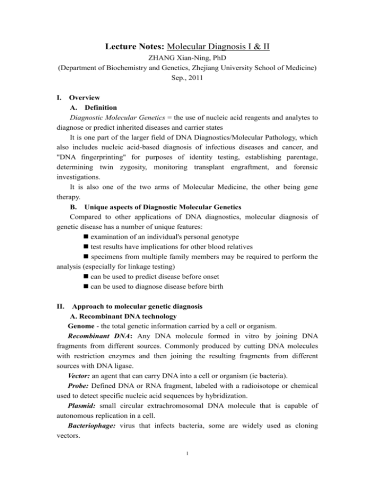
Lecture Notes: Molecular Diagnosis I & II ZHANG Xian-Ning, PhD (Department of Biochemistry and Genetics, Zhejiang University School of Medicine) Sep., 2011 I. Overview A. Definition Diagnostic Molecular Genetics = the use of nucleic acid reagents and analytes to diagnose or predict inherited diseases and carrier states It is one part of the larger field of DNA Diagnostics/Molecular Pathology, which also includes nucleic acid-based diagnosis of infectious diseases and cancer, and "DNA fingerprinting" for purposes of identity testing, establishing parentage, determining twin zygosity, monitoring transplant engraftment, and forensic investigations. It is also one of the two arms of Molecular Medicine, the other being gene therapy. B. Unique aspects of Diagnostic Molecular Genetics Compared to other applications of DNA diagnostics, molecular diagnosis of genetic disease has a number of unique features: examination of an individual's personal genotype test results have implications for other blood relatives specimens from multiple family members may be required to perform the analysis (especially for linkage testing) can be used to predict disease before onset can be used to diagnose disease before birth II. Approach to molecular genetic diagnosis A. Recombinant DNA technology Genome - the total genetic information carried by a cell or organism. Recombinant DNA: Any DNA molecule formed in vitro by joining DNA fragments from different sources. Commonly produced by cutting DNA molecules with restriction enzymes and then joining the resulting fragments from different sources with DNA ligase. Vector: an agent that can carry DNA into a cell or organism (ie bacteria). Probe: Defined DNA or RNA fragment, labeled with a radioisotope or chemical used to detect specific nucleic acid sequences by hybridization. Plasmid: small circular extrachromosomal DNA molecule that is capable of autonomous replication in a cell. Bacteriophage: virus that infects bacteria, some are widely used as cloning vectors. 1 Restriction endonuclease (enzyme): An enzyme that recognizes and cleaves a specific short sequence in double stranded DNA. Northern blot-technique used to detect mRNA molecules after separation by gel electrophoresis. Southern blot- technique to detect genomic DNA after digestion with a restriction enzyme and separation by gel electrophoresis. Expression vector- modified plasmid or virus carrying a gene or cDNA into a suitable host cell where it directs expression of the encoded protein. B. Identify Changes in DNA sequence Southern blot o Can detect specific genes in the complex mixture of genomic DNA o Can detect large deletions, insertion or rearrangements o Can only detect point mutations which alter restriction enzyme recognition site o Can detect gene amplification Polymerase Chain Reaction (PCR) o Used as the basis for many sophisticated diagnostic and forensic tests C. Measuring Gene Expression RT-PCR Northern blot o Based upon design of Southern o Can be used to compare levels of mRNA among samples o Can detect expression of a gene in specific cells or tissues o Can detect changes in transcript size o Can detect changes in the level of expression DNA Microarrays o Allows analysis of thousands of genes simultaneously o Based on cDNA or oligonucleotides bound to glass slides o one important use is the molecular classification of tumors. D. Use of recombinant DNA to produce medicine Use of bacterial expression systems o Inexpensive and easy to grow large quantities o Lack enzymes to catalyze post-translational modifications o May fold proteins incorrectly o Production of recombinant growth hormone stopped the transmission of a deadly virus o Example G-CSF for supportive care in cancer Eukaryotic expression systems o Inexpensive and easy to grow o Yeast can perform simple modifications such as glycosylation 2 o Early vaccines used attenuated viruses that could revert to wild type o Example- hepatitis B vaccine Mammalian cells o Expensive and difficult to use o Sometimes they provided the only way to produce an active drug o Production of recombinant Factor VIII was a great success storyprevious hemophiliac patients were infected with HIV from the Factor VIII preparations Future likely to find more “designer” drugs- small molecules o Screened by high throughput techniques o Synthesized by chemists o Key is to validate the targets and identify appropriate biological endpoints III. Molecular diagnosis A. Classification of genetic disease The type of approach chosen to diagnosis a genetic disease at the molecular level is usually based on what is currently known about the causative gene. In increasing order of diagnostic difficulty, the molecular classes of genetic disease are: those for which both the gene and causative mutation are known those for which the gene is known, but not the mutation those for which neither gene nor the mutation is known those caused by more than one gene interacting with environmental factors (polygenic/multifactorial disorders) Molecular genetic diagnosis is generally concerned with detection of inherited (germline) as opposed to acquired (somatic) mutations. B. Types of clinical molecular genetic applications Molecular genetic tests may be performed for any of a number of quite different clinical purposes: Clinical diagnosis/confirmation Carrier screening Prenatal diagnosis Newborn screening Presymptomatic diagnosis/predisposition screening C. Sample collection Since germline mutations are present in every cell of the body, site-specific biopsy is not required and simple phlebotomy will suffice for most purposes. Alternatively, the great sensitivity of PCR allows for genetic testing to be performed on minute samples of any readily accessible tissue or body fluid, including saliva, urine, etc. For prenatal diagnosis, several sample types are possible, depending on the 3 clinical situation: Amniocentesis: 15th to 16th weeks Chorionic villus samples (CVS): 10th to 12th weeks Cordocentesis (PUBS): 19th to 21th weeks Embryonic blastomeres for preimplantation diagnosis (PGD): before the fertilization Fetal cells circulating in maternal blood D. Types of mutations and methods of detection (Table 1) Usually the laboratory technique chosen will depend on the nature of the mutation(s) being tested for. Mutation mechanisms to be analyzed in genetic disease include: Point mutations - single nucleotide changes, causing missense or nonsense coding alterations, or RNA splicing defects Microdeletions and insertions - of one or a few nucleotides, causing an amino acid loss or insertion or (if the change is not an even multiple of three nucleotides) a frameshift mutation Deletions - of large portions of a gene Trinucleotide repeat expansions - increase in number of a tandem repeat of three nucleotides, which may be within an exon, an intron, or a promotor region It is important to keep in mind that many genetic diseases can be caused by more than one mutation, by more than one mutational mechanism, or even by more than 4 one gene. This phenomenon is called genetic heterogeneity, and it makes otherwise straightforward molecular genetic screening tests much more labor-intensive, expensive, and difficult. Some techniques are better suited for detecting particular gene alterations than others. In general, PCR is advantageous because of its extreme sensitivity, its ability to hone in on small segments of a complex gene, its cost savings in labor and reagents, and its adaptability to batching of large numbers of specimens. It is not reliable for examining very large regions of DNA, however, an application for which Southern blot hybridization is still often required. To detect point mutations and microdeletions/microinsertions, appropriate techniques include: Southern blot or dot blot hybridization with allele-specific oligonucleotide (ASO) probes Southern blot analysis or PCR amplification, subjecting the products to digestion with a particular restriction endonuclease whose natural cleavage sequence is either destroyed or created by the mutation in question DNA sequencing To detect large deletions, use: Southern blot Fluorescence in situ hybridization (FISH) Routine cytogenetics, if deletion is big enough to be visualized by light microscopy (Note that the last two techniques are formally based in cytogenetics labs rather than molecular genetics labs.) To detect trinucleotide repeat expansions, use: Sizing by PCR if the expansion is of moderate length Sizing by Southern blot if the expansion is of large size (too large to be amplified reliably by PCR) To detect many possible mutations in diseases with marked genetic heterogeneity, use: PCR of several gene regions followed by serial hybridization with a panel of ASO probes Dot blot hybridization with a pooled cocktail of ASO probes for several rare mutations at once PCR followed by reverse dot blot hybridization to an array of ASO probes bound to a paper strip or other solid support 5 Manual or automated DNA sequencing Mutation scanning techniques - Denaturing Gradient Gel Electrophoresis (DGGE) - Single Strand Conformation Polymorphism (SSCP) - Protein Truncation Test (PTT) Hybridization to a complex oligonucleotide array ("DNA chip") All of the techniques listed above are direct mutation detection methods, which can only be used for disorders whose gene has been identified and cloned. Testing for diseases whose genes have not yet been identified but have at least been mapped to a particular chromosome requires linkage analysis, the use of polymorphic DNA markers to follow segregation of the mutant chromosome in a family group. E. Resources • Gene Tests: www.genetests.org • American College of Medical Genetics: www.acmg.net • National Society of Genetic Counselors: www.nsgc.org • OMIM: http://www.ncbi.nlm.nih.gov/omim • The journals: Molecular Diagnosis, Diagnostic Molecular Pathology, Journal of Molecular Diagnosis, Genetic Testing and Molecular Biomarkers, Prenatal Diagnosis, … IV. Sources of Technical and Interpretive Errors Molecular genetic testing is generally considered to be extremely accurate, but errors and misinterpretations can and do occur. The most common sources of false-negative, false-positive, or indeterminate results are: Suboptimal DNA quantity and purity Incomplete restriction endonuclease digestion PCR amplicon contamination Specimen mislabelling and clerical errors False paternity Recombination between the probe hybridization site and the linked disease gene (for linkage analysis only) Incomplete penetrance or inconsistent genotype-phenotype correlations compromising clinical predictive value V. Ethical issues in molecular genetic testing Abortion Eugenics Stigmatization Insurance discrimination 6 Ethnicity issues Testing of children Genetic privacy and confidentiality Informed consent and patient autonomy Inappropriate test ordering and reporting DNA banking and access to stored specimens Patenting of genes, techniques and DNA sequences 7
