Chapter 16 – Lecture Outline
advertisement
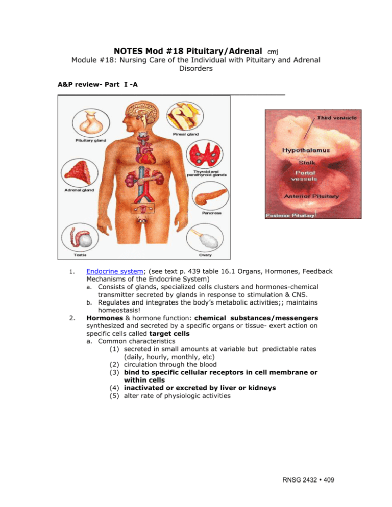
NOTES Mod #18 Pituitary/Adrenal cmj Module #18: Nursing Care of the Individual with Pituitary and Adrenal Disorders A&P review- Part I -A __________________________________________________ 1. 2. Endocrine system; (see text p. 439 table 16.1 Organs, Hormones, Feedback Mechanisms of the Endocrine System) a. Consists of glands, specialized cells clusters and hormones-chemical transmitter secreted by glands in response to stimulation & CNS. b. Regulates and integrates the body’s metabolic activities;; maintains homeostasis! Hormones & hormone function: chemical substances/messengers synthesized and secreted by a specific organs or tissue- exert action on specific cells called target cells a. Common characteristics (1) secreted in small amounts at variable but predictable rates (daily, hourly, monthly, etc) (2) circulation through the blood (3) bind to specific cellular receptors in cell membrane or within cells (4) inactivated or excreted by liver or kidneys (5) alter rate of physiologic activities RNSG 2432 409 3. 4. 5. 6. 7. Hypothalmus: integrative center for endocrine and autonomic (involuntary nervous system) a. Controls some endocrine glands by neural and hormonal pathways Negative feedback (negative feedback system) a. Regulates endocrine system by inhibiting hormone overproduction b. Can be simple or complex system Dysfunction can result from 1) Defects in gland 2) Release of trophic (gland stimulating hormones) or effector hormones 3) Hormone transport 4) In target tissue such as adrenal cortex *Examples of negative feedback: low serum calcium have increased PTH; increased serum calcium, have decreased PTH Hormone to hormone regulation: dec. thyroid hormone (T3 & T4 > release of TRH by hypothalamus and TSH by anterior pituitary; inc. T3 and T4 levels inhibit TSH release (review thyroid and parathyroid). Hypothalmus and pituitary gland a. Form a complex called the hypothalamic pituitary axis (HPA) b. Integrates communication from nervous and endocrine systems c. Examples, what hormone is released under stress? Can this hormone make you fat? (p. 459) (cortisol) (click here for more) Endocrine disorders due to a. Hypersecretion or hyposecretion of hormones b. Hyporesponsiveness of hormone receptors c. Inflammation of glands d. Gland tumors 1) Hypofunction or hyposecretion due to congenital defects, gland destruction, aging, atrophy 2) Hyperfunction due to hyperplasia, tumors 410 RNSG 2432 Part B Components endocrine system: (great link) 1. Pituitary gland (hypophysis, master gland) a. Parts: (see diagram page #1) 1) anterior pituitary (adenohypophysis): composed of cells that secrete protein hormones 2) posterior pituitary (neurohypophysis): not really an organ-extension of hypothalamus: composed mostly of axons of hypothalamic neurons-extend downward as large bundle behind anterior pituitarylso forms so-called pituitary stalk-appears to suspend anterior gland from hypothalamus. 3) intermediate (pars intermedia) *secretes MSH (melanocytes for skin pigmentation b. Components of Pituitary Gland 1) Anterior portion: adenohypophysis **know this!! Name & Source ACTH (adrenocorticotrophic hormone; corticotrophin) TSH (thyroid stimulating hormone; thyrotropin; thyrotrophic hormone) GH (growth hormone; Somatotropin, STH) *an anabolic hormone, promotes protein synthesis and mobilizes glucose and free fatty acids; stimulates the liver to produce insulin-like growth factor-1 (IGF-1) also known as somatomedin C; which stimulates growth of bones and soft tissues. FSH (follicle stimulating hormone) LH (lutenizing hormone) Prolactin (PRL); LTH (Prolactin; luteotropic hormone; luteotrotropin; lactogenic hormone: mammotropic hormone; mammotropin MSH (Melanocyte-stimulating hormone: interdin) Functions Stimulates production of hormones from adrenal cortex , especially glucocorticoids**Stimulate secretion of adrenal cortex hormones (release cortisol); Stimulates synthesis and release of thyroid hormones by thyroid: Stimulates uptake of iodine and release of T3 & T4 (* Calcitonin from thyroid; reduces serum calcium levels by decreasing bone resorption and resorption of calcium in the kidneys) Stimulates growth of tissues and bone; also protein synthesis; stimulates growth of body (epiphyseal plates of long bones in youth; promotes increased mitosis; increase in size of cells; decrease CHO utilization in striated muscle and adipose tissue; increase mobilization of stored fat; increase use of fats for energy Stimulates growth of ovarian follicles and spermatogenesis in males Regulates growth of gonads and their reproductive activities; female, ovulation and formation of corpus luteum; male, called Interstial cell-stimulating hormone (ICSH), stimulates testes to produce male sex hormones Promotes mammary gland growth and milk production Stimulates melanocytes causing pigmentation * If too much secretion of prolactin what would occur? Milk secretion!! * If too much release of LH what would occur? Enlarged reproductive organs; not enough…undeveloped reproductive organs! RNSG 2432 411 * Identify source of problem: Primary: organ itself: secondary; defect is outside of gland itself) 2) Posterior portion: neurohypophysis *Be sure to listen to Kelly’s & her Pituitary tumor and remember this… Stores and releases hormones produced by hypothalamus: Name & Source Functions 1. ADH; Vasopressin; Antidiuretic; Hormone 2. Oxytocin 3) II. Promotes H2O retention by way of the renal tubules and stimulates smooth muscle of the blood vessels and digestive tract. Decreased urine formation Stimulates the release of milk and contraction of smooth muscles in the uterus. Sucking stimulates increased secretion of oxytocin Intermediate (pars intermedia) *secretes MSH (melanocytes for skin pigmentation Thyroid gland (review only): produces Thyroid hormone (TH) composed of: Name & Source Functions 1. Triiodothyronine (T3); (more rapid and potent;action - shorter duration 2. Thyroxine (T4) 3. Calcitonin Aid in growth and development. Increase in basal metabolic rate (BRR) associated with increase in 02 consumption and heat production; shorter acting; more rapid and potent action than T4 As above; slower action Lowers serum calcium and serum phosphate by inhibiting bone resorption *Decreases excessive calcium by slowing calcium release by bone cells III. Parathyroid gland: (review only) produces PTH hormone: (increases renal excretion of phosphate, decreases excretion of CA, releases calcium from bone) Name & Source 1. 412 RNSG 2432 Parathyroid Hormone (PTH) Functions Regulates CA & PO4 metabolism as a result of its effects on three target organs: Bone,Kidney, GI IV. Adrenal Gland: 2 parts: cortex and medulla a. Parts: 1) Inner medulla: source of catecholamines- epinephrine and norepinephrine- innervated by preganglionic sympathetic fibersextension of sympathetic nervous system. 2) Outer cortex: secretes several classes steroid hormones (glucocorticoids and mineralocorticoids); a few others b. Components 1) Adrenal Medulla: Hormones: catecholamines Name & Source 1. Epinephrine (15%) 2. Norepinephrine (85%) Functions Inc. blood glucose, stimulate ACTH, glucocorticoids; inc. rate and force of cardiac contractions; constricts blood vessels in skin, mucous membranes, kidneys; dilates blood vessels in skeletal muscles, coronary and pulmonary arteries; * Acts on beta –adrenergic receptors Inc.heart rate and force of contractions; constricts blood vessels throughout body; * Acts on alphaadrenergic receptors 2) Adrenal Cortex (Salt, sugar and sex)*can’t live without! Hormones: corticoids …Know the function=nursing problems! Name & Source 1. Mineralocorticoids: aldosterone 2. Glucocorticoids: Corticol, cortisone *can’t live without it! *Stress makes you fat! Functions Retains sodium and water to inc. blood volume and blood pressure; excretes potassium** Carbohydrate metabolism-regulating glucose use in body tissue, mobilizing fat, shifting energy source for muscle cells from glucose to fat **Responds to stress Depresses inflammatory response, inhibits immune system *Affects carbohydrate, protein and fat metabolism 3. Sex hormones Androgens & Estrogens RNSG 2432 413 V. Pancreas (endocrine portion) (review only) Name & Source Functions 1. Glucagon (alpha cells) Increases blood glucose 2. Insulin (beta cells) Decreases blood glucose 3. Somstostatin (delta cells) Inhibits secretion of glucagons and insulin a. Gonads (review only) Name & Source Functions 1. Androgens (mainly testosterone) Male sex hormone 2. Estrogen and progesterone Female sex hormone (several types of estrogens) Part C-Keys to Assessment of Endocrine Function (review only) Signs and symptoms of dysfunction, often nonspecific a. Health assessment interview inc. medical history, family history, changes in size or functioning of organs, skin, hair; changes in thirst, appetite, weight, energy, sleep; use of medications that may affect hormones; changes in reproductive functioning, secondary sex characteristics b. Physical Assessment including: palpation of thyroid; inspection of skin, hair, nails, facial appearance; reflexes, musculoskeletal system; height, weight, vital signs; assessment for hypocalcemia c. Abnormal findings 1) Skin assessment a) Pigmentation: hyper or hypo with adrenocorticodysfunction b) Rough, dry skin, yellow cast with hypothyroidism c) Smooth, flushed skin with hyperthyroidism d) Purple striae (stretch marks) e) Skin lesions on extremities: diabetes mellitus 2) Hair and nails a) Pigmentation with hypoadrenocorticofunction b) Dry, thick, brittle nails and hair with hypothyroidism c) Thin, brittle nails, thin soft hair with hyperthyroidism d) Excessive hair growth with hyperadrenocorticofunction 3) Facial Assessment a) Abnormal growth, symmetry with excess growth hormone b) Exophthalmoses (protruding eyes) with hyperthyroidism 4) Thyroid assessment a) Enlargement of thyroid gland or goiter b) One or multiple palpable nodules 5) Motor function assessment a) Increased deep tendon reflexes with hyperthyroidism b) Decreased deep tendon reflexes with hypothyroidism 6) Sensory function assessment 414 RNSG 2432 Peripheral neuropathy or paresthesias with diabetes, hypothyroidism, excess growth hormone 7) Musculoskeletal assessment Size and proportion, insufficient or excess growth hormone 8) Hypocalcemic tetany (possible thyroid, parathyroid abnormalities) a) Trousseau’s sign (carpal spasm with inflation of blood pressure cuff b) Chvostek’s sign (tap front of client’s ear in angle of jaw to elicit facial muscle contraction) ___________________________________________________________________ Part IIAdrenal Cortex Dysfunction A. Etiology/Pathophysiology: see above- two glands, located on top of each kidney; composed of: Cortex (80-90% of gland) and medulla Cannot survive without function of cortex! Produce steroids, amines, epinephrine, and norepinephrine Hyposecretion or hyposecretion > disorders and complications that range from psychiatric and sexual problems to coma and death! Think “Salt-Sugar-Sex” problems! 1. Mineralocorticoids: (cortex) regulate fluid and electrolytes balance (Na and H2O retention and K excretion) (SALT) a. Aldosterone; mineralocorticoid - regulate reabsorption of sodium and excretion of potassium by kidneys and excretion of hydrogen ions. Aldosterone synthesis and secretion- stimulated by antiotensin II, hyponatremia and hyperkalemia-inhibited by atrial natriuretic hormone and hypokalemia. What is atrial naturiuetic hormone? *Think…What is the usual physiologic response when an individual is dehydrated (think aldosterone and kidney)? (ref. p. 82) 2. Glucocorticoids: cortisol, a glucocorticoid (SUGAR) a. Stimulation of gluconeogenesis (formation of glycogen from noncarbohydrate sources)-occurs in liver in response to low CHO intake or starvation <inc. glucose> b. Breakdown of protein and mobilization of free fatty acid c. Suppression of immune response d. Assistance with stress response >inc stress = inc. cortisol> e. Assistance with maintenance of blood pressure and cardiovascular response f. *Note: Cortisol secreted in diurnal pattern: major control by negative feedback i. involves secretion of corticotrophin-releasing hormone (CRH) from hypothalamus ii. CRH stimulates secretion of ACTH by anterior pituitary iii. Cortisol levels also inc. by surgical stress, burns, infection, fever, psychosis, acute anxiety, and hypoglycemia Remember: (must understand!*) *Prednisone or Solucortef = glucocorticoids! a) *Release of glucocorticoids controlled by ACTH- released by anterior pituitary! RNSG 2432 415 b) ACTH levels affected by circulating levels of cortisol: Dec. cortisol levels inc. ACTH; inc. cortisol levels dec. ACTH levels c) Never suddenly stop steroids! d) ACTH levels highest 2 hours before awakening & just after awakening; dec. rest of day! (diurnal pattern) e) *Stress inc. cortisol production and secretion f) *Stress > adrenal medulla to release the catecholamines (epinephrine and norepinephrine!) 3. Androgens: (SEX) third class of steroids-synthesized and secreted by adrenal cortex stimulate pubic and axillary hair growth and sex drive In female-androgens converted to estrogens in peripheral tissue; post-menopausal women, source of estrogen from peripheral conversion of adrenal androgen to estrogen Negligible effects of adrenal androgen in men compared to testosterone secreted by testes. ______________________________________________________________ Dysfunction- Adrenal #1 Cushing’s Syndrome (understand this one!) A. Etiology/Pathophysiology: Hypercortisolism: Hypercortisolism (Cushing’s Syndrome) (click for more!) **too much of a good thing…cortisol!!! (text does not differentiate between disease and syndrome)…basic problem is too much cortisol, corticotropin, but due to different causes: Cushing’s Disease-primary origin of problem>pituitary Cushing’s Syndrome- problem originates from other sources as adrenal, ectopic sites, etc.) “Syndrome”-group of signs and symptoms due to too much cortisol. 1. Cushing’s syndrome due to: a. Pituitary form: (as above-Cushing’s disease if primary origin- pituitary) Due to ACTH hypersecretion from pituitary adenoma Persistent, random overproduction of ACTH 416 RNSG 2432 2. 3. Inc ACTH= Inc cortisol b. Ectopic form due to ACTH-secreting tumors (corticotrophin) Small-cell lung cancers, random and episodic ACTH Production Tumor=inc ACTH=inc cortisol c. Adrenal cause: excessive porduction cortisol >negative feedback to pituitary Suppresses pituitary ACTH production Results in atrophy of uninvolved adrenal cortex (*Adrenal tumor >inc cortisol > dec ACTH > adrenal cortex atrophy) do you understand “why”? d. Iatrogenic Cushing’s syndrome:: due to long-term steroids Steroid use >inc cortisol > dec ACTH > adrenal cortex atrophy Basic problem= **EXCESSIVE amounts of cortisol More common in females between the ages of 30 and 50 B. Common Manifestation/Complications (see text p. 460 Fig. 17-3) Cushing’s Syndrome/Disease) 1. Signs and symptoms **related to adrenal cortex functions ie effect functions of adrenal cortex “sugar, sex, and salt”: a. Altered glucose metabolism, secondary sex characteristics, and mineralcorticoid levels (sodium and water retention) b. Obesity & redistribution of body fat: central obesity, fat pads under clavicles, upper back (“buffalo hump”), rounded face due to altered fat metabolism and fatty acid mobilization c. Glucose and electrolyte abnormalities: hyperglycemia; sodium retention; hypokalemia, hypertension d. Thinning of skin, bruises easily, abdominal striae (due to inc. protein catabolism with muscle wasting, loss of collagen support, etc) e. Altered immunity, delayed healing, prone to infection; dec WBC, f. Altered calcium absorption inc. osteoporosis; risk for fractures g. Inc. gastric acid secretion inc. risk for ulcers h. Emotional changes from depression to psychosis i. Changes in secondary sexual characteristics due to excess androgen secretion: excessive hair growth; acne; change in voice: receding hairline; Menstrual irregularities Before and after treatment (tumor of pituitary tumor that secreted excess ACTH) RNSG 2432 417 Before (Cushionoid) & After (right) post tumor producing cortisol removed Multiple wide purplish striae on the Moon face of patient with Cushing syndrome abdomen of a patient with Cushing's 418 RNSG 2432 C. Therapeutic Interventions/Collaborative Care/Diagnostic Tests 1. Goals of Collaborative Care: (*identify underlying cause!) a. Collaborative Care -due to long-term Steroid Therapy 1) long term steroid therapy for “another condition”-be aware of potential problems; careful follow-up 2) maintain at lowest level of steroids adequate treatment; efforts to minimize untoward effects 3) Always be tapered off steroids!!!!* Do you know why?? 2. Diagnostic Tests (see text p. 461 Table 17-3 Laboratory findings in Cushings Syndrome) *Remember what adrenal cortex function and its relationship to pituitary and ACTH RNSG 2432 419 a. *Measurement of plasma/serum cortisol, ACTH: Alterations in normal diurnal alteration: higher in mornings, lower in afternoons and evenings b. *24-hour urine collections for measurements of hormones: 1) 17-ketosteroids and 17-hydroxycorticosteroids; elevated 2) Critical-collections done properly with correct additives in specimens c. *Electrolytes, calcium, and glucose levels (elevated Na, glucose; decreased K, Ca) Why? d. ACTH suppression: synthetic cortisol (dexamethasone) (suppress ACTH production) and plasma cortisol levels measured If ACTH is not suppressed with cortisol > adrenal tumor If very high levels of cortisol needed to suppress ACTH = adrenal cortex hyperplasia *What will typical serum cortisol levels be if you draw AT 7AM AND 7PM? Inc. from 7-10 am, dec. from 7-10 p; Increased URINARY LEVELS OF STEROID METABOLITES: inc 17-OHCS (hydroxycorticoid steroid) and inc 17-KS (ketosteroid) (normal) Recall diurinal pattern. Hormonal Diagnosis: (go to this site more information) Also—not in text 1 2 3 4 Confirm presence of excessive cortisol secretion (Cushing's syndrome)…perform a low-dose dexamethasone suppression test or a 24-Hour urine collection to quantitate cortisol levels Then determine source of excess cortisol … to be determined: either from an adrenal gland tumor, an ectopic ACTH-producing tumor or a pituitary ACTH-producing adenoma…use high dose dexamethasone test, ACTH levels, metyrapone test, and/or sometimes a CRH test are used for this determination…. Petrosal Sinus Sampling: an angiographic and endocrinological test to distinguish between ectopic ACTH production or pituitary ACTH production (Cushing's disease.. If lab tests suggest pituitary adenoma as cause of Cushing's, then pituitary MRI is performed to confirm the diagnosis ….. 3. Treatment of Cushings: surgery, radiation, medications, or combination a. Surgery 1) Adrenalectomy: (see text p. 461 Nursing Care of the Patient Having Arenalectomy) removal of adrenal gland-if both glands removed, client requires *lifelong hormone replacement (at risk for Addisonian Crisis & hypovolemic shock…do you know why??) 2) Hypophysectomy (removal of pituitary gland): removal of pituitary gland through transphenoidal (through nostril) route or craniotomy 420 RNSG 2432 3) Ectopic: removal of source of ACTH secretion lung or pancreas tumors b. Post-operatively, clients being treated for adrenal or pituitary surgeryICU (ref to surgery-craniotomy) 1) life-long hormone replacement; wear medical identification bracelet 2) must not abruptly stop hormone replacement-develop Addisonian crisis (medical follow-up critical) c. Medications: if adrenal or pituitary tumors not operable 1) *Suppress adrenal cortex> dec. cortisol synthesis; use Mitotane, Metyrapone, Ketoconazole; (Somatostatis analog octeotide suppresses ACTH secretion in some cases) (*read text...know/understand effect of each drug; how do they achieve their effect?) 4. Nursing Diagnoses/Nursing Priorities (understand pathophysiology & problems) a. Fluid Volume Excess (One liter fluid retention corresponds to about 2 lb (.9 kg) body weight); HTN, edema b. Risk for Injury: potential for falls, fractures (skin thin, easy bruising, etc) c. Risk for Infection: immune suppressed, elevated blood sugar, poor wound healing, decreased protein synthesis d. Disturbed Body Image (changes revert when Cushing’s syndrome is treated) #2 Hyperaldosteronism “Conn’s Syndrome” ( Too Much Aldosterone..not in text) A. Etiology/Pathophysiology: Too much aldosterone secretion due to adrenal adenoma (70% or bilateral adrenal hyperplasia (30%) > to Na and H20 retention > inc. blood volume, HTN, headache, dec. K (hypokalemia); muscle RNSG 2432 421 weakness, cardiac dysrhythmias, metabolic alkalosis; rare peripheral edema unless cardiac problems (Do you understand why this develops?) B. Common Manifestation/Complications/Diagnosis/Treatment 1. Manifestations a. Hypokalemia >muscle weakness, cardiac weakness, usually no peripheral Edema (p. 99+) b. Elevated urine K levels (24 hour urine collection)-excessive K loss c. Inc. plasma aldosterone level with low rennin levels (Why?) d. Adrenal scan/CT scan to visualize adenomas e. EKG changes due to dec. K; ventricular dysrhythmias 2. Interventions: treat disease underlying cause: a. Surgical intervention-treat tumor: must dec. BP, use aldactone, (spironolactone…potassium sparing) to inc. Na b. Correct hypokalemia c. Adrenalectomy (partial or total depending on tumor size!) 1) Keys points-Pre-op stabilize hormonally; correct electrolyte imbalance; cortisol evening prior to surgery, AM of surgery and during surgery. 2) Post-op: ICU; BP, fluid and electrolyte mgt; IV cortisol preparation 1st 24 hours; IM cortisol 2nd post-op day then po steroids 3rd day; have inc. susceptibility to infection, poor wound healing. Unilateral adrenalectomy steroids eventually weaned. (same as above) #3 Addison’s Disease-Hypofunction of Adrenal Cortex (know this one!!) A. Etiology/Pathophysiology: dysfunction of adrenal cortex; chronic deficiency of cortisol, aldosterone, adrenal androgens; more common in women ,adults under 60 (Deficiency of salt sex, sugar!) 1. Autoimmune destruction of adrenal-accounts for 80% of spontaneous cases; occurs alone or with polyglandular autoimmune syndrome 2. Untoward effect of anticoagulant, trauma in which client has bilateral adrenal hemorrhage (iatrogenic causes) 3. Pituitary dysfunction due to tumors, surgery, radiation, exogenous steroid 4. Abrupt withdrawal from long-term, high-dose corticosteroid therapy (iatrogenic causes) (*What does iatrogenic mean? a. *Primary Addison’s disease 1) originates within adrenal glands 2) characterized by decreased mineralocorticoids, glucocorticocorticoids, and androgen secretions b. *Secondary Addisons’ disease 1) due to disorder outside adrenal gland such as pituitary tumor with corticotrophin deficiency 2) aldosterone secretion may be unaffected. B. Common Manifestation/Complications (see text p. 465 Manifestation of Addison’s Disease) Which famous President had Addison’s Disease??? 422 RNSG 2432 1. Slow onset (dec. levels of cortisol and aldosterone) a. Relates to lack of functions of adrenal cortex-decrease in “sugar, salt and sex” 1) Hyponatremia, hyperkalemia, low circulating blood volume 2) Postural hypotension (muscle weakness due to lack of cortisol), syncope, and possibly hypovolemic shock 3) Dizziness, confusion, cardiac dysrhythmias 4) Hypoglycemia, nausea, vomiting, weakness, lethargy, diarrhea 5) Hyperpigmentation (good link here) due to inc. ACTH levels (bronzed appearance in Caucasians); small black freckles (*Dec. plasma cortisol reduces feedback inhibition of pituitary ACTH and plasma ACTH rises….in primary adrenal disease) RNSG 2432 423 *Primary Addison’s…common findings: Poor coordination Dry skin and mucous membranes Sparse axillary and pubic hair in women Skin- typically deep bronze especially in creases of hands and on knuckles, elbows and knees; skin shows darkening of scars, areas of vitaligo o Abnormal coloration due to dec. secretion of cortisolglucorticoid causes pituitary gland to secrete excessive amounts of melanocyte-stimulating hormone (MSH) and corticotrophin. *Note: Secondary Addison’s adrenal hypofunction doesn’t cause hyperpigmentation as corticotrophin and MSH levels are low. 2. * Major complication-Addisonian Crisis a. *Life-threatening response to acute adrenal insufficiency > dec blood volume b. Occurs in clients with Addison’s disease who don’t respond to treatment or who has stress & without medication! c. Occurs with clients with Addisons disease who are undiagnosed & are exposed to stress! d. Patient use of steroids that are discontinued without tapering! e. *Major symptoms (Why?) high fever dehydration decreased serum sodium increased potassium decreased glucose confusion, headache pallor weakness, abdominal pain, diarrhea severe hypotension, circulatory collapse, shock, coma renal shut down, death! f. *Treatment - rapid intravenous replacement of fluids and glucocorticoids until signs/symptoms disappear (*know this!!) Check VS and urine output frequently Monitor EKG Usually - adm hydrocortisone 100 mg IV bolus Then hydrocortisone diluted with dextrose in NS given IV until condition stabilizes May require up to 300 mg/day hydrocortisone and 3/5 L of IV NS in acute stage (may require 4-6 hours! ) Also try to decrease anxiety May require vasopressors such as Dopamine or Epinephrine; avoid additional stress C. Therapeutic Interventions/Collaborative Care/Diagnostic Tests 1. Diagnostic Tests: (see Cushing’s) a. Serum cortisol and urine 17-ketosteroids and 17hydroxycorticosteroids are decreased b. Plasma ACTH is inc. if cause from adrenal dysfunction c. ACTH stimulation test d. Electrolytes - hyponatremia, hyperkalemia e. Serum glucose –dec. f. Hematocrit and hemoglobin are elevated; BUN (dehydration) 424 RNSG 2432 g. CT scan of head (R/O intracranial lesion affecting pituitary) 2. Medications/diet/Collaborative Interventions (see text p. 466 Medication Administration) *know this! a. Hydrocortisone; require life long hormone replacement: primaryoral cortisone 20-25mgs in AM and 10-12mg in PM; change dose PRN for stress also mineralocorticoid-(FLORINEF) b. Flurocortisone (Florinef), a mineralcorticoid replacement c. Diet with increased sodium: Salt food liberally ( 5-8 gm/day; 1 tsp salt = 2 gm Na; do not fast or omit meals; eat between meals and snack; eat diet high in carbohydrates and proteins; wear medic- alert bracelet; kit of 100mg hydrocortisone IM d. Avoid cold temperatures and infections (stress) e. Teaching Continue medications Signs and symptoms of insufficient hormone levels Special care required during times of increased stress (surgery, serious illness) Why is this necessary? 3. Nursing Diagnoses (See text p. 468 Nursing Care Plan; A Client with Addison’s Disease) a. Deficient Fluid Volume b. Risk for Ineffective Therapeutic Regimen Management #4 Pheochromocytoma (Tumor of Adrenal Medulla) A. Etiology/Pathophysiology: Tumors of adrenal medulla (Pheochromocytoma) 1. Definition: adrenal medulla produces catecholamines (epinephrine, norephinephrine) (rare) 2. Tumors of adrenal medulla produce excessive levels of catecholamines; typically benign, encapsulated, unilateral and solitary. 3. *Secretion of excessive catecholamines > severe hypertension; if undiagnosed and untreated pheochromocytoma > death! RNSG 2432 425 Pheochromocytoma is a tumor or the adrenal gland that Causes excess release of epinephrine and norepinephrine, hormones that regulate heart rate and blood pressure B. Common Manifestation/Complications 1. Paroxysmal severe hypertension (episodic) (systolic: 220 – 300; diastolic 150 – 175) with tachycardia 2. Can be life-threatening; stressor induced 3. Deep breathing; pounding heart; headache; moist cool hands & feet; visual disturbances C. Therapeutic Interventions/Collaborative Care/Diagnostic Tests 1. Diagnostic Tests: a. Catecholamine levels (serum and urine) are elevated b. 24 hour urine-VMA (metabolite of Epinepherine)…can have false negatives! c. Plasma catecholamines d. CT and MRI to locate tumor e. Adrenal biopsy (definitive) 2. Treatment: Adrenalectomy to remove tumor (focus =management of dangerously high BP); post adrenalectomy= adrenal crisis and long term steroids!! a. Pre-op: Sympathetic blocking agents= Minipress (prazosin), Hytrin (terazosin), Cardura (doxazosin) to reduce BP and other symptoms of of catecholamine excess Since change in BP sudden, client may experience orthostatic hypotension Use Beta blocking agents such as Inderal to dec. heart rate, BP and force of contraction and calcium channel blocking agents also used. b. General management Diet: high in vitamin, mineral, calorie, no caffeine Sedatives; Monitor BP 426 RNSG 2432 Eliminate attacks; If attack- complete bedrest and HOB 45 degrees c. Surgery via laparoscopic adrenalectomy or open abdominal incision; complete removal of the tumor cures hypertension in 1030% of the cases May require REGITINE AND NIPRIDE TO PREVENT HYPERTENSIVE CRISIS in surgery (How do these drugs work?) BP may be elevated initially, BUT CAN BOTTOM OUT May require volume expanders, vasopressors Hourly I and O Observe for hemorrhage d. *See cautions re adrenalectomy (typically only tumor is removed); if entire adrenal gland removed; Addisons crisis risk and long term steroids. e. If not a candidate for surgery: 1) Use Demser (drug which inhibits catecholamine synthesis) 2) Avoid opiates, histamines, reglan, anti-depressants (stimulate SNS) ___________________________________________________________________ #5 Pituitary Gland (refer to introduction) Anterior Pituitary Gland (Hyperfunction) A. Etiology/Pathophysiology: Hyperfunction of anterior pituitary gland 1. Pathophysiology: Most often- benign adenoma producing excess hormones; growth hormone (GH), Prolactin (PRL), or ACTH; 10% OF ALL BRAIN TUMORS 2. Specific Conditions a. Gigantism: Growth hormone hypersecretion occurs prior to puberty b. resulting in person becoming excessively tall (over 7 feet tall) c. Acromegaly: Growth hormone hypersecretion (somatotropin) occurs after puberty d. Causes bone and connective tissue continuing to grow > enlargement of face, hands, and feet e. Overproduction of prolactin secretion > dec. reproductive and sexual function f. Cushing’s Disease (inc ACTH due to pituitary adenoma) 3. **Recall anterior pituitary hormones (refer to chart with hormones produced) B. Common Manifestation/Complications 1. *Manifestations depend upon which hormone(s) is/are produced in excess: 2. What would happen if you had too much growth hormone secretion??? Which goolish character on the Addam’s Family may have had too much GH secretion? RNSG 2432 427 a. Giantism in children: skeletal growth; may grow to 8 ft. tall and > 300 lbs b. Acromegaly in adults: enlarged feet/hands, thickening of bones, prognathism, diabetes, HTN, wt. gain, H/A, visual disturbances, diabetes mellitus Clinical features develop slowly …aged 40-60 years…Symptoms…arthralgia, increased sweating and physical weakness…apparent increase in the size of hands and feet, coarsening of the facial features mainly of the brow, widening of gaps between teeth, thickening of skin, headache… carpal tunnel syndrome and other peripheral neuropathies, visual problems, colon polyps and sleep apnoea…. women, ovulatory disorders, amenorrhoea and galactorrhoea…men, decreased libido and hypogonadism….hypertension, heart disease and diabetes…Signs enlarged tongue, stomach, heart, liver and spleen, hypertension, glucose intolerance or type 2 diabetes mellitus. If before puberty - gigantism with abnormal height. After puberty - normal Hands of individual with acromegaly; normal hands. 428 RNSG 2432 Acromegaly: Facial changes secondary to elevated growth hormone levels. Note in particular prominent supra-orbital ridge, jaw, and generally enlarged facial features. C. Therapeutic Interventions/Collaborative Care/Diagnostic Tests 1. Diagnostic Tests: a. Key; history and physical exam b. Evaluation of GH levels; and GH response to oral glucose challenge c. MRI to identify pituitary hormone; CT scan with contrast d. Opthalmologic exam and visual fields due to pressure on optic chiasm or optic nerves. 2. Treatment: a. Medications: Parlodel (bromocriptine)= reduce prolactin & GH levels. b. Radiation therapy: external radiation reduce GH levels 80% of time (usually given with medications); usually develop hypopituitarism with radiation, need replacement therapy c. Surgical removal (hypophysectomy) is treatment of choice; cure if tumor is smaller than 10 mm; usually accomplished with **transsphenoidal approach; goal to remove only tumor that is causing the GH secretion; procedure produces an immediate reduction in IGF-1 levels within a few weeks. (see previous notes) RNSG 2432 429 *Incision made thru floor of nose into sella turcica *In some cases entire pituitary gland removed surgery (hyposectomy) > permanent absence of pituitary hormones; rather than replacing the pituitary (tropic) hormones, which requires parenteral administration, essential hormones produced by target organs (glucocorticoids, thryroid hormone and sex hormones) given orally- must be continued throughout life!! 1) Pre-op hypophysectomy: a) Anxiety r/t body changes, fear of unknown, brain involvement, chronic condition; requiring life-long care b) Sensory-perceptual alteration r/t visual field cuts, diplopia and secondary to pressure on optic nerve. c) Alteration in comfort (headache) r/t tumor growth/edema 2) Post-op (was entire pituitary removed or only tumor) a) Knowledge deficit: post-op teaching including pain control, ambulation, hormone replacement. Activity b) Require use of hormone patch; activity restricted, NO straining/ bending for 2 months, use stool softners avoid coughing, saline mouth rinses (no tooth brushing as risk of meninitis) as can have CSF leak where sella turcica was entered *test any clear nasal drainage for glucose to see if it is glucose; notify physician; elevate HOB, bedrest as CSF usually resolves within 72 hours; spinal taps to relieve pressure!! c) Periocular edema/ecchymosis d) **Monitor and treat for post-op complications as diabetes insipitus: lead to hypovolemic shock; very thirsty, urinate a lot!! **Due to ADH insufficiency!! If develops-must be replaced through hormone replacement (DDAVP (Desmopressin, synthetic ADH, give by spray or pitressin IM)!** e) Dec. ACTH > require cortisone replacement due to decrease glucocorticoid production. Can you live without glucocorticoids???? f) Dec. in sex hormones >infertility due to decrease production of ova & sperm #6 Anterior Pituitary (Hypofunction) A. Etiology/Pathophysiology: Etiology (rare disorder) may be due to disease, tumor, or destruction of the gland. B. Common Manifestation/Complications: Have signs and symptoms of dec. hormones: GH, FSH/LH, Prolactin; ACTH; TSH C. Therapeutic Interventions/Collaborative Care/Diagnostic Tests 1. Diagnostic Tests: CT Scan; Serum hormone levels 2. Treatment: a) neurosurgery: removal of tumor b) radiation: tumor size c) hormone replacement: cortisol, thyroid, sex hormones 3. Assessment of S & S of hypo or hyper: functioning hormone levels 4. Teaching-Compliance with hormone replacement therapy: Counseling and referrals and support medical interventions 430 RNSG 2432 #7 Posterior Pituitary Gland (SIADH) (**Important) A. Etiology/Pathophysiology: Excessive or deficiency in antidiuretic hormone (ADH) *What hormones are released by the posterior pituitary? ADH (vasopressin) which is secreted by cells in the hypothalamus and stored in the posterior pituitary and acts on distal and collecting tubules of nephrons making them more permeable to H20 thus decreasing water excreted! Oxytocin controls lactation and stimulates uterine contraction 1. ADH-secreted in response to changes in serum osmolality (hypothalamus) 2. Specific Conditions: Syndrome of Inappropriate ADH Secretion (SIADH) Too much ADH! What is SIADH? (p. 95) a. **Occurs when ADH released despite normal or low normal plasma osmolarity: results from abnormal production or sustained secretion of ADH; characterized by fluid retention, serum hypo-osmolality, dilutitional hyonatremia, hypochoremia, concentrated urine in presence of normal or inc. intravascular volume and normal renal function b. Under what conditions is ADH released; does it have vasocontrictive or vasodilative action? ** released in response to decrease blood volume, increase concentration of Na+ or other substances, pain, stress; ADH has vasocontrictive properties c. Results in hyponatremia and water intoxication d. Due to: (too much ADH) 1) Malignant tumors (e.g. oat cell or small cell lung cancer) which secret 2) ADH 3) Post head injury, side effect of some medications including diuretics 4) and anesthetics such as morphine 5) Ca duodenum/pancreas, trauma, pulmonary disease, CNS 6) disorders, drugs -- Vincristine, nicotine, general anesthetics, tricyclic antidepressants B. Common Manifestation/Complications (SIADH) 1. Signs and Symptoms: neurologic symptoms including dec. level of consciousness, confusion, muscle twitches, seizures 2. Signs/symptoms hypotnatremia: lethargy, decrease tendon reflexes, seizures RNSG 2432 431 C. Therapeutic Interventions/Collaborative Care/Diagnostic Tests **1. Diagnostic tests: decreased Serum Na+ <135meq/l decreased Serum osmolality <275 OSM/kg H2O increased urine specific gravity decreased or normal BUN 2.***Treatment: correction of Na deficit, restriction of fluids, treat underlying cause a. ***FLUID RESTRICTION: LIMIT TO 1000ML/24HRS b. IV 3% NaCl to replace Na c. IF CHF -- Lasix (temporary fix) d. Treat underlying problem --Chemo, radiation e. **Declomycin 600 po-1200mg/day to inhibit ADH f. Fluid restriction may be as little as 500-600ml/24hrs g. Daily weights...1 lb. weight = 500ml fluid retention h. Accurate I & Os i. F & E imbalances; monitor fluid intake j. High risk for injury r/t complications of fluid overload (seizures #8 Diabetes Insipidus (Posterior Pituitary Gland) (see Kelly’s video) A. Etiology/Pathophysiology: Diabetes Insipidus ((too little ADH) 1. ADH insufficiency from neurogenic or nephrogenic origin 2. Pathophysiology: Brain tumors, closed head trauma, other brain conditions, renal failure; 50% idiopathic a. central (neurogenic -- i.e. brain tumors; sudden onset! b. nephrogenic - inability of tubules to respond to ADH c. psych (dispogenic DI) less common; can be a structural lesion or a psychological disorder leading to water intoxication, is it true DI? B. Common Manifestation/Complications 1. Signs and Symptoms: excretes large amounts of dilute urine; client at risk for dehydration and hypernatremia 2. **Polydipsia; Polyuria (10L in 24 hours); have low urine specific gravity less than 1.005 and urine osmolality of < 100mOsm/kg. *Serum osmolality is elevated as a result of hypernatremia due to pure water loss in the kidney 3. Severe fluid volume deficit a. wt loss b. tachycardia c. constipation d. shock C. Therapeutic Interventions/Collaborative Care/Diagnostic Tests 1. Must differentiate among different causes of DI; requires complete history and physical a. Dehydration test: 2 units of Vasopressin (ADH) mixed in saline administered over 2 hrs then check urine osmolality levels 432 RNSG 2432 b. Water deprivation-confirm diagnosis of central DI; get baseline weights, pulse, urine and plasma osmolalities, specific gravity, urine and BP; withhold all fluids for 8 to 16 hours; *potential risk due to fluid volume deficit; during test, patient assessed hourly for BP, weight, urine osmolality; test continues until urine osmolalities stabilizes or body weight declines by 5% or orthostatic hypotension develops. ADH then given and urine osmolality is measured 1 hour later; in central DI the rise in urinary osmolality after vasopressin exceeds 9%.observed c. What is the expected urine specific gravity; serum Na and serum osmolality without treatment? 2. *Treatment: administer intravenous hypotonic fluids, oral fluids and replace DH hormone (Desmopressin acetate) a. Identification of etiology, H & P b. Tx of underlying problem c. **DDAVP(desomopressin acetate) (nasal spray); Pitressin s.c. IM, nasal spray d. Assess for F & E imbalances e. High risk for sleep disturbances f. Increase po/IV fluids 3. Nursing diagnosis: a. RF Injury (hypovolemic shock) b. Knowledge deficit c. High risk for ineffective coping RNSG 2432 433

