Supplementary Information (doc 118K)
advertisement
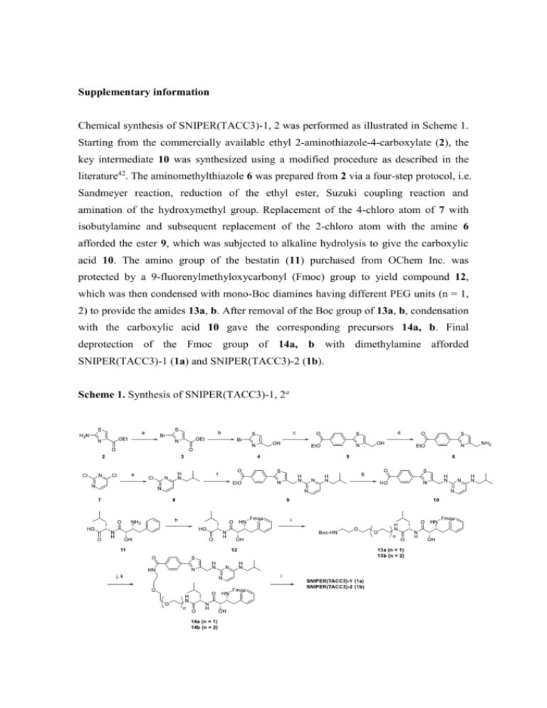
Supplementary information Chemical synthesis of SNIPER(TACC3)-1, 2 was performed as illustrated in Scheme 1. Starting from the commercially available ethyl 2-aminothiazole-4-carboxylate (2), the key intermediate 10 was synthesized using a modified procedure as described in the literature42. The aminomethylthiazole 6 was prepared from 2 via a four-step protocol, i.e. Sandmeyer reaction, reduction of the ethyl ester, Suzuki coupling reaction and amination of the hydroxymethyl group. Replacement of the 4-chloro atom of 7 with isobutylamine and subsequent replacement of the 2-chloro atom with the amine 6 afforded the ester 9, which was subjected to alkaline hydrolysis to give the carboxylic acid 10. The amino group of the bestatin (11) purchased from OChem Inc. was protected by a 9-fluorenylmethyloxycarbonyl (Fmoc) group to yield compound 12, which was then condensed with mono-Boc diamines having different PEG units (n = 1, 2) to provide the amides 13a, b. After removal of the Boc group of 13a, b, condensation with the carboxylic acid 10 gave the corresponding precursors 14a, b. Final deprotection of the Fmoc group of 14a, b with dimethylamine afforded SNIPER(TACC3)-1 (1a) and SNIPER(TACC3)-2 (1b). Scheme 1. Synthesis of SNIPER(TACC3)-1, 2a aReagents and conditions: (a) CuSO4, NaBr, NaNO2, H2SO4, water, 0 °C, 50%; (b) LiBH4, THF, MeOH, 0 °C, 84%; (c) (4-(ethoxycarbonyl)phenyl)boronic acid, Pd(PPh3)4, DME–MeOH, 80 °C, 59%; (d) diphenylphosphoryl azide (DPPA), 1,8-diazabicyclo[5.4.0]undec-7-ene (DBU), THF, rt then PPh3, water, rt, 79%; (e) isobutylamine, EtOH, 80 °C, 77%; (f) compound 6, N,N-diisopropylethylamine (DIEA), n-BuOH, reflux, 67%; (g) 1 M NaOH, MeOH, rt, 99%; (h) 9-fluorenylmethyloxycarbonyl chloride (Fmoc-Cl), K2CO3, THF–water, rt, 81%; (i) tert-butyl (2-(2-(2-aminoethoxy)ethoxy)ethyl)carbamate [for 14a], tert-butyl (2-(2-(2-(2-aminoethoxy)ethoxy)ethoxy)ethyl)carbamate [for 14b], 1-ethyl-3-(3-dimethylaminopropyl)carbodiimide (EDCI), 1-hydroxybenzotriazole hydrate (HOBt·H2O), THF, rt, 86–87%; (j) 4 M HCl in cyclopentyl methyl ether (CPME), THF, rt; (k) compound 10, 1-[bis(dimethylamino)methylene]-1H-1,2,3-triazolo[4,5-b]pyridinium 3-oxid hexafluorophosphate (HATU), DIEA, 0 °C, 87–99%; (l) 2M dimethylamine in MeOH, rt, 84–89%. Physicochemical Data on SNIPER(TACC3)-1, 2 The proton nuclear magnetic resonance (1H NMR) spectra were determined on a Bruker AVANCE II (300 MHz) spectrometer. Chemical shifts for 1H NMR were reported in parts per million (ppm) downfield from tetramethylsilane (δ) as the internal standard in deuterated solvent and the coupling constants (J) are in Hertz (Hz). The following abbreviations are used for spin multiplicity: s = singlet, d = doublet, dd = doublet of doublet, m = multiplet, and br s = broad singlet. Mass spectra (MS) were acquired using a Shimadzu UFLC/MS (Prominence UFLC high pressure gradient system/LCMS-2020) operating in electron spray ionization mode (ESI+). The column used was an L-column 2 ODS (3.0 x 50 mm I.D., 3 μm, CERI, Japan) with a temperature of 40 °C and a flow rate of 1.5 mL/min. Mobile phase A was 0.05% trifluoroacetic acid (TFA) in ultrapure water. Mobile phase B was 0.05% TFA in acetonitrile (MeCN) which was increased linearly from 5% to 90% over 2 minutes and 90% over the next 1.5 minutes, after which the column was equilibrated to 5% for 0.5 minutes. Analytical HPLC for purity determination was performed using a Shimadzu LC-20AD, equipped with an L-column2 ODS (3.0 x 30 mm I.D., 2 μm-particle size, CERI, Japan), elutied with 0.1% TFA in ultrapure water (Mobile phase A), and 0.1% TFA in MeCN (Mobile phase B), using the following elution gradient of 5% B to 90% B over 2.0 min followed by 90% B isocratic over 1.3 min at a flow rate of 1.2 mL/min (detection at 220 nm). N-((11S,14S,15R)-15-Amino-14-hydroxy-11-isobutyl-10,13-dioxo-16-phenyl-3,6-dio xa-9,12-diazahexadecyl)-4-(4-(((4-(isobutylamino)pyrimidin-2-yl)amino)methyl)thi azol-2-yl) benzamide (SNIPER(TACC3)-1, 1a). 1H NMR (CD3OD) δ 0.76 – 0.99 (12H, m), 1.52 – 1.86 (4H, m), 2.64 (1H, dd, J = 13.3, 7.8 Hz), 2.88 (1H, dd, J = 13.3, 6.9 Hz), 3.08 (2H, d, J = 6.6 Hz), 3.25 – 3.38 (3H, m), 3.47 – 3.71 (10H, m), 3.92 (1H, d, J = 3.0 Hz), 4.37 – 4.47 (1H, m), 4.69 (2H, d, J = 0.8 Hz), 5.80 (1H, d, J = 6.0 Hz), 7.13 – 7.33 (6H, m), 7.60 (1H, br s), 7.88 – 8.08 (4H, m). MS m/z 804.7 (M+H)+. Purity 99.3% (HPLC). N-((14S,17S,18R)-18-Amino-17-hydroxy-14-isobutyl-13,16-dioxo-19-phenyl-3,6,9-t rioxa-12,15-diazanonadecyl)-4-(4-(((4-(isobutylamino)pyrimidin-2-yl)amino)methy l)thiazol-2-yl)benzamide (SNIPER(TACC3)-2, 1b). 1H NMR (CD3OD) δ 0.79 – 0.98 (12H, m), 1.53 – 1.86 (4H, m), 2.65 (1H, dd, J = 13.4, 7.8 Hz), 2.89 (1H, dd, J = 13.4, 6.9 Hz), 3.08 (2H, d, J = 6.3 Hz), 3.26 – 3.37 (3H, m), 3.44 – 3.72 (14H, m), 3.93 (1H, d, J = 3.0 Hz), 4.35 – 4.48 (1H, m), 4.69 (2H, d, J = 0.8 Hz), 5.80 (1H, d, J = 6.0 Hz), 7.11 – 7.34 (6H, m), 7.60 (1H, br s), 7.88 – 7.97 (2H, m), 7.99 – 8.08 (2H, m). MS m/z 848.8 (M+H)+. Purity 98.8% (HPLC). Supplementary Figure Legends Fig. S1. Schematic model of the TACC3-APC/CCDH1 interaction. (A) Inherent interaction that is not affected by KHS108 or Me-BS. (B) SNIPER(TACC3)-mediated interaction that is impaired by KHS108 or Me-BS. Fig. S2. SNIPER(TACC3) did not affect cell cycle distribution. HT1080 cells were treated with 30 M of SNIPER(TACC3)-1 for 4 hr and analyzed in a flow-cytometer. Cell-cycle distribution was quantified by MultiCycle software. Fig. S3. Selective induction of cancer cell death by SNIPER(TACC3) determined by annexin V and PI staining. Cells were treated with or without 10 M of SNIPER(TACC3)-1 for 48 hr, stained with annexin V and PI, and analyzed in a flow-cytometer. Double negative cells (lower left) represent living cells, whereas annexin V positive (lower right), PI positive (upper left) or double positive (upper right) cells represent dead cells. Fig. S4. Selective induction of cancer cell death by SNIPER(TACC3) determined by flow-cytometric analysis. Cancer cells (A) and normal cells (B) were treated with or without 10 M of SNIPER(TACC3)-1 for 48 hr. Cells were stained with propidium iodide and DNA contents were analyzed in a flow-cytometer. Fig. S5. Depletion of TACC3 protein by siRNA did not induce cell death. (A) Depletion of TACC3 protein by siRNA was analyzed by Western blot. (B) Cell viability was measured by WST-8 cell proliferation assay. (C, D) Cancer cells (C) and normal cells (D) were stained with PI and analyzed in a flow-cytometer. Cells were transfected with siRNA against TACC3 for 72 hr. Fig. S6. Suppression of SNIPER(TACC3)-induced cell death by downregulation of APC/CCDH1. HT1080 cells were transfected with the indicated siRNA for 24 hr and treated with 10 M of SNIPER(TACC3)-1 for 30 hr. The cells were stained with annexin V and PI, and analyzed in a flow-cytometer.


