Epoxy sections of osmicated renal tissue were subjected to
advertisement
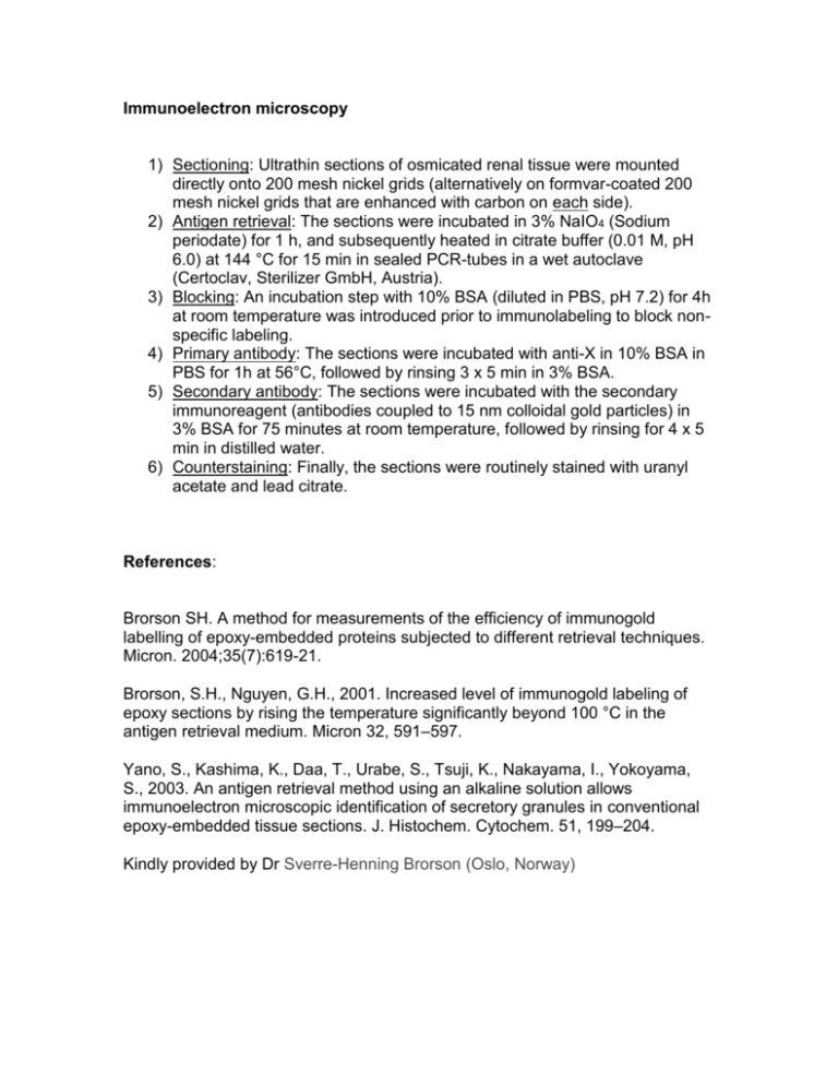
Immunoelectron microscopy 1) Sectioning: Ultrathin sections of osmicated renal tissue were mounted directly onto 200 mesh nickel grids (alternatively on formvar-coated 200 mesh nickel grids that are enhanced with carbon on each side). 2) Antigen retrieval: The sections were incubated in 3% NaIO4 (Sodium periodate) for 1 h, and subsequently heated in citrate buffer (0.01 M, pH 6.0) at 144 °C for 15 min in sealed PCR-tubes in a wet autoclave (Certoclav, Sterilizer GmbH, Austria). 3) Blocking: An incubation step with 10% BSA (diluted in PBS, pH 7.2) for 4h at room temperature was introduced prior to immunolabeling to block nonspecific labeling. 4) Primary antibody: The sections were incubated with anti-X in 10% BSA in PBS for 1h at 56°C, followed by rinsing 3 x 5 min in 3% BSA. 5) Secondary antibody: The sections were incubated with the secondary immunoreagent (antibodies coupled to 15 nm colloidal gold particles) in 3% BSA for 75 minutes at room temperature, followed by rinsing for 4 x 5 min in distilled water. 6) Counterstaining: Finally, the sections were routinely stained with uranyl acetate and lead citrate. References: Brorson SH. A method for measurements of the efficiency of immunogold labelling of epoxy-embedded proteins subjected to different retrieval techniques. Micron. 2004;35(7):619-21. Brorson, S.H., Nguyen, G.H., 2001. Increased level of immunogold labeling of epoxy sections by rising the temperature significantly beyond 100 °C in the antigen retrieval medium. Micron 32, 591–597. Yano, S., Kashima, K., Daa, T., Urabe, S., Tsuji, K., Nakayama, I., Yokoyama, S., 2003. An antigen retrieval method using an alkaline solution allows immunoelectron microscopic identification of secretory granules in conventional epoxy-embedded tissue sections. J. Histochem. Cytochem. 51, 199–204. Kindly provided by Dr Sverre-Henning Brorson (Oslo, Norway)
