Protocol
advertisement
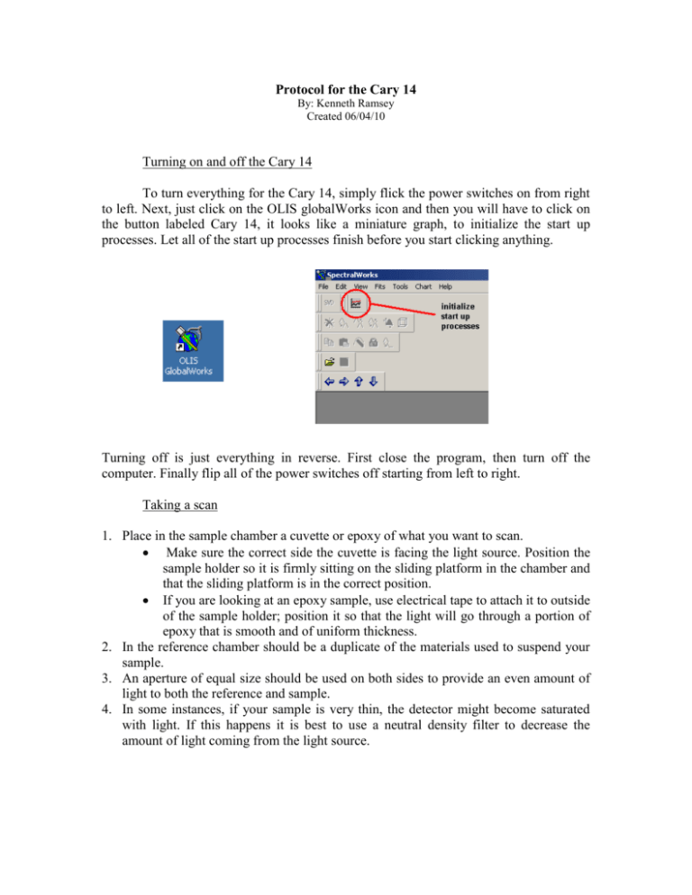
Protocol for the Cary 14 By: Kenneth Ramsey Created 06/04/10 Turning on and off the Cary 14 To turn everything for the Cary 14, simply flick the power switches on from right to left. Next, just click on the OLIS globalWorks icon and then you will have to click on the button labeled Cary 14, it looks like a miniature graph, to initialize the start up processes. Let all of the start up processes finish before you start clicking anything. Turning off is just everything in reverse. First close the program, then turn off the computer. Finally flip all of the power switches off starting from left to right. Taking a scan 1. Place in the sample chamber a cuvette or epoxy of what you want to scan. Make sure the correct side the cuvette is facing the light source. Position the sample holder so it is firmly sitting on the sliding platform in the chamber and that the sliding platform is in the correct position. If you are looking at an epoxy sample, use electrical tape to attach it to outside of the sample holder; position it so that the light will go through a portion of epoxy that is smooth and of uniform thickness. 2. In the reference chamber should be a duplicate of the materials used to suspend your sample. 3. An aperture of equal size should be used on both sides to provide an even amount of light to both the reference and sample. 4. In some instances, if your sample is very thin, the detector might become saturated with light. If this happens it is best to use a neutral density filter to decrease the amount of light coming from the light source. Computer settings Here is most of the information you will need to know about the computer settings. 1. To take either the absorption or transmittance, first go into the operations mode tab and set the data reduction mode to what you need to scan. 2. When inputting the bounds of your scans, it is best to scan the absorption spectra starting from the longest wavelength and ending at the shortest wavelength. You can set when the Cary 14 will change from the IR detector to the visible detector. The visible detector is good on a range of 300 - 900 nm. The IR detector is good on a range of 800 - 2500 nm. You want to change detectors at a wavelength where this is no activity. 3. The resolution of your spectra is defined be the number of increments the Cary 14 takes. For example, to obtain a resolution of .1 nm, all you have to do is divide the total range of wavelengths you will be looking at by the resolution you want, and this will give you the number of steps you should put in. the max resolution of the Cary 14 is .05 nm. It is best to put the number of increments in first and then the range of wavelengths you want to scan, otherwise the computer might change the range you’re scanning on its own without you noticing. 4. Reads per datum is just the number of times that the detector is read at a specific wavelength, and then that information is averaged. In the reads per datum input, make sure it is set to 4 or 5. Saving Data 1. To save your data, first select the data so that it is showing on the screen. 2. Next left click on the graph so that the data changes from red to yellow. 3. Now you can right click and select the save as ASCII option. To calibrate the Cary 14, refer to the manual.

