Monocyte Separation Protocol - the CFAR Virology Core Laboratory
advertisement
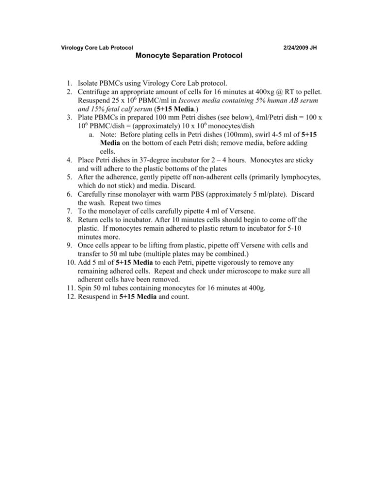
Virology Core Lab Protocol 2/24/2009 JH Monocyte Separation Protocol 1. Isolate PBMCs using Virology Core Lab protocol. 2. Centrifuge an appropriate amount of cells for 16 minutes at 400xg @ RT to pellet. Resuspend 25 x 106 PBMC/ml in Iscoves media containing 5% human AB serum and 15% fetal calf serum (5+15 Media.) 3. Plate PBMCs in prepared 100 mm Petri dishes (see below), 4ml/Petri dish = 100 x 106 PBMC/dish = (approximately) 10 x 106 monocytes/dish a. Note: Before plating cells in Petri dishes (100mm), swirl 4-5 ml of 5+15 Media on the bottom of each Petri dish; remove media, before adding cells. 4. Place Petri dishes in 37-degree incubator for 2 – 4 hours. Monocytes are sticky and will adhere to the plastic bottoms of the plates 5. After the adherence, gently pipette off non-adherent cells (primarily lymphocytes, which do not stick) and media. Discard. 6. Carefully rinse monolayer with warm PBS (approximately 5 ml/plate). Discard the wash. Repeat two times 7. To the monolayer of cells carefully pipette 4 ml of Versene. 8. Return cells to incubator. After 10 minutes cells should begin to come off the plastic. If monocytes remain adhered to plastic return to incubator for 5-10 minutes more. 9. Once cells appear to be lifting from plastic, pipette off Versene with cells and transfer to 50 ml tube (multiple plates may be combined.) 10. Add 5 ml of 5+15 Media to each Petri, pipette vigorously to remove any remaining adhered cells. Repeat and check under microscope to make sure all adherent cells have been removed. 11. Spin 50 ml tubes containing monocytes for 16 minutes at 400g. 12. Resuspend in 5+15 Media and count.
