Table 1 - BioMed Central
advertisement
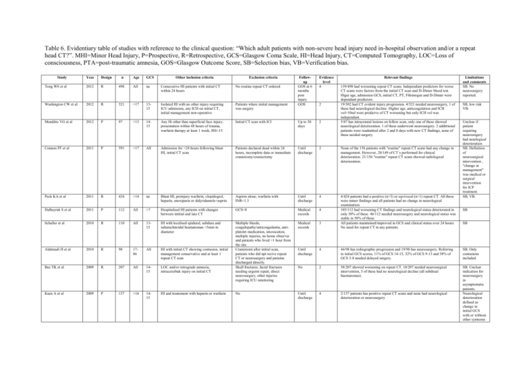
Table 6. Evidentiary table of studies with reference to the clinical question: “Which adult patients with non-severe head injury need in-hospital observation and/or a repeat head CT?”. MHI=Minor Head Injury, P=Prospective, R=Retrospective, GCS=Glasgow Coma Scale, HI=Head Injury, CT=Computed Tomography, LOC=Loss of consciousness, PTA=post-traumatic amnesia, GOS=Glasgow Outcome Score, SB=Selection bias, VB=Verification bias. Study Year Design n Age GCS Tong WS et al 2012 R 498 All na Consecutive HI patients with initial CT within 24 hours Other inclusion criteria No routine repeat CT ordered Exclusion criteria Followup GOS at 6 months post injury GOS Evidence level 4 Washington CW et al 2012 R 321 >17 1315 Isolated HI with no other injury requiring ICU admission, any ICH on initial CT, initial management non-operative Patients where iniital management was surgery Menditto VG et al 2012 P 97 >13 1415 Any Hi other than superficial face injury, presentation within 48 hours of trauma, warfarin therapy at least 1 week, ISS<15 Initial CT scan with ICI Up to 30 days 2 Connon FF et al 2011 P 591 >17 All Admission for >24 hours following blunt HI, initial CT scan Patients declared dead within 24 hours, incomplete data or immediate craniotomy/craniectomy Until discharge 2 None of the 156 patients with "routine" repeat CT scans had any change in management. However, 28/149 of CT´s performed for clinical deterioration. 21/156 "routine" repeat CT scans showed radiological deterioration. Peck KA et al 2011 R 424 >14 na Blunt HI, preinjury warfarin, clopidogrel, heparin, enoxiparin or didyridamole+asprin Aspirin alone, warfarin with INR<1.3 Until discharge 4 All >7 Hospitalised HI patients with changes between intitial and late CT GCS<8 Medical records 4 110 All 1315 HI with localised epidural, subdura and subarachnoidal heamatomas <5mm in diameter Medical records 3 R 98 1786 All HI with initial CT showing contusion, initial management conservative and at least 1 repeat CT scan Until discharge 4 44/98 has rediographic progression and 19/98 has neurosurgery. Referring to initial GCS scores, 11% of GCS 14-15, 32% of GCS 9-13 and 58% of GCS 3-8 needed delayed surgery. SB. Only contusions included. 2009 R 207 All 1415 LOC and/or retrograde amnesia, intracererbak injury on initial CT Multiple bleeds, coagulopathy/anticoagulantia, antiplatelet medication, intoxicaiton, multiple injuries, no home observer and patients who lived >1 hour from the site Craniotomi after initial scan, patients who did npt recive repeat CT or neurosurgery and patietns discharged directly. Skull fractures, facial fractures needing urgernt repair, direct neurosurgery, other injuries requiring ICU minitoring 4/424 patients had a positive (n=3) or eqvivocal (n=1) repeat CT. All these were minor findings and all patients had no change in neurological examination. 103/112 had worsening CT findings and neurological status deteriorated in only 30% of these. 46/112 needed neurosurgery and neurological status was stable in 50% of these. All patients maintained/improved in GCS and clinical status over 24 hours. No need for repeat CT in any patients. Dalbayrak S et al 2011 P 112 Schaller et al 2010 R Alahmadi H et al 2010 Bee TK et al No 2 58/207 showed worsening on repeat CT. 18/207 needed neurosurgical intervention, 5 of these had no neurological decline (all subdrual haematomas). 2009 P 137 >16 1415 HI and treatement with heparin or warfarin No Until discharge 4 2/137 patients has positive repeat CT scans and none had neurological deterioration or neurosurgery SB. Unclear indication for neurosurgery in asymptomatic patients. Neurological deterioration defined as change in initial GCS with or without other symtoms Kaen A et al 2 Relevant findings 139/498 had worsening repeat CT scans. Independant predictors for worse CT scans were factors from the initial CT scan and D-Dimer blood test. Higer age, admission GCS, initial CT, PT, Fibrinogen and D-Dimer were dependant predictors. 19/302 had CT evident injury progression. 4/321 needed neurosurgery, 1 of these had neurological decline. Higher age, anticoagulation and ICH vol>10ml were predictve of CT worsening but only ICH vol was independant. 5/87 has intracranial lesions on follow scan, only one of these showed neurological deterioration. 1 of these underwent neurosurgery. 2 addittional patients were readmitted after 2 and 8 days with new CT findings, none of these needed surgery. Limitations and comments SB. No neurosurgery reported. SB, low risk VB. Unclear if patient requiring neurosrugery had neurlogical deterioration SB. Definition of neurosurgical intervention , "change in management" was medical or surgical intervention for ICP treatment. SB, VB. SB SB Tauber et al 2009 P 100 >64 15 Regular los-dose aspirin therapy, initial negative CT, no hypertenisve irregularities Anticoagulants, moderate-severe HI. Patients with pathology on initial CT Until discharge 4 4/100 has positive repeat CT scans, all without neurological deterioration. 1 patient died (age 84) after neurological deterioration and 1 patient need neurosurgery but first after neurological deterioration. Turedi S et al 2008 P 240 All 1315 Blunt HI, LOC < 15 min or post-traumatic amnesia <1 hr No No 2 Brown CV et al 2007 P 274 All All Blunt HI and ICH on initial CT Immediate neurosurgery and death within 24 hours Until discharge 2 Sifri ZC et al 2006 P 130 >17 1315 HI and intracranial bleed or contusion on initial CT Prior brain surgery or cerebral pathology, chronic neurological condition, spinal cord injury, coagulopathy, anticoagulation, immediate or planned neurosurgery after the initial CT and patients who never had a follow-up CT GOS in discharge 4 Repeat CT scans in 120 patients with high risk criteria (GCS 14-15 and LOC, amnesia, vomiting, suspected skull fracture, multiple trauma, severe/increasing headache, aymmetric pupils, focal neurology, posttraumatic seizures or anticoagulant/coagulopathy) showed abnormalities in 3 and none of these needed neurosurgery. 163/274 underwent repeat CT scans. 17/45 of repeat CT scans for neurological change led to a medical or surgical intervention vs 2/196 routine scans led to an intervention, The 2 cases of intervention after routine scans were in patients with severe head injury (GCS <9). 99/130 patients had normal neurological findings at repeat CT and none had neurosurgery or deterioration. 31/130 had abnomral neurological findings and 2 needed immediate neurosurgery. In patients with normal neurological exam, no change or improvement in 87% of repeat CT scans but no change in management. Fior the 12 CT´s that were worse, these patients all had favourable outcome. Itshayek E et al 2006 R 4 6586 15 No 2006 R 179 All 1315 GOS up to 26 months Medical records 4 Velmahos GC et al HI patients with anticoagulation with normal initial CT and delayed acute subdural haematoma LOC, short-term amnesia, headache, emesis or dizziness Sifri ZC et al 2004 R 202 >15 1415 HI with LOC/amnesia and positive initial CT scan No 4 Fainardi E et al 2004 R 141 All All Hi with traumatic subarchnoid haemorrahge on initial CT History of brain injury or coagulopathy. Patients who required immediate neurosurgery after initial CT Brain death on admission, hypotension due to extracranial injuries and penetrating injuries not due to traffic accidents GOS at 6 months post injury 2 Brown CV et al 2004 P 100 >17 All Consecutive blunt HI patients with abnormal initial CT Until discharge 2 68 patients underwent 90 repeat CT scans. 81/90 CT scans were routine, none of these led to any intervention. 9/81 ST scans were due to neurological deterioration and 3 of these needed intervention. Livingston DH et al 2000 P 2152 >15 1415 LOC or posttraumatic amnesia Isolated skull fracture/pneumocephalus. Patients who underwent immediate craniotomy and patients declared brain dead or died. GCS<14, focal neurological deficit, open skull fracture, clinical basilar skull fracture, anticoagulantia, cirrhosis, emergency operation before CT, severe heart disease, bleeding disorder, low platelet count, renal dialysis 4-8 hours, 20 hours and at discharge 3 Patients with GCS 14-15 and LOC/posttraumatic amnesia with an initial normal CT scan kan be safely discharged in absence of persistent neurological findings and other body system injuries Nagy KK et al 1999 P 1170 All 15 Blunt HI with LOC/amnesia No Shortterm hospital 2 Admission of patients with GCS 15 and LOC/amnesia and with normal initial CT results is unneccessary No routine repeat CT ordered 4 4 patients with minimal HI (GCS 15, no LOC/amnesia) and normal initial CT all showed delayed subdrual haematoma after 9 hours to 3 days posttrauma. 37/179 patients had progress of CT injury and 7 of these needed medical or neurosurgical intervention. All of these 7 patients had clinical deterioration before repeat CT. Lower GCS and higher age were predictors of worse repeat CT. 22/151 patients with normal/improved neurological examination at 24 hours had worse CT scans, none (of the 151) needed surgery. 18/51 patients with abnormal/worsening neurological examination at 24 hours has worse CT scans, 5 needed surgery. 83 patients had worse repeat CT. 30 of these patients had GCS 14-15 , 32 had GCS 9-13 and 21 had GCS 3-8. 38 patients had significant Ct worsening (worse CT and change in Marshall category). Of these, 7 were GCS 14-15, 18 were GCS 9-13 and 13 were GCS 3-8. SB. Unclear if neurological deterioration could have been used as test for repeat CT. VB SB. CT change classified as improved, worse or unchanged by neurosurgical team. Neurosurgical intervention defined as craniotomy or ICP monitor. SB. Case series. SB. NS = medical or neurosurgical intervention SB SB. Only patients with evidence of traumatic subarachnoid blood on initial CT. Intervention defined as medical or surgical SB. Definition of neurosurgical intervention includes intubation, anticonvulsives and antieodema treatment SB followup
