Exchange Assay (measuring tryp fluorescence)
advertisement
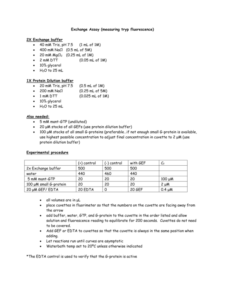
Exchange Assay (measuring tryp fluorescence) 2X Exchange buffer 40 mM Tris, pH 7.5 (1 mL of 1M) 400 mM NaCl (0.5 mL of 5M) 20 mM MgCl2 (0.25 mL of 1M) 2 mM DTT (0.05 mL of 1M) 10% glycerol H2O to 25 mL 1X Protein Dilution buffer 20 mM Tris, pH 7.5 200 mM NaCl 1 mM DTT 10% glycerol H2O to 25 mL (0.5 mL of 1M) (0.25 mL of 5M) (0.025 mL of 1M) Also needed: 5 mM mant-GTP (undiluted) 20 μM stocks of all GEFs (use protein dilution buffer) 100 μM stocks of all small G-proteins (preferable…if not enough small G-protein is available, use highest possible concentration to adjust final concentration in cuvette to 2 μM (use protein dilution buffer) Experimental procedure 2x Exchange buffer water 5 mM mant-GTP 100 μM small G-protein 20 μM GEF/ EDTA (+) control 500 440 20 20 20 EDTA (-) control 500 460 20 20 0 with GEF 500 440 20 20 20 GEF Cf 100 μM 2 μM 0.4 μM all volumes are in μL place cuvettes in fluorimeter so that the numbers on the cuvette are facing away from the arrow add buffer, water, GTP, and G-protein to the cuvette in the order listed and allow solution and fluorescence reading to equilibrate for 200 seconds. Cuvettes do not need to be covered. Add GEF or EDTA to cuvettes so that the cuvette is always in the same position when adding. Let reactions run until curves are asymptotic Waterbath temp set to 20ºC unless otherwise indicated *The EDTA control is used to verify that the G-protein is active *If the volume of the G-protein must be adjusted to adjust the concentration, compensate adjusting the volume of water so that final volume equals 1 mL. *295 nm ex, 335 nm em (bigger excitation slit = smaller signal, bigger emission slit = bigger signal)
