Blood
advertisement
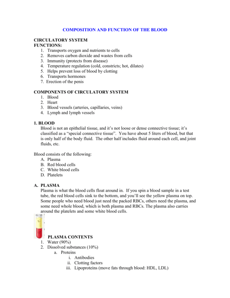
COMPOSITION AND FUNCTION OF THE BLOOD CIRCULATORY SYSTEM FUNCTIONS: 1. Transports oxygen and nutrients to cells 2. Removes carbon dioxide and wastes from cells 3. Immunity (protects from disease) 4. Temperature regulation (cold, constricts; hot, dilates) 5. Helps prevent loss of blood by clotting 6. Transports hormones 7. Erection of the penis COMPONENTS OF CIRCULATORY SYSTEM 1. Blood 2. Heart 3. Blood vessels (arteries, capillaries, veins) 4. Lymph and lymph vessels 1. BLOOD Blood is not an epithelial tissue, and it’s not loose or dense connective tissue; it’s classified as a “special connective tissue”. You have about 5 liters of blood, but that is only half of the body fluid. The other half includes fluid around each cell, and joint fluids, etc. Blood consists of the following: A. Plasma B. Red blood cells C. White blood cells D. Platelets A. PLASMA Plasma is what the blood cells float around in. If you spin a blood sample in a test tube, the red blood cells sink to the bottom, and you’ll see the yellow plasma on top. Some people who need blood just need the packed RBCs, others need the plasma, and some need whole blood, which is both plasma and RBCs. The plasma also carries around the platelets and some white blood cells. PLASMA CONTENTS 1. Water (90%) 2. Dissolved substances (10%) a. Proteins i. Antibodies ii. Clotting factors iii. Lipoproteins (move fats through blood: HDL, LDL) b. Nutrients i. Glucose (main energy source) ii. Amino Acids (builds proteins) c. Wastes (urea) d. Gases (O2, CO2, Nitrogen) e. Electrolytes = ions (Na+, K+, Cl-, Ca++) A. RED BLOOD CELLS (ERYTHROCYTES) These are small red biconcave discs. They are among the smallest cells in the body. There are about 5 million of them in each of us. Their structure is simple; like a doughnut with the hole not fully cut out. a. They have no nucleus b. Filled with a red pigment called hemoglobin, which carries O2 throughout the body. Oxygenated Hb is bright red, deoxy Hb is dull red. Blood in the veins only looks blue because you are seeing the dull red color through a yellow fat layer in the skin and subdermal tissue. c. Average life span is 120 days. They are made in the red bone marrow, and the old ones are destroyed in the spleen and liver, and Hb is recycled. During your lifetime, about 250 billion of these cells are destroyed, and 250 billion are made. B. WHITE BLOOD CELLS (LEUKOCYTES) There are different kinds; all fight infection. They seep out of the blood vessels whenever they sense bacteria nearby. C. PLATELETS When a platelet encounters a broken blood vessel it releases a substance that clots blood. Platelets are responsible for clot formation. HEMOPHILIA is a hereditary disease of males, where they are unable to clot properly. When they get even a slight bump or bruise they have to have an intravenous infusion of clotting factors or they will bleed to death. This is probably the disease that was in the genes of Henry VIII, which caused all of his male children to become weak and die in infancy. STEM CELLS: A cell that has not matured and differentiated yet. An embryo has lots of stem cells which have not decided to become a nerve cell, muscle cell, liver cell, etc. Stem cells become the type of cell the body needs. The placenta of a newborn infant has many of these stem cells, too, but not as many as an embryo. That’s why people want to research stem cells on embryos; there are more stem cells there. The first step for a stem cell is to DIFFERENTIATE, which is to decide what system of cells it will belong to. A stem cell that matures in the bone marrow will become a blood cell. Adults don’t have too many stem cells that are so immature that they have not yet decided what system of cells to belong to. Most of our stem cells have matured to the next step, which is that they have decided what system to evolve into. An adult has stem cells that will ONLY become blood, nerve tissue, organs, etc. BONE MARROW Most blood cells mature in the red bone marrow. When they are mature, they are released into the bloodstream. When they are old, they are destroyed in the spleen. ANEMIA: If the body makes too few erythrocytes. a. Causes of anemia include lack of iron, lack of hemoglobin, hemorrhage, lack of vitamin B12 (needed for cell division). b. Characteristic sign of anemia: pale skin and fatigue. LEUKEMIA: Cancer of the blood is called leukemia. It actually only involves the white blood cells. Something goes wrong in one stem cell, and it starts making huge amounts of clones of itself which don’t work right and not enough normal white blood cells are made. Therefore, the body cannot fight infection. There are many types of leukemias. BLOOD TYPING: The ABO SYSTEM Blood typing is the technique for determining which specific protein type is present on RBCs. Only certain types of blood transfusions are safe because the outer membranes of the red blood cells carry certain types of proteins that another person’s body will think is a foreign body and reject it. These proteins are called antigens (something that causes an allergic reaction). There are two types of blood antigens: Type A and Type B. A person with Type A antigens on their blood cells have Type A blood. A person with Type B antigens have Type B blood. A person with both types has type AB blood. A person with neither antigen has type O blood. If a person with type A blood gets a transfusion of type B antigens (from Type B or Type AB, the donated blood will clump in masses (coagulation), and the person will die. The same is true for a type B person getting type A or AB blood. Type O blood is called the universal donor, because there are no antigens, so that blood can be donated to anyone. Type AB blood is considered the universal acceptor, because they can use any other type of blood. This blood type is fairly rare. RH FACTOR There is another term that follows the blood type. The term is “positive” or “negative”. This refers to the presence of another type of protein, called the Rh factor. A person with type B blood and has the Rh factor is called A-positive. A person with type B blood and no Rh factor is called B-negative. The reason this is so important is that if an Rh- mother has an Rh+ fetus in her womb (from an Rh+ father), her antibodies will attack the red blood cells of the fetus because her body detects the Rh protein on the baby’s red blood cells and thinks they are foreign objects. This is called Hemolytic Disease of the Newborn (HDN). This can be prevented if the doctor knows the mother is Rh- and the father is Rh+, because that means the baby has a 50% chance of being Rh+ like the father. Therefore, anytime a mother is Rh-, they will ask if the father is Rh-. If so, they will give her an injection of a medicine that will prevent her immune system from attacking the baby.
